Ultrasound not only passes through tissue, but, reflected from blood cells, sends an image of the vessel to the screen, which allows one to evaluate the patency and degree of narrowing of the vessel.
There are several types of Doppler:
- Ultrasound Dopplerography (Doppler ultrasound) is a study of the vessels of the neck, head, brain or other organs, which allows you to determine the patency of the vessel, i.e. his anatomy.
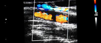
- USDS - (Duplex Ultrasound Scanning) combines two functions: In this case, the vessel is already visible on the monitor, and an image of the tissue around it is obtained, as with a conventional ultrasound. It turns out that this method, unlike Doppler ultrasound, helps in diagnosing the cause of poor vessel patency. It helps to visualize plaques, blood clots, tortuosity of blood vessels, and thickening of their walls.
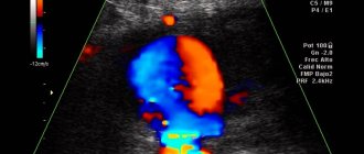
- With Triplex scanning, the vessel is visible on the monitor against the background of the image of the tissues in the thickness of which it passes. In this case, the vessel is painted in different colors depending on the speed of blood flow in it.
Ultrasound scanning of head and neck vessels is recommended for the following diseases:
- congenital anomalies of the location, course or branching of blood vessels
- atherosclerosis
- injury to an artery or vein
- inflammatory change in the walls of arteries and capillaries (vasculitis)
- diabetic, hypertensive, toxic angiopathy
- encephalopathy
- vegetative-vascular dystonia.
Ultrasound of the vessels of the head and neck helps to understand:
- causes of repeated transient ischemic attacks, strokes
- the degree of damage to these particular arteries due to metabolic or antiphospholipid syndromes
- the degree of impairment of the patency of blood vessels in the arterial bed due to atherosclerosis, diabetes mellitus, and smoking.
Knowledge of the condition of extra- and intracranial arteries and veins, obtained using duplex scanning, helps in prescribing the correct treatment, objective monitoring of its effectiveness, and drawing up an individual prognosis.
Similarities and differences between methods
Ultrasound using Dopplerography and duplex examination also have common features:
- do not cause any discomfort in patients during the procedure;
- safe for health (used during pregnancy);
- do not use invasive methods;
- Ultrasound reflection is used to image vessels.
The similarities end at these four points, because ultrasound and ultrasonic ultrasound scans have a number of differences.
Carrying out ultrasound examination
Ultrasound Dopplerography (Dopplerography):
- for diagnosis, the doctor installs the device’s sensor almost blindly, analyzing the location of the vessel, based on the general rules of the anatomical structure of the body;
- allows you to visualize only the valves;
- vein screening can only suggest the possibility of pathology in the form of varicose veins;
- determines only the patency of blood vessels;
- can scan the perforating veins that connect the venous collectors of the deep and superficial systems.
Ultrasound with duplex scanning:
- makes it possible to install the device’s sensor in the required location, since all adjacent tissues are displayed on the monitor;
- establishes the exact cause of vascular obstruction;
- visualizes venous valves;
- examines perforating veins anywhere;
- allows you to determine the cause of recurrence of varicose veins;
- can determine the stage of thrombosis.
All these parameters should be taken into account, since the information content and accuracy of the methods differ significantly.
Who needs to examine cerebral vessels
Duplex scanning (or at least ultrasound scanning) of intracranial arteries and veins (that is, those located in the cranial cavity) is indicated in cases of such complaints:
- headache, noise in the ears or head
- heaviness in the head
- dizziness
- visual impairment
- attacks of impaired consciousness such as fainting or inadequacy
- unsteadiness of gait
- lack of coordination
- impairment of speech production or understanding
- limb weakness
- numbness of hands.
The examination is also performed when pathology is detected during ultrasound examination of the vessels of the neck, when pathology of the neck organs is detected using CT, scintigraphy, MRI (for example, enlargement of the thyroid gland). In this case, in order to prescribe adequate therapy, a neurologist needs to know how all these diseases affect the brain, and whether its nutrition may suffer from this.
Vein scanning: duplex and triplex - what is the difference
This is how the doctor sometimes wraps up such a word, and in a tongue twister, and the patient stands in thought, what kind of study is this?.. duplex and triplex scanning refer to just such terms. The main thing is that it is not clear what to expect - either they will pierce the leg, or this is a type of x-ray.
Now we will decipher it for you, translate it from “medical to human.” In general, in medicine, scanning (from the English scan - to consider) is the controlled movement of a beam of radiation across the body, in our case - ultrasonic.
When pulses are reflected, they are recorded on the monitor screen. This is the essence of an ultrasound scan; its purpose is to obtain a picture of the internal organs or veins in order to determine the extent of the disease.
So we found out that this is all a regular ultrasound examination, without damaging the skin. The skin is smeared with a special gel, the doctor slides a sensor over the body, and a picture of the insides is obtained on the screen in real time. Not only the lumen of the vein and the condition of its walls are visible, but also the speed of blood flow and other characteristics of the blood.
But it was not always so. Previously, vessels were examined using X-rays - they injected a radiopaque substance into the vessel and watched how it filled the vein. The most noticeable violations, of course, could be seen, but, firstly, the method was invasive (that is, puncture of the skin and injection of a substance), and secondly, it did not show everything.
As for the names, when duplex scanning
The device operates in 2 modes at once (normal and Doppler).
The usual (traditional) ultrasound scanning mode allows you to see the vein in a two-dimensional image, its structure, and developmental anomalies.
The Doppler mode allows you to determine the “fluidity” of blood, the direction of its movement and the speed of blood flow.
The Doppler mode allows you to determine the “fluidity” of blood, the direction of its movement and the speed of blood flow.
Duplex scanning is an improved diagnostic method that combines the already familiar ultrasound and Doppler ultrasound.
It has an advantage over the last two, which is expressed in the ability to diagnose vascular pathologies at the earliest stage of development. This is achieved due to the fact that with the help of duplex it is possible to evaluate not only external characteristics, but also to examine the internal structure of blood vessels and identify their internal pathology.
Duplex ultrasound examination is a three-dimensional echography method that has a high degree of information content, which is important in determining the causes of diseases and making a diagnosis.
With all its research capabilities, this procedure is characterized by absolute safety and painlessness and the absence of contraindications and side effects. To carry out the diagnostic procedure, the patient does not need special preparation. However, it requires a highly qualified doctor to obtain reliable results.
This research provides an opportunity to:
- reliable determination of any pathology of the vascular bed;
- determination of blood flow speed;
- identifying the causes of changes and disturbances in blood flow.
Triplex scanning
is not any other research method. The same ultrasound machine already works in 3 modes - color Doppler is added to the first two.
Triplex scanning of blood vessels allows you to examine:
- the anatomy of the vessel, the degree of its tortuosity, which is very important to know during operations;
- structure of the vascular wall;
- speed and direction of blood flow;
- vein patency in color mode (presence of narrowings or blood clots).
Color scanning today provides doctors with the most accurate information about vascular diseases (which is very important not only for conservative treatment, but also before surgery).
Color scanning is performed in the following cases:
- Thrombophlebitis, its early diagnosis is especially valuable.
- Varicose veins, especially in the moderate and severe stages.
- Thrombosis.
- The method helps to accurately determine whether surgery is necessary for these diseases and where exactly the greatest destruction of blood vessels is.
- If deep veins are affected, triplex scanning is a must!
- You will be surprised, but when preparing operations in the abdominal cavity, doctors often scan blood vessels. This helps reduce the risk of postoperative complications (thrombosis).
Indications for examination of the vascular bed of the head and neck
Ultrasound duplex scanning of those arteries and veins that supply blood to the brain, but are located in the neck (that is, extracranial - outside the cranial cavity) should be carried out in the following cases:
- headache
- dizziness
- unsteadiness of gait
- impaired memory, attention
- coordination problems
- when planning operations on blood vessels and heart muscles
- when identifying pathology of the neck organs, due to which the vessels passing there may be compressed
- visible contraction of the blood vessels of the heart.
Where can I do duplex scanning?
A multidisciplinary medical center in St. Petersburg offers ultrasound color duplex scanning of the great vessels of the upper and lower extremities. Our specialists have at their disposal modern, expert-class ultrasound equipment that provides high-resolution images, simplifies research and allows for a quick and accurate diagnosis.
To make an appointment for a diagnostic appointment, find out the current price of duplex scanning of the veins of the upper and lower extremities, as well as find out other questions of interest, call the phone number provided on the page.
| Name of service (price list incomplete) | Price, rub.) | In installments* |
| Ultrasound scanning of the veins of the lower extremities | 2 400 | — |
| Ultrasound scanning of the veins of the upper extremities | 2 200 | — |
| Ultrasound scanning of the arteries of the lower extremities | 2 600 | — |
| Ultrasound scanning of the arteries of the upper extremities | 2 600 | — |
* You can read more about the conditions here - Treatment on credit or in installments.
When is routine Doppler sonography necessary?
Doppler of both extra- and intracranial arteries and veins should be performed at least once a year as a routine study (even before the appearance of any complaints):
- all women over 45 years old
- all men over 40 years old
- those who have close relatives suffering from hypertension, diabetes, coronary artery disease
- for diabetes
- smoking
- antiphospholipid syndrome
- for osteochondrosis of the cervical spine
- metabolic syndrome
- arterial hypertension
- if you have had a stroke or transient cerebrovascular accident
- if a person suffers from rhythm disturbances (the likelihood of cerebral thromboembolism with subsequent stroke is increased)
- increased levels of cholesterol, triglycerides, low-density lipoproteins in the blood (signs of atherosclerosis)
- had surgery on the spinal cord or brain
- before planned heart surgery.
Ultrasound Dopplerography (USDG)
In Doppler ultrasound, ultrasound responds to moving red blood cells in the blood, sending signals to a monitor and giving an idea of the speed and pattern of blood flow. Doppler ultrasound mainly gives an idea of the patency of the veins and the ability to identify their damage and the causes that provoked vascular changes. As a result of the ultrasound examination procedure, a one-dimensional image is obtained.
Advantages of ultrasound scanning
The main advantage of Doppler ultrasound is the availability of the method, since scanning in Doppler mode is much cheaper. Doppler ultrasound also allows us to identify stenoses and their degree, atherosclerotic plaques and blood clots.
Ultrasound scanning of the vessels of the lower extremities
A doctor may recommend ultrasound scanning of the vessels of the lower extremities for the following diseases and conditions:
- Atherosclerosis, endarteritis and diabetic angiopathy of the vessels of the lower extremities
- Atherosclerosis of the visceral branches of the abdominal aorta (vessels supplying the gastrointestinal tract, liver, spleen and kidneys)
- Aneurysm of the abdominal aorta and other vessels
- Varicose veins of the lower extremities
- Vasculitis (inflammatory vascular disease)
- Vascular diseases of the brain and neck
- Control of performed surgical intervention on blood vessels
- Postthrombophlebitic disease
- External vessel compression syndrome
- Screening examination (a study to identify asymptomatic forms of the disease)
- Thrombophlebitis and phlebothrombosis of the veins of the extremities
- Thrombosis of intestinal vessels
- Vascular trauma and its consequences
How is the examination carried out?
When examining the head and neck, ultrasound examination is performed in a sitting position.
All other types of examination are performed lying down. In some cases, when examining the veins of the lower extremities, the patient will be asked to stand or place the leg in a certain position. A gel is applied to the area under study to facilitate better penetration of ultrasound into the vessels. Next, a sensor is passed over this place several times in different directions, which transmits data to the monitor.
Based on the data obtained, a conclusion is formed. Based on it, a neurologist, phlebologist, cardiologist or other specialized specialist makes a diagnosis and prescribes treatment
Preparation
The simplicity and safety of Doppler ultrasound eliminates the need for lengthy preparation of the patient for the procedure. However, it is recommended to listen to the following recommendations:
- pain in the head, neck;
- stop drinking coffee, tea, energy drinks and nicotine, which affect the tone of blood vessels (on the day of the procedure);
- free the examination areas from foreign objects in the form of chains, hairpins, jewelry, tight and narrow collars, large earrings (immediately before the examination).
Carrying out the procedure
A referral from the attending physician is intended for Doppler ultrasound in the neurology departments of clinics at the patient’s place of residence. The examination standards include the following manipulations:
- position lying on your back (on the couch);
- a cushion or small pillow is placed under the head;
- the head turns in the direction opposite to the one being examined;
- a water-soluble gel is applied to the area under study, increasing the conductivity of ultrasonic waves and facilitating the sliding of the transducer over the area under study;
- the study begins from the neck, moving to the head
- the examination lasts from 20 to 30 minutes
- the gel is washed off with napkins
Analysis of examination results
The venous bed is analyzed according to the following parameters:
- permeability;
- anatomical structure;
- pathological formations inside blood vessels;
- blood flow speed.
The condition of arterial vessels is assessed by processing digital information, which compares the data obtained with normal parameters.