Rheovasography is a method for studying arterial and venous blood flow in the upper and lower extremities using a rheograph apparatus.
The procedure is painless and comfortable and takes 10–15 minutes. Two pairs of electrodes are placed on the limbs - above and below along the blood vessels. The electrical resistance between these areas is measured. The data is processed by a computer program and decrypted by a doctor.
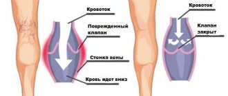
Electrical resistance depends on the degree of blood filling of the vessels, their expansion or narrowing. The stronger the blood flow, the lower this indicator. An increase in resistance indicates difficulty in blood flow. Based on the nature of the diagram, the angiologist judges the characteristics of the arterial supply and venous outflow in the extremities.
Indications for the procedure
Rheovasography of the lower extremities can be prescribed for the following symptoms:
- Numbness of the legs.
- Cramps.
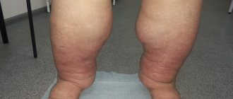
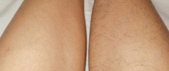
- Change in skin color of the lower extremities.
- Loss of sensation.
- Edema.
This diagnostic procedure is mandatory for diseases such as:
- Atherosclerosis.
- Thrombophlebitis.
- Rheumatic diseases.
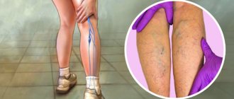
- Diabetes.
- Phlebeurysm.
- Blockage of the bloodstream.
- Obliterating endarteritis, etc.
Limitations of the rheovasography method of the lower extremities
Unlike duplex scanning, which does not require any preparation or special conditions, rheovasography of the lower extremities requires compliance with a number of conditions and careful attention to external factors.
The rheovasography schedule of the lower extremities may be inaccurate in the presence of heart failure, vein occlusion (vein obstruction), or local thrombosis. Also, these rheograms are distorted when exposed to external factors: the presence of pressing force (tight bandage or plaster on the leg, tight, tight clothing), low temperature in the office, patient mobility during the examination.
For rheovasography of the legs to be informative, a number of conditions must be met.
- A day or more before, stop taking any vascular medications that can affect blood circulation;
- Two hours before the test, smoking patients should quit nicotine;
- Immediately before rheovasography of the lower extremities, the patient should rest for approximately 15 minutes;
- During vein diagnostics, the patient should remain as still as possible, unless the doctor asks otherwise.
Key Indicators: What They Measure
The RVG results show a curve. Wave interpretation is based on qualitative and quantitative values. These include: the steepness of descents and ascents, intervals, amplitude, peaks.
Based on the values, control indices are derived, which are:
- rheographic - it controls the strength of blood flow in the arteries;
- arterial walls shows IE;
- IPS – peripheral resistance index;
- numerical value of venous outflow.
The most basic indicator is considered to be RI - rheographic index.
Decoding the results
Obtaining research results does not take much time.
Within 30 minutes, a specialist can decipher the received line and display the indices. RVG decoding is output in Ohms. As mentioned above, RI shows the total volume of filling of blood vessels. With readings of 0.05 Ohm and above, we can say that their condition is normal.
With deviations of one unit, mild deficiency is observed. If RI is sharply reduced, the patient is diagnosed with a blood supply disorder.
Standards for vascular tone data without deviations are calculated from 0.4. Values that do not reach the specified units are already considered violations. The lower the indicator, the lower the tone of the vascular wall.
The outflow index shows the speed of blood movement in the venous canal. RVG should range from 0.2 to 0.5. Anything that exceeds this line indicates serious illness.
The IPI responsible for counteracting vascular channels should be in the range of 0.2-0.45. In case of obvious blood flow pathologies, the index may be higher or lower than the norm.
Additional samples
Violations that were recorded during RVG do not always indicate damage.
The resulting index may have a functional nature of deviations. Often such results are obtained when preliminary preparation is not followed. There are two additional samples. They help determine what is really happening, a temporary spasm or pathology:
- Nitroglycerin. Using the drug, the rheovasography procedure must be completed several times. The first occurs before the drug is used, and the second after its action. The doctor receives data from two studies and conducts an analysis. If comparable results do not differ significantly, then we can conclude that there is a pronounced pathology on the face. In the case where the drug had an effect and the indicators improved, one-time disturbances caused are assumed.
- Compression. The essence of the procedure is to use a cuff that compresses the required area of study. As in the case of the RVG drug, the patient undergoes it twice. Before and after applying the cuff to the limbs. This test helps determine how quickly blood supply is restored when the deep veins are blocked.
The latest rheovasography devices have a large number of positive aspects. They greatly simplify the work of specialists. The system itself calculates the results, which is much faster than routine calculations.
At the same time, the patient does not receive any radiation or other negative effects on his body. The method is more than safe and informative. Due to this, this diagnosis is relevant in medical practice.
How to prepare
The procedure itself is quick and does not require special preparation.
The patient should sit on the couch, on his back. The doctor attaches sensors in the form of suction cups to the area being examined. Then observes the indicators for 15 minutes.
Before performing RVG it is recommended:
- Before starting the examination, you need to relax your muscles and lie down for 20-30 minutes;
- stop taking medications that affect blood flow and blood vessels at least one day before the test;
- do not drink alcoholic beverages several days before the procedure;
- those who smoke should abstain for a couple of hours before rheovasography;
- on the day of the examination, do not overwork and try to avoid emotional stress.
This preparation guarantees the most accurate result of the readings obtained during rheovasography.
If you ignore the rules of preliminary organization, then a single ailment can be confused with a serious pathology. The specialist must take this into account.
In case of violation of the rules, an additional test must be carried out. This will allow you to compare the results of the indices and make the correct diagnosis.
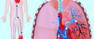
Central rheography
Varieties of rheography cover a huge number of proposals. But the central variation is consistently in high demand, as it is aimed at accurately assessing the condition of the heart.
The manipulation is based on the study of blood flow coming from the pulmonary artery and aorta. It is these two large vessels that allow you to familiarize yourself in detail with the health of the cardiac region.
Taking into account the blood flow of the lung and right ventricle, it is possible to evaluate the contractile function of the heart muscle.
Normal values in clinical practice are as follows:
- flat ascending part;
- round top with a small depression;
- round top with wave;
- smooth descent.
Carrying out central rheography involves dividing rheograms into certain types, depending on the data obtained:
- hypervolemic;
- hypovolemic;
- hypertensive.
The first version indicates an increased volume of incoming blood. The main indicators indicating such a deviation cover a high and at the same time peaked curve with a steep downward part.
Hypovolemic conditions are no less dangerous, but not all patients know what it is.
The scenario involves a reduced volume of blood flow, which is visible on visualization due to:
- decreasing the height of the curve;
- serifs on the ascending part;
- flat top;
- gently descending parts.
No less dangerous are the biophysical manifestations of the hypertensive subtype. This will indicate increased pressure recorded in the lungs. In the image, deviations can be recognized due to the steep rise of the curve, as well as the gentle descent and round top.
How it goes
The room where the procedure itself takes place must be quite warm, since the patient is forced to take off his clothes, freeing his limbs for examination.
In order for the fixed electrodes to show accurate data, the person must lie in a motionless position. For convenience, a soft couch is available.
Before fixing the sensors, the surface fatty layer is removed from the skin. They are installed clearly according to the principle of longitudinal or transverse placement. This depends on the limbs being examined.
If the mechanical activity of the vessels of the arms is examined, then the sensors are placed in four places throughout the limb. On the legs these are the feet, legs and thighs.
What the procedure will show
Having figured out what the essence of the diagnostic method is, people begin to be interested in how they can independently decipher the result they receive. But without the proper knowledge, this is quite problematic, because for most people, a rheogram is some kind of mathematical graph, and the impedance of biological tissues does not mean anything at all.
The basis of the picture is based on two aspects:
- sinusoids, which are presented as a steep rise, which describes arterial blood flow;
- a smooth descent, which is a reflection of the venous blood flow.
But not only this makes it possible to assess the condition of the vascular network in terms of volumes and other features of the incoming blood. It will be necessary to take into account many curves and use additional calculations, where Kedrov’s formula plays an important role.
The diagnostician will definitely take into account the regularity of the verified curve, which implies similarity between several curves. The shape and the number of secondary curves that are in a descending phase are also taken into account.
For example, it is worth analyzing the indicators characteristic of vegetative-vascular dystonia or arrhythmia. The presented ailments will be indicated by adjacent curves with different shapes.
Next, a special index is calculated, which must fit into the average statistical interval. Falling out of it threatens with suspicion of serious pathologies.
The following values specifically help identify diseases:
- level of venous outflow;
- amplitude-frequency indicator;
- measurements of the time interval for pulse wave propagation.
An experienced doctor will take into account various functional tests in order to get a detailed picture of what is happening.
Preparatory measures
Performing rheography involves preparation similar to the procedure for recording an electrocardiogram.
Beforehand, the patient is not allowed to smoke or consume large quantities of caffeine-containing products.
If you are taking any medications, it is recommended to stop therapy one day before.
This point is especially important in the case of medications that change the blood circulation process.
Immediately before the examination, the person takes a sitting or lying position, relaxes as much as possible, nervousness is unacceptable.
Our services
Our neurological clinic diagnoses a wide range of diseases and also accepts adult patients:
- with epileptic and non-epileptic paroxysmal conditions;
- with osteochondrosis;
- with pain syndromes of various origins;
- with vegetative dystonia;
- with consequences of cerebral strokes;
- with consequences of traumatic brain injuries;
- with panic and anxiety states.
We successfully examine young patients in the following cases:
- perinatal damage to the central nervous system;
- epileptic and nonepileptic paroxysms;
- manifestations of the consequences of neuroinfections;
- cerebral palsy;
- speech development disorders;
- hyperactivity, inattention, decreased memory, mental functions, emotional disturbances;
- delayed development of communication functions, autism, behavioral disorders;
- hereditary and chromosomal pathology.
Suspicion of diseases
Some symptoms may indicate obvious diseases that can be identified through examination:
- atherosclerotic lesion due to stenosis or complete occlusion of the supply artery;
- autoimmune pathology with abnormal hemodynamics in the hands;
- diabetes mellitus with complications;
- inflammation of the walls of arteries from the inside;
- varicose veins
In addition, using RVG it is possible to determine the etiology of the disease. Understand what influenced the subsequent development of the pathology, the structure of the canals or reasons caused by lifestyle.
About the impact of the procedure
The technique does not harm health, there is no damage, there is no outside interference in the operation of internal systems.
There is no damage to the tissues or skin through which the current is passed, because it is characterized by insignificant regularity and magnitude.
Despite this, the study is highly sensitive.
Rheography allows you to assess cardiac function, evaluate the blood flow system and identify deviations in its functioning. It helps to diagnose the pathological process in the vascular system as accurately as possible.
A little information about the rheograph
The device is based on a generator that generates current. Using a special attachment, measurements are converted into graphic form.
Using appropriate electrodes attached to certain areas of the body, a rheogram is recorded.
What is rheography? At the beginning of the procedure, an electrophoresis-treated napkin is placed on the patient’s skin, through which the electrode reacts better with the body.
The skin is first wiped with medical alcohol; the presence of a fatty layer worsens the result of the process.