Causes
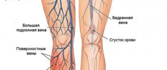
When it comes to vascular damage, we talk about primary (vascular) intermittent claudication. It has been established that in 80–90% of cases, the pathology occurs as a result of atherosclerotic damage to the peripheral arteries, that is, in fact, it is a symptom of atherosclerosis.
In other cases, the causes of the pathology are diabetic angiopathy and vascular damage of autoimmune origin.
Risk factors for developing PC:
- hereditary predisposition;
- inactive lifestyle;
- obesity;
- smoking;
- excessive alcohol consumption;
- hypercholesterolemia (high cholesterol in the blood);
- hypertonic disease;
- diabetes;
- diseases of the immune system;
- elderly age;
- injuries.
A separate category is neurogenic intermittent claudication (or caudogenic) - a syndrome of spinal canal stenosis (pinched nerve roots), mainly in the lumbar region. Usually caused by degenerative changes in the spine and is diagnosed mainly in people over 50 years of age.
History of the development of ideas about KHOZANK
If we turn to history, we will notice that gangrene of the limb was known to doctors earlier than the disease leading to it. Intermittent arterial claudication in humans was described only a century and a half ago by the French neurologist Jean-Martin Charcot. It was he who in 1858 presented a description of the syndrome of intermittent claudication (claudicatio intermittens) [7].
However, intermittent lameness (or claudication, from the Latin claudicatio - “the effect of lameness”, claudus - “lame” [7]) was known to veterinarians long before Charcot, because similar cases of the disease were found in horses. According to Charcot, intermittent claudication syndrome was first described by French authors - veterinary student G. Bouley and, somewhat later, Goubeaux as a disease of horses. Thus, in 1831, Bouley presented a case of lameness in a horse without an understandable cause, healthy at rest and beginning to limp only when trotting. Later lameness affected both hind legs. J. Bouley performed a necropsy on the deceased horse and found: “... the aorta contains a fibrinous and floating clot, 4 to 5 inches long. The thigh muscles were pale, discolored, much firmer than in their natural state. The femoral artery was very dilated, filled with approximately 18 cm of fibrin clot, which completely closed the artery...” Based on this, Bouley explains the symptoms of lameness: “... when the animal was resting, the numerous anastomoses that existed were sufficient to maintain life. But as soon as the blood circulation accelerated when walking, these anastomoses ceased to supply a sufficient amount of blood...” [7]. In 1838, the German veterinarian Rademacher described the same case as J. Bouly, apparently unaware of his work. In 1846, A. Goubault of the Veterinary School of Alfort published data on 10 other cases and confirmed Boulley's interpretation.
Charcot in 1835 described a case of obliteration of the aorta in a 51-year-old woman (Barth, General Medical Archives) long before Leriche. Until the end of his life, Charcot insisted on using the term “intermittent claudication” and complained that he “did not meet a single doctor who would take into account my observations.” Sabourin completed his dissertation under the guidance of Charcot, noting that it was the doctors’ ignorance of the syndrome and the lack of examination of the pulsation of the arteries in the lower extremities during the interview of the patient that were the main reasons for the lack of observations (consideration of intermittent claudication of arterial origin, 5 cases, Paris, 1873) [7].
Subsequently, despite the data presented by Bouley and the observations of Charcot, Sabourin, Delaunay (1890), when discussing the pathogenesis of intermittent claudication, it was suggested that neurological lesions and vasospasm (Dutil, Lamy, 1893; Brissaud, 1899) play a more significant role in occurrence of symptoms. The theory of the occurrence of intermittent claudication and vasospasm was developed in the works of Leriche (1917) [7].
In 1952, the 1st Congress of the European Society of Cardiovascular Surgery was held in Strasbourg, where the problems of surgical treatment of lesions of the iliac artery of the lower extremities were discussed. It was determined that intermittent claudication is the second stage of the development of limb ischemia (there are only two stages - compensated and decompensated) [7].
Symptoms of intermittent claudication
The first sign of a disorder is pain of a different nature - dull, sharp, burning, throbbing, aching. Pain with intermittent claudication occurs during physical activity - during prolonged walking, climbing stairs, etc. Its localization depends on the location of the artery lesion; it can be the toes, feet, legs, thighs, buttocks. Pain and weakness in the legs force the patient to stop and take breaks to rest. Unpleasant symptoms subside and the person can continue to move.
As the pathological process progresses, the nutrition of the leg tissues deteriorates, which causes trophic changes:
- dryness and flaking of the skin of the feet;
- pale or bluish skin;
- change in the appearance and color of nails;
- hair loss;
- muscle spasms;
- decreased sensitivity and temperature of the feet;
- formation of ulcers;
- development of necrosis and gangrene.
The disease is characterized by a chronic progressive course.
Caudogenic intermittent claudication is characterized by back pain that occurs during walking and radiates to the legs along the surface of the thighs and legs. The pain is relieved when sitting or squatting more than when standing, and may intensify when the patient lies down. Due to the position of the spine, which mechanically narrows the neural canal, paresthesia and dysesthesia occur in the legs. When moving, neurological disorders worsen - muscle weakness, sensory disturbances. Men often complain of urinary problems and erectile dysfunction.
Lack of full rehabilitation
Incorrect behavior in the postoperative period often leads to a decrease in muscle strength and gradual muscle atrophy. Because of this, a person begins to limp even with the normal functioning of the installed prosthesis.
Main mistakes in rehabilitation:
- late activation and incomplete load on the operated limb;
- reduction of hospital stay after surgery;
- insufficient or excessive load on the joint in the late postoperative period;
- neglect of orthopedic devices (cane, crutches);
- refusal of rehabilitation in the first months after surgery.
In the Russian Federation, the lack of rehabilitation is a real problem. Almost all public hospitals and private clinics discharge their patients within a few days after surgery. As a rule, doctors give recommendations to their patients and let them go “free swimming”. Naturally, no one controls a person and checks whether he is doing everything correctly. This leads to impaired muscle functioning and persistence of lameness.
Minimally invasive endoprosthetics in the Czech Republic: doctors, rehabilitation, terms and prices.
Find out more
Treatment
If the diagnosis of “intermittent claudication” is confirmed, medications and other methods of therapy are selected by the doctor depending on the degree of the disease and the characteristics of the clinic. The main goal of treatment is to improve the patient’s quality of life and reduce the risk of complications.
In the initial stages, the patient is shown lifestyle changes: quitting smoking and drinking alcohol, a low-fat diet, eliminating tight shoes, tight clothing and other risk factors. To improve blood circulation, a set of physical therapy exercises is recommended. Physiotherapeutic procedures - radon and hydrogen sulfide baths, mud therapy, diathermy, UHF - have a good effect.
If lifestyle changes are not effective enough, it is advisable to use cilostazol, a drug with vasodilating, disaggregant and metabolic effects. Regular use of the medicine allows an average of 50% increase in pain-free walking distance.
If necessary, medications are prescribed that lower blood cholesterol levels, anticoagulants, and vasodilators.
Dietary supplements that improve peripheral blood circulation are effective, for example, Bilobil based on ginkgo biloba.
Patients with atherosclerosis are prescribed antiplatelet agents to reduce the risk of cardiovascular events, for arterial hypertension - antihypertensive drugs, etc.
In severe cases, surgery is required.
Diagnostics
The causes of lameness are determined by orthopedic traumatologists. In case of vascular diseases, patients are examined by a vascular surgeon, in case of neurological pathologies - by a neurologist. The examination plan includes the following diagnostic procedures:
- Survey
. The doctor clarifies under what circumstances the lameness appeared, what nature it has (constant, periodic, intermittent), what symptoms are accompanied. - Visual inspection
. The specialist evaluates the patient’s gait, measures the length of the legs, identifies deformities, examines joint mobility, and determines the condition of the skin and soft tissues. - Radiography
. The images show fractures, arthritic changes, inflammatory processes, cysts and tumors in the bones and joints. - Ultrasound
. Recommended for vascular pathologies and soft tissue diseases. Allows you to assess the patency of blood vessels, the cause and degree of blood flow disturbance, and the severity of pathological changes. - and MRI
. Prescribed to clarify the results of radiography and sonography of the extremities. They are used to determine the severity of damage in cases of spinal circulation disorders. - Electrophysiological techniques
. Electromyography, electroneurography, evoked potentials are used to assess the level of neuromuscular transmission and localize the level of nerve damage. - Invasive Research
. If joints are affected, puncture or arthroscopy can be performed. In the first case, the nature of the synovial fluid is assessed, in the second, the joint is examined, tissue samples are taken, if necessary, and therapeutic measures are carried out. - Lab tests
. Necessary for confirming inflammatory changes in the blood, identifying specific markers for autoimmune diseases, identifying pathogens in infectious pathologies, studying the morphological structure of biopsy samples.
Applying a plaster cast
FAQ
Why is intermittent claudication dangerous?
Progressive pathology can lead to gangrene of the limb. Intermittent claudication, which develops against the background of atherosclerosis, increases the risk of stroke and heart attack.
How to identify intermittent claudication?
The first symptom of the disease is pain in the legs, which occurs with prolonged walking and other physical activity. At rest it goes away.
Which doctor treats intermittent claudication?
It is worth contacting an angiologist or vascular surgeon.
Discussion
Thus, the results of the study demonstrated the existence of various clinical forms of NPH in patients with PDS. Noteworthy is the relatively high incidence of painless forms of NPH among the observed patients (7.2%), despite the fact that pain is a key manifestation of the disease, and there are practically no descriptions of the painless form of NPH in the literature. Silent NPH can cause difficulties in diagnosing PDS and NPH and require differential diagnostic measures. We did not find a connection between clinical manifestations and the nature of changes in MRI studies and the type of NPH, while the form of clinical manifestations of NPH did not affect the outcome of surgical treatment. We noted a symptom of pacing, manifested by a relatively short period of increase in the clinical manifestations of NPH during walking, followed by their complete regression, despite non-stop walking. A typical manifestation of NPH is an increase in radicular symptoms and pain when walking, while the phenomenon of pacing is noted much less frequently and is not discussed in the literature. The possibility of the existence of such an atypical symptom, which may be the reason for the incorrect identification of the nature of NPH, seems important. It should also be noted that the presence of this phenomenon in the observed patients did not affect the outcome of the disease.
The data presented indicate a variety of (often atypical) clinical manifestations of PDS, which requires both a thorough analysis of the clinical picture of the disease and an imaging examination to clarify the cause of NPH and select the optimal treatment tactics.
The authors declare no conflict of interest.
The authors declare no conflicts of interest.
What is coxarthrosis
Arthrosis of the hip joint or coxarthrosis is a chronic, slowly progressive degenerative disease, accompanied by the destruction of cartilage and bone tissue of the joint, the growth of bone osteophytes that deform the joint, as well as the involvement of periarticular tissues in the pathological process. All this gradually leads to complete loss of leg function and disability. Women suffer from this type of arthrosis more often.
For a long time, the disease was considered age-related, but it has now been proven that in many people this pathology of the legs begins to develop after 20 years, and sometimes in childhood, remaining asymptomatic for a long time, appearing after 40 - 50 years. In terms of incidence, coxarthrosis is second only to arthrosis of the knee joint. But in terms of the duration of periods of incapacity, it is much ahead of it, since it is more severe. Coxarthrosis of both joints and one joint develop equally often.
In some cases, the disease is detected in late stages, but this is not a death sentence: conservative and surgical treatment methods have been developed for any stage. The code for coxarthrosis according to ICD 10 (International Classification of Diseases, 10th revision) is M16.