Postcholecystectomy syndrome (PCES) is not the most common phenomenon in gastroenterology. It is generally accepted that PCES belongs to the group of gallbladder diseases. In fact, this is not even a disease, but a collective name for a set of symptoms that appear immediately or shortly after surgery on the bile ducts or removal (resection) of the gallbladder.
Until now, both therapists and surgeons find it difficult to clearly determine the causes of the development of this syndrome. Doctors tend to use the term PCES only to make a preliminary diagnosis in operated patients1.
Symptoms of postcholecystectomy syndrome
At its core, PCES is a consequence of surgery for resection (removal) of the gallbladder. This means that after resection the patient may experience unpleasant symptoms, such as:
- dyspepsia or disruption of the normal functioning of the stomach, manifested in the form of bitterness in the mouth, nausea, bloating and intestinal upset;
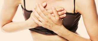
- pain in the right hypochondrium with transition to the right collarbone or shoulder. The intensity of pain can vary, from unexpressed aching to acute burning;
- general weakness, pale skin (appears against the background of poor absorption of food and developing vitamin deficiency).
With PCES, other symptoms are possible due to exacerbated diseases:
- exacerbation of cholangitis - inflammation of the bile ducts - is expressed in a long-lasting temperature in the range of 37.1-38.0 ° C;
- Cholestasis (stagnation of bile in the liver tissue) can cause severe jaundice2.
Tracheostomy in the practice of a resuscitator
Indications for performing tracheostomy and tracheotomy are:
1. Stenosing laryngotracheobronchitis grade III and IV after 1-2 days of unsuccessful treatment with nasotracheal intubation,
2. Foreign bodies of the trachea if it is impossible to remove them during direct laryngoscopy or upper tracheobronchoscopy.
3. When draining the respiratory tract in patients with coughing disorders. 4. In cases of obviously long-term (more than 5 days) artificial ventilation. 5. For cicatricial stenoses and tumors of the larynx when it is impossible to perform radical operations.
6. For injuries of the larynx and trachea with disturbances in external respiration. 7. In case of traumatic brain injury with the need for systematic drainage of the tracheobronchial tree or long-term artificial ventilation.
8. Extensive wounds with damage to the facial skeleton.
Advantages of tracheotomy over tracheal intubation:
- facilitating artificial ventilation;
- facilitating the sanitation and toilet of the tracheobronchial tree;
- preventing aspiration of gastric contents;
- reducing the so-called dead space by 30-40%.
According to current literature reviews, well-known benefits of tracheostomy include less need for deep sedation, faster weaning from the respirator, and decreased length of stay in the ICU and hospital. First of all, patients with severe concomitant trauma, especially with traumatic brain injury and/or impaired consciousness, will benefit from early tracheostomy. With the exception of a few reports, there were no differences in mortality rates between patients with tracheostomy or prolonged transglottic intubation. Often, patients with a tracheostomy can be transferred earlier from the ICU to a specialized department, so the issue of early tracheostomy should be considered as soon as suitable conditions arise. In patients with a tracheostomy, tracheostomy tube-related incidents may be more frequent and have more severe consequences, so patient safety must be a primary consideration when deciding whether to transfer a patient from the ICU.
Of course, tracheostomy is not without serious disadvantages associated with an increased risk of infectious complications, the need for constant careful care of the tracheostomy and the likelihood of complications during this operation (esophageal trauma, bleeding, acute tracheal stenosis, etc.). of tracheostomy care is fundamentally important , so in addition to the constant removal of tracheobronchial secretions, active prevention of purulent tracheobronchitis, skin maceration, secondary infection of soft tissues around the postoperative wound, etc. is necessary.
This is achieved by regularly carrying out the following set of measures: 1. Patients with tracheostomy must be kept in a room with high air humidity. For this purpose, a special humidifier is installed. Increased air humidity in the summer can be achieved by hanging wet sheets, and in winter by drying them on steam or water heating radiators;
2. In addition to regular rinsing of the respiratory tract with solutions of furatsilin, novocaine, and the use of proteolytic enzymes, it is necessary to periodically carry out inhalations with antibiotics and alkaline-oil mixtures;
3. The skin around the tracheostomy should be systematically thoroughly dried and lubricated with zinc ointment or Lassar paste;
4. The tracheostomy should be treated as an absolutely sterile wound - manipulate only with carefully cleaned hands, boiled instruments, drainage tubes and solutions. When changing dressings, use only sterile dressings and prohibit work without gauze masks and special gowns;
5. In the ward where a patient with a tracheostomy is located, it is advisable to periodically sanitize the air with ultraviolet irradiation;
6. With such patients, it is necessary to regularly engage in active breathing exercises and, if possible, provide them with a motor regime.
It should be remembered that the operation itself and the tracheostomy cannula are far from indifferent to the respiratory tract. Already 2-3 hours after the placement of a tracheostomy, a belt of loose fibrinous plaque appears on the mucous membrane around the tracheostomy opening. In some cases, this plaque spreads to the mucous membrane of the larynx, epiglottis and down to the bifurcation of the trachea and even the main bronchi. Directly around the cannula, a necrotic zone, tissue edema, hyperemia, and leukocyte infiltration develop very quickly (within a few hours).
At a later date, erosions or (much less often) deep ulcers are detected. On the 5th-7th day, metaplasia of the prismatic epithelium into multilayered squamous epithelium occurs. All this indicates that the content of the cannula in the lumen of the trachea is far from indifferent to the patient. Therefore, the question arises not only about strict indications for tracheostomy, but also about the most rational timing of cannula removal (decannulation).
The following rules exist: if within 1-2 days after the operation the victim’s breathing remains within normal limits, and there is no threat of relapse of respiratory failure, the cannula should be removed.
Serious changes in the tissues surrounding the tracheostomy also indicate that in cases where artificial ventilation is presumably necessary for a relatively short period (up to 24 hours), tracheostomy should and can be replaced by intubation.
If the expected duration of mechanical ventilation is more than 14 days, a tracheostomy should be considered. However, unfortunately, clinicians' ability to predict the duration of mechanical ventilation at an early stage of the disease is limited. However, it should be expected that more severe disease over a longer period of time is a predictor of longer stay on mechanical ventilation. One study (Rumbak) also noted that patients with a higher APACHE II score (>25) required longer intubation.
Tracheostomy techniques are constantly being developed and improved. An increasing number of tracheostomies are being performed in the ICU at the bedside using the percutaneous dilatational tracheostomy (PDT) technique.
In our clinic, CCT surgery is performed quite regularly. Experience in performing percutaneous dilatational tracheostomy allowed us to be convinced of the advantages of this technique (in particular, Griggs tracheostomy under endoscopic control), as the simplest, least economically expensive, with the least number of complications.
Reasons for the development of postcholecystectomy syndrome
The causes of postcholicystectomy syndrome and its development are often associated with disruption of the normal functioning of the sphincter of Oddi (orbicularis muscle). The sphincter of Oddi is a smooth muscle that is located in the lower part of the duodenum and is responsible for regulating the supply of bile and pancreatic juice to the duodenum3.
If we consider that PCES most often affects patients who have undergone surgery to remove the gallbladder4, then the mechanism of occurrence of this syndrome can be explained as follows:
- after the operation, the sphincter, which normally opens when the gallbladder is full, does not receive a signal about filling, as a result of which it is almost constantly under tension;
- due to the absence of a bladder, bile enters the duodenum in a diluted state, which increases the pressure inside the intestinal walls. In addition, bile itself has a bactericidal effect and a change in its composition can lead to intestinal infection5.
But sphincter of Oddi dysfunction may not always be caused by removal of the gallbladder. Sometimes the cause of the syndrome is advanced gastrointestinal diseases* (chronic colitis or gastritis, peptic ulcer) and chronic pancreatitis) - a disease of the pancreas, as well as errors in preoperative examination.
Take a deep breath: learning to breathe correctly with a Moscow psychologist
Human breathing is an everyday and imperceptible process that we usually don’t think about. But as soon as you are deprived of oxygen for a minute, all other problems fade into the background. Often during anxiety, people experience a feeling of difficulty breathing, the inability to breathe deeply, or a sudden or increasing feeling of lack of air. Konstantin Zherdev , a specialist at the Moscow Psychological Assistance Service, told us how to cope with this illness
There are several types of shortness of breath:
- True shortness of breath is associated with diseases (usually chronic) in organs and systems.
- Functional shortness of breath indicates physical stress; a person experiences it, for example, when running. It goes away after the end of the load or as the body adapts to such work. This ability to adapt is sometimes called "second wind."
- Psychogenic shortness of breath is associated with the experience of acute or chronic psycho-emotional stress: anxiety, depression, neurosis, stress and even intense joy. Episodes associated with joyful difficulty breathing are well known to us: “the breath stole from the goiter from joy.”
“Often, hidden anxiety manifests itself through difficulty breathing, nighttime and daytime episodes of suffocation, which go away on their own. True, at the moments of these experiences they are seriously frightening. Such experiences were clearly illustrated by G. Fusli in a series of paintings, which he called “Nightmare”. As a rule, shortness of breath during anxiety goes away when attention is switched from it to something extraneous. Warm drinks, conversations, and simply getting out of an alarming situation have a positive effect on breathing,” notes the psychologist.
Breathe out your anxiety
Frequent shallow breathing, which often accompanies anxiety, leads to an increase in oxygen levels in the blood and a decrease in carbon dioxide. This shift in balance is accompanied by various unpleasant manifestations: dizziness, a feeling of suffocation, cold extremities, a feeling of impending loss of consciousness, and increasing tinnitus. Often such complaints are the reason for seeking help from doctors.
Of course, in order to rule out various types of diseases, you need to seek advice from medical specialists and undergo an examination. But it often happens that no diseases are detected. In this case, you should pay attention to your psychological state.
“To overcome these moments, it will be useful to reduce excess muscle tone, as well as normalize respiratory cycles. That is, allow the body to independently manage important processes, in other words, do not interfere. Using the power of breathing to create a comfortable psychological environment is the right way. Thinking that the body won’t cope without us and will forget to breathe is wrong,” advises Konstantin.
Moderate sports and physical activity, reducing caffeine and alcohol consumption, and limiting negative information load will also be useful.
Workshop from a psychologist
During an acute state of anxiety, the whole consciousness “screams” that there is not enough air and you want to inhale as deeply and often as possible - to get air into your chest.
Try to do the following: relax the muscles of the shoulders, neck, abdomen, relax the chest muscles, facial muscles, base of the tongue. At the same time, exhale calmly and slowly. Help yourself with your stomach by pulling your abdominal muscles inward as you exhale. Make room inside yourself for a short but full breath. Relax your stomach and take a calm breath (1-2 seconds) and exhale long again (4-6 seconds). Sometimes it is recommended to take a short pause between each cycle: inhale - pause for 2-4 seconds, exhale - pause, etc. If it is difficult for you to hold a pause, then at the beginning you can not include it in the exercise. Repeat this cycle of inhalation and exhalation 6 times. Practice breathing like this in a calm state. Do not hurry.
There is no need to try to exhale all the way and there is no need to take a sharp breath.
This leads to irritation by the air flow of the upper respiratory tract and causes a feeling of additional tension in the throat, and can also provoke coughing and tickling. The entire exercise is performed in a comfortable mode. The task is to breathe calmly and without jerking. Breathing cycles can be accompanied mentally with the phrase: “My breathing is smooth and calm.” We say the first part of the phrase - “My breath” while inhaling, the second part - “even and calm” - we say as we exhale. You can sign up for free classes on self-regulation and training in breathing techniques at the Moscow Service for Psychological Assistance to the Population
www.msph.ru. Source
Press service of the Department of Labor and Social Protection of the Population of Moscow
Diagnosis of postcholicystectomy syndrome
Difficulties in accurately determining the causes that led to the development of PCES and the vagueness of the very definition of the syndrome require a thorough examination of the patient. In order to choose the right treatment, it is necessary to clearly establish what led to the appearance of PCES.
That is why effective diagnosis of postcholicystectomy syndrome includes several methods:
- collection of anamnesis data - the doctor carefully studies old medical reports and records, paying close attention to preoperative diagnostics and the protocol of the operation;
- clinical examination of the patient;
- laboratory tests - clinical and biochemical blood test, stool test for protozoa and worm eggs, general urine test;
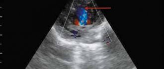
- ultrasonography;
- endoscopy of the bile ducts;
- Magnetic resonance imaging or computed tomography of the abdomen6.
Treatment of postcholicystectomy syndrome
Since PCES is not an independent disease, treatment of the syndrome is always determined by its causes. Not knowing how to properly treat postcholicystectomy syndrome can only aggravate the condition and increase unpleasant symptoms.
The principles of treatment for PCES include two key points:
- collection of anamnesis data - the doctor carefully studies old medical reports and records, paying close attention to preoperative diagnostics and the protocol of the operation;
- eliminating the causes of the syndrome;
- prevention and treatment of suspected complications.
Treatment is mainly based on:
- diet therapy;
- drug treatment;
- surgery (according to indications)7.
Together with comprehensive treatment, these measures can reduce the severity of symptoms of PCES8.
Treatment of the disease
Only doctors, based on diagnostics and a comprehensive examination of the body, can make the correct conclusion - chest neurosis. Treatment methods differ depending on the course of the disease. They mean relieving pain and spasms, strengthening the nervous system, and improving blood circulation. There are several ways to treat intercostal neurosis:
- Blockade. It does not eliminate the main cause of neurosis, but this method allows you to relieve pain for several days. A blockade is done when non-steroidal anti-inflammatory drugs do not help.
- Injections. They are used only in extreme cases. For example, for radiculopathy, spinal pathologies and myelopathy, steroid injections are prescribed.
- Physiotherapy. It also relieves pain symptoms due to chest neurosis. This category includes electrophoresis, UHF and other thermal procedures. Doctors prescribe such treatment methods for incarceration, arthropathy and spondyloarthrosis.
- Training. To prevent and treat the disease, doctors recommend physical exercise. Among them are hyperextension, which serves to strengthen the back muscles, and stretching to relieve tension.
- Massage. This method helps relax muscles and also helps relieve pain. Massage is prescribed for the initial stage of intercostal neurosis, osteochondrosis, displacement of the vertebrae and spinal discs, as a preventive measure or as a method of treatment in combination with medications.
- Taking medications. B vitamins and ascorbic acid nourish nerve fibers with useful elements and strengthen the immune and nervous systems.
At the first sensation of pain, it is recommended to go to the doctor. This will allow you to prescribe timely treatment and eliminate the disease at the initial stage. Contacting a specialist will help you completely get rid of the disease. Save your health with I.G. Gernet is a psychotherapist with 20 years of experience!
To the list of articles
Other articles
- Throat neurosis: symptoms, treatment
- How to relieve a panic attack at home?
The use of enzyme preparations in postcholicystectomy syndrome
In some cases, PCES may be accompanied by disorders of the digestive system. This is due to the fact that food intake becomes a signal for the production of bile and pancreatic enzymes. If the signal does not arrive or arrives intermittently, subsequent events are also disrupted. As a result, food is not processed properly, and the body does not receive enough nutrients. This can affect the general condition of the body and manifest itself as heaviness after eating, discomfort, bloating or diarrhea.
Enzyme preparations have been developed to support digestion; they deliver enzymes from the outside to compensate for their lack in the body. The flagship among such drugs is Creon®.
The drug is available in the form of capsules containing hundreds of small particles - minimicrospheres. They do not exceed 2 mm in size, which is recorded as recommended in world and Russian scientific works9,10. The small particle size allows Creon® to recreate the digestive process as it was intended in the body, and thereby cope with unpleasant symptoms. Dosages of the drug are usually selected by a doctor, however, in accordance with modern recommendations, the starting dosage is considered to be 25,000 units10.
To learn more
An important stage in the treatment of PCES is enzyme therapy, because In many cases of PCES, pancreatic function is impaired and enzyme deficiency occurs11. Because of this, digestive problems arise, manifested in the form of unpleasant symptoms such as abdominal pain, heaviness in the stomach, flatulence and diarrhea. In addition, the lack of enzymes affects the quality absorption of nutrients: the body does not receive the necessary vitamins and minerals.
Creon® is a modern enzyme preparation that replenishes the deficiency of its own enzymes and promotes better absorption of food and improved digestion. More information about the drug can be found here.