Systematization of manifestations of aortic aneurysm
Currently, there is no unified approach to systematizing the manifestations of peritoneal aortic aneurysm. Most often, doctors and authors of medical works use the method of A. Pokrovsky and R. Ermolyuk, according to which aneurysms are divided:
- by etiology
– acquired (non-inflammatory or inflammatory) and congenital; - according to morphology
- into false (traumatic origin), true and stratifying; - by shape
- diffusion and saccular; - according to the course of the clinical process
- into diseases with an uncomplicated course, complicated and dissecting; - by type and location
- on the proximal segment of the peritoneal aorta, on the infrarenal section, as well as with total damage to the entire section of the abdominal aorta.
According to medical statistics, up to 95% of aneurysms are localized in the infrarenal region.
Lifestyle and Home Remedies
The best way to prevent an aortic aneurysm is to keep your blood vessels as healthy as possible. To do this, you can take the following steps:
- quit smoking;
- keep your blood pressure under control;
- get checked regularly;
- reduce the amount of cholesterol and fat in your diet.
If you think you may have an abdominal aortic aneurysm, or are concerned about your risk of an aneurysm due to a family history, see your doctor. If an aneurysm is detected early, treatment may be easier and more effective.
Causes of development of abdominal aortic aneurysm
- atherosclerosis, in which the appearance of cholesterol plaques on the wall of the aorta gradually reduces its strength, which contributes to protrusion in one of the areas;
- congenital predisposition, transmitted through the male line, confirmed by many years of observations: the presence of an aneurysm in the father indicates a 50% chance of this disease occurring in his son;
- an inflammatory process of a chronic, sluggish nature that occurs in the aortic wall itself or in the adipose tissue that surrounds the vessel;
- traumatic damage to the wall due to trauma or injury to the abdomen, surgery or endovascular intervention.
Atherosclerotic manifestations become the cause in 85-90% of cases of the development of acquired aneurysm in the abdominal aorta. Signs of predisposition to the development of the disease are smoking, arterial hypertension and chronic pulmonary diseases.
What it is
We should start by defining the aorta. The aorta is the largest vessel in our body, and accordingly its importance is high. An aneurysm is an enlargement of a blood vessel. An aortic aneurysm can form in the thoracic or abdominal regions. Moreover, aneurysm of the abdominal aorta is much more common. This condition of the vessel may not cause any harm for a long time, however, an aneurysm is very dangerous due to the unpredictable course of expansion and the threat of rupture of the vessel walls when they become thinner - and this condition already poses a threat to life due to severe internal bleeding. In addition, blood clots can form at the site of vessel expansion due to changes in blood flow - the danger of this condition is the risk of the blood clot breaking off and clogging a smaller vessel.
Symptoms of aortic aneurysm
In approximately a quarter of cases, an abdominal aortic aneurysm develops completely asymptomatically and is discovered by chance during an ultrasound or x-ray examination of the abdominal cavity. If the disease is not detected in time, there is a high probability of a sudden rupture of the aneurysm, which is externally accompanied by sudden pallor and loss of consciousness. The life of a patient with a ruptured aneurysm depends on how quickly he is taken to the hospital and placed on the operating table.
However, the asymptomatic course of the disease is not very common. As a rule, the development of an abdominal aortic aneurysm is indicated by:
- dull, aching pain in the epigastrium (in the upper abdomen) and mesogastrium (near the navel), often acquiring the character of attacks and even radiating to the lower back;
- a pulsating sensation reminiscent of a heartbeat and felt in the epigastric or mesogastric area.
These symptoms appear individually or in combination, depending on the type of aneurysm.
Aortic aneurysm
The aorta is the largest, most powerful blood vessel in the human body. Powerful, therefore, it seemed that nothing “took” him. However, aortic aneurysm is the scourge of modern cardiovascular surgery. In normal conditions, in adult women and men, the diameter of the lumen of the ascending aorta is about 3 cm, the descending part is 2.5 cm, the abdominal segment of this large vessel is even smaller - 2 cm. The diagnosis of an aneurysm is announced only if the diameter of the affected aorta increases by 2 or more times compared to the norm.
An aneurysm is an abnormal bulge that occurs in the walls of an artery. The walls of the arteries are quite thick and strong, the muscle fibers of which they are composed allow them to withstand intense blood pressure. However, if there is a weak area in the artery wall, the pressure causes the area to bulge, thereby forming an aneurysm.
An aortic aneurysm can develop in two parts of this artery:
- the abdominal part passing through the lower part of the abdominal cavity is an abdominal aortic aneurysm;
- A thoracic aortic aneurysm developing in the chest cavity. This type of aneurysm is less common, but both types are equally dangerous to human health and life.
Depending on the appearance, the aneurysm can be: 1. fusiform 2. saccular.
Small aneurysms usually pose no threat. However, they can increase the risk of: the formation of atherosclerotic plaques at the site of the aneurysm, which cause further weakening of the artery walls; formation and separation of a blood clot, therefore increasing the risk of stroke; an increase in the size of the aneurysm, which means compression of nearby organs, which causes pain; aneurysm rupture. The main complication of aneurysms of any location is their dissection followed by possible rupture (mortality rate - 90%).
Causes and risk factors
The main causes of aneurysm are diseases and conditions that reduce the strength and elasticity of the vascular wall:
- atherosclerosis of the aortic wall (according to various sources, from 70 to 90%); inflammation of the aorta (aortitis) of a syphilitic, giant cell, mycotic nature;
- traumatic injury;
- congenital systemic connective tissue diseases (for example, Marfan or Ehlers-Danlos syndrome);
- autoimmune diseases (nonspecific aortoarteritis);
- iatrogenic causes caused by medical manipulations (reconstructive operations on the aorta and its branches, cardiac catheterization, aortography).
Risk factors for the development of atherosclerosis and aneurysm formation:
- male gender (the incidence of aneurysms in men is 2–14 times higher than in women);
- smoking (during screening diagnostics of 455 people aged 50 to 89 years in the Department of Vascular Surgery of the Moscow Regional Research Clinical Institute, it was revealed that 100% of patients with abdominal aortic aneurysms had a smoking history of more than 25 years, and as a result of the Whitehall study it was proven that life-threatening complications of aneurysms occur 4 times more often in smokers than in non-smokers);
- age over 55 years;
- family history;
- long-term arterial hypertension (blood pressure above 140/90 mm Hg);
- physical inactivity;
- excess body weight;
- increased blood cholesterol levels.
They also talk about a dissecting aneurysm, which is formed as a result of rupture of the inner membrane with its subsequent dissection and the formation of a second false channel for blood flow.
Depending on the location and extent of the dissection, 3 types of pathology are distinguished: 1. Dissection begins in the ascending aorta and moves along the arch (50%). 2. Dissection occurs only in the ascending aorta (35%). 3. Dissection begins in the descending aorta and moves down (more often) or up (less often) along the arch (15%). Depending on the duration of the process, a dissecting aneurysm can be: acute (1–2 days from the appearance of the endothelial defect); subacute (2–4 weeks); chronic (4–8 weeks or more, up to several years).
SYMPTOMS OF AN AORTIC ANEURYSM
Aortic aneurysm manifests itself in different ways - it mainly depends on the size of the aneurysmal sac and its location (below is a visual clinical picture using the example of an aneurysm of the sinus of Valsalva). In some cases, no symptoms are observed at all (in particular, before the aneurysm ruptures, but this will be a different diagnosis), which makes early diagnosis difficult. The most common complaints from patients with an aneurysm of the ascending aorta: pain in the chest (in the area of the heart or behind the sternum) - due to the fact that the aneurysmal protrusion presses on nearby organs and tissues, as well as due to the pressure of the blood flow on thinned and weak wall; shortness of breath, increasing over time; feeling of palpitations (“As if something is pounding in the chest” - a comment from patients); dizziness; with large aneurysms, attacks of headaches, swelling of the soft tissues of the face and upper half of the body are disturbing - due to the development of the so-called superior vena cava syndrome (because the aneurysm presses on the superior vena cava).
An aortic arch aneurysm is characterized by:
- difficulty swallowing (due to pressure on the esophagus);
- hoarseness of the voice, sometimes coughing - if the aneurysm puts pressure on the recurrent nerve, which is “responsible” for the voice;
- suddenly increased salivation and slow pulse - if pressure spreads to the vagus nerve, which controls salivation and pulse rate;
- strained breathing, and later shortness of breath if the trachea and bronchi are compressed by a huge aneurysm;
- unilateral pneumonia - if an aneurysm, pressing on the root of the lung, interferes with its normal ventilation, then, as a result, congestion occurs in the lungs, which, when an infection occurs, develops into pneumonia.
With an aneurysm of the descending aorta, the following appear:
- pain in the left hand (sometimes up to the fingers) and shoulder blade;
- with pressure on the intercostal arteries, a lack of oxygen supply to the spinal cord may develop, which is why paresis and paralysis are inevitable;
- in case of constant long-term pressure of a large aneurysm on the vertebrae, they may even be displaced;
- in milder cases, due to pressure on the intercostal nerves and arteries - pain, as with radiculitis or neuralgia.
The most common complaints with an aneurysm of the abdominal aorta:
- a feeling of fullness in the stomach and heaviness in the epigastrium (upper floor of the abdomen), which the patient initially tries to explain by overeating or stomach pathology;
- belching;
- in some cases, reflex vomiting (appears as a reaction to the pressure of the aortic aneurysm on nearby organs and tissues);
- When palpated, a tense, tumor-like pulsating formation is felt. Sometimes patients can independently detect this pulsation.
DIAGNOSIS OF AORTIC ANEURYSMS AND ITS COMPLICATIONS
An aortic aneurysm in the period before rupture has rather meager clinical manifestations: murmurs that are heard on auscultation; the doctor listens not only to the chest, but also to the abdominal cavity; a tumor-like pulsating formation, which is found with deep but careful palpation (sometimes it is actually regarded as a tumor, since it is quite dense to the touch); incomprehensible discomfort at the site of aneurysmal protrusion formation. Therefore, to clarify the pathology before it “gives birth” to dangerous complications, instrumental diagnostic methods are used: fluoroscopy and radiography of the chest and abdominal cavity - they visualize a tumor-like formation (its pulsation is visible during fluoroscopy); echocardiography - if an aneurysm of the ascending aorta is suspected; Doppler ultrasound (USDG) - for signs of aneurysm in other parts of the aorta; CT and MRI.
TREATMENT AND SURGERY FOR AORTIC ANEURYSM
If an aneurysm is diagnosed, but its progression is not observed, doctors adopt conservative tactics: further careful observation by a vascular surgeon and cardiologist - monitoring the general condition, blood pressure, pulse, repeated electrocardiography and other more informative methods to monitor the possible progression of the aneurysm and notice in time the prerequisites for complications of an aneurysm; antihypertensive therapy - in order to reduce blood pressure on the thinned wall of the aneurysm; anticoagulant treatment - to prevent the formation of blood clots and possible subsequent thromboembolism of medium and small vessels; reducing the amount of cholesterol in the blood (using both drug therapy and diet). Surgical intervention is resorted to in the following cases: large aneurysms (at least 4 cm in diameter) or with a rapid increase in size (by half a centimeter in six months); complications that threaten the patient’s life - aneurysm rupture and others; complications that, although not critical from the point of view of death, sharply reduce the patient’s quality of life - for example, pressure on nearby organs and tissues, which causes pain, shortness of breath, vomiting, belching and similar symptoms.
PROGNOSIS FOR AORTIC ANEURYSM
Aortic aneurysm is a nosology that should be constantly under close monitoring by doctors. The reason is possible complications, which in most cases threaten human life. Over time, the aneurysm progresses morphologically (the altered wall becomes thinner and thinner, the protrusion increases). The life and health of a patient can be saved only through careful monitoring of the course of the disease and, if necessary, immediate surgical intervention.
PREVENTIVE MEASURES
Prevention, thanks to which it is possible to prevent the occurrence of aortic aneurysm in healthy people, is nonspecific (that is, effective not only in the case of this pathology) and includes: complete cessation of smoking; reducing alcohol standards to the level of “only for holidays”, or better yet, a complete refusal; physical education and sports; elimination of factors that cause a rise in blood pressure (stress, kidney disease); treatment and prevention of pathology that contributes to the formation of aortic aneurysm (atherosclerosis); immediate alertness in the event of a sudden, at first glance inexplicable appearance of interruptions in the functioning of the heart, gastrointestinal tract and respiratory system and immediate examination by specialized specialists to exclude an aortic aneurysm; regular, high-quality, not just for show, medical examinations with a vascular surgeon and cardiologist. If an aortic aneurysm is already present, preventive measures are indicated in order to prevent complications of this disease: well-chosen anticoagulant therapy to prevent the formation of blood clots in the lumen of the aneurysm; a significant reduction in physical activity - otherwise they can cause overstrain of the thinned wall of the aneurysm, which will result in its rupture; sometimes a complete refusal of physical activity is necessary until the doctor clarifies the diagnosis and assesses the risk; antihypertensive treatment - thanks to it, it is possible to avoid an increase in the pressure of the blood flow on the thinned wall of the aneurysm, which can rupture at any moment; careful psychological control - in some patients, even minor stressful situations pushed the aortic aneurysm to rupture.
Who is in more danger?
- Age - 50-79 years
- Gender – male (3 times more often)
- Smoking (increases risk 4-5 times)
- Patients with uncontrolled arterial hypertension (high blood pressure);
- Overweight patients;
- Patients with atherosclerotic lesions of blood vessels supplying the brain;
- Patients with impaired blood cholesterol metabolism (dyslipidemia);
- Patients whose relatives also had an aneurysm (the risk is doubled).
Men who smoke in adulthood are at greatest risk (statistically, every 10 have an aneurysm).
Risk factors
Risk factors for abdominal aneurysm include:
- Age.
Abdominal aortic aneurysms occur most often in people over 65 years of age. - Smoking tobacco.
Tobacco smoking is a significant risk factor for the development of abdominal aortic aneurysm. The longer you smoked or chewed tobacco, the more likely you were to develop an aneurysm. - Atherosclerosis.
Atherosclerosis, the deposition of lipids and other substances that can damage the inner wall of a blood vessel, increases the likelihood of an aneurysm. - Male gender.
Men develop abdominal aortic aneurysms much more often than women. - Family history.
People who have a family history of abdominal aortic aneurysm are at increased risk. People who have a family history of aneurysms tend to develop aneurysms at a younger age and are at increased risk of aneurysm rupture.
Signs of RUPTURE of an abdominal aortic aneurysm:
- The appearance of abdominal pain, or a change (intensification) of pre-existing pain;
- The appearance of pain in the lower back, radiating to the groin, thighs, genitals;
- Possible clinical picture of myocardial infarction;
- Hypotension;
- Enlargement of a pre-existing pulsating mass in the abdominal cavity;
- Anemia;
- Possible bloody vomiting, etc.
More than 70 percent of patients with complicated abdominal aortic aneurysms were misdiagnosed during hospitalization.
Causes
Most aortic aneurysms appear in the part of the aorta that belongs to the abdominal cavity. Although the exact cause of abdominal aortic aneurysms is unknown, many factors may play a role, including:
- Smoking tobacco.
Cigarette smoking and other forms of tobacco use increase the risk of aortic aneurysms. In addition to the harmful effects that smoking has directly on the arteries, smoking contributes to the accumulation of lipid plaques in the arteries (atherosclerosis) and high blood pressure. Smoking can also cause rapid growth of the aneurysm and subsequent damage to the aorta. - Hardening of the arteries (atherosclerosis).
Atherosclerosis occurs when lipids and other substances deposit on the inner wall of a blood vessel, increasing the risk of an aneurysm. - Infection of the aorta (vasculitis).
In rare cases, an abdominal aortic aneurysm may be caused by an infection or inflammation that weakens part of the aortic wall. - Aneurysms can develop anywhere along the aorta, but when they form in the upper part of the aorta, they are called thoracic aortic aneurysms. Most often, aneurysms form in the lower part of the aorta and are called abdominal aortic aneurysms.
Abdominal aortic aneurysm - main diagnostic methods:
- Physical examination - includes palpation of the abdomen, percussion, i.e. tapping, listening to the peritoneum using a phonendoscope, measuring pulse and blood pressure.
- Laboratory tests of physiological fluids - urine and blood. They help diagnose and determine the cause of the aneurysm.
- Duplex ultrasound examination is the “gold standard” for screening patients (detection and follow-up);
- CT angiography is the “gold standard” of preoperative examination and in cases where information from ultrasound scanning is insufficient;
- X-ray of the abdominal organs;
- Contrast-enhanced MRI angiography;
- X-ray contrast aortography.
Treatment tactics for abdominal aortic aneurysm
- If the size of the aortic aneurysm is less than 5.0 cm in diameter, the patient needs correction of risk factors under the dynamic supervision of a cardiologist, cardiovascular surgeon, and regular monitoring studies;
- If the size of the aortic aneurysm is ≥ 5.0 cm, then the patient requires surgical treatment to eliminate the risk of aneurysm rupture and other life-threatening complications.
- If the aneurysm is more than 3.0 cm and its size increases ≥ 6 mm per year, then the patient also needs surgical treatment.
An established diagnosis of an abdominal aortic aneurysm is an indication for surgery (at any age).
Contraindications for surgery:
- acute disorders of coronary and cerebral circulation with severe neurological deficits,
- circulatory failure stage IIB-III.
Diagnostics
Due to the absence of clear symptoms and gradual expansion of the aorta, most often the suspicion of an aneurysm appears to the doctor during an examination for another reason. The diagnosis is confirmed using ultrasound or CT scan of the abdominal cavity, angiography - which examination is suitable for a particular patient is decided by the attending physician. Examinations will provide information about the size of the aneurysm, the presence of a blood clot in this location, and the condition of the vessel walls.
In many patients, the identified dilatation of the aorta does not require surgical intervention by a specialist for a long time - the main task will be observation. In this case, the patient will be given a list of recommendations so as not to worsen the condition of the blood vessels (first of all, quitting smoking, if such an addiction is present), and will also be offered to undergo periodic examinations to monitor the condition of the aorta. But in any case, the doctor must exclude the risk of aortic rupture at the site of expansion. For this purpose, examination data is used: the width of the aorta and the rate of its expansion over time. If there is a threat of rupture, the doctor may decide to perform an operation, and this will require additional tests of blood, blood vessels, and urine to prepare for the operation.
Important!
- A myocardial infarction suffered 3 months ago - with stable ECG readings, as well as a stroke - in the absence of a pronounced neurological deficit are not a contraindication to surgery.
- If there is severe coronary insufficiency, then coronary angiography is performed and the state of coronary blood flow is determined to decide on the priority implementation of coronary revascularization.
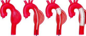
Abdominal aortic aneurysm surgery
Abdominal aorta replacement
This is a standard open surgical procedure. In our Center, this operation is performed through a mini-access - an incision on the abdominal wall 5-7 cm long (while in the standard version the incision is 15-20 cm long). After processing the surgical field and preparing a vascular prosthesis of the required length for the time required for the operation, the abdominal aorta is clamped above and below the aneurysm. The aneurysm is excised and a prepared vascular graft is sewn in place of the removed area. After checking the tightness of the seams and installing drains, the wound is sutured.
At the Clinic of High Medical Technologies named after. N.I. Pirogov St. Petersburg State University in the treatment of an aneurysm of the abdominal aorta uses vascular prostheses impregnated with silver, which differ from conventional ones in being particularly resistant to infection. The operations last on average 3-4 hours, after which the patient is transferred to the intensive care unit for observation. In the standard course of the early postoperative period, the next morning the patient is transferred to the ward of the specialized department. The total length of hospitalization for such patients is about 7 days.
It should be noted that there are various forms of the disease that complicate the standard course of treatment, which in some cases may require longer hospitalization.
Aortic endoprosthetics
- A more modern method of treating abdominal aortic aneurysm, related to hybrid surgery. This method combines open surgical technologies with endovascular ones; it represents the replacement of an aneurysmally dilated section of the aorta from the inside using a special prosthesis, made in most cases “to order” (this explains its high cost). The vascular prosthesis is specially placed in the delivery system. It is straightened directly into the cavity of the aneurysm, under the control of an X-ray machine. Thus, the prosthesis eliminates the impact of systemic arterial blood flow on the weak, stretched walls of the aorta.
This method allows you to achieve results comparable to the open surgical technique, only with fewer complications, reducing the length of hospitalization and rehabilitation of the patient by 2 times! The use of this modern method is limited only by some anatomical parameters of the aorta itself and the high cost of the endoprosthesis.
Control and treatment
- aneurysms are often asymptomatic;
- Abdominal aortic aneurysms are often discovered by chance during examination of the abdomen during radiography, computed tomography, ultrasound, performed for another purpose;
- if the aneurysm is small (4.5 cm in diameter) and there are no symptoms, annual duplex ultrasound examination is recommended;
- quitting smoking and controlling hypertension is the optimal prevention of the appearance and growth of aneurysms;
- It is recommended to operate on aneurysms with a diameter greater than 5 cm in women and 5.5 cm in men, or if the aneurysm has increased by 5 mm (or more) in less than 6 months.
The procedure is performed through small incisions in the femoral arteries. After puncture of the femoral artery, the guidewire is passed through the dilated part of the aorta, then the stent is advanced along the guidewire. Once the stent is positioned correctly, the balloon expands and the stent pushes the vessel wall apart to prevent the aneurysm from spreading below the renal arteries.
To ensure proper sealing between the implanted stent and the aorta, most currently available stents require that the aneurysm have a proximal isthmus at least 1.0 to 1.5 cm below the renal arteries. However, surgery can be done in patients with aneurysms whose aneurysm isthmuses are shorter or absent altogether, by implanting a stent with multiple holes and branches into the arteries of the kidneys or intestines.
Suitable iliac artery conditions are required for guidewire placement, but the potential placement of a stent through a small retroperitoneal incision has increased the number of candidates for endovascular surgery
Compared to traditional open surgery, endovascular surgery has several advantages:
- reduction of operation time;
- reducing blood loss and the need for infusions;
- reducing the time of treatment in the intensive care unit and hospital stay;
- reduced risk of complications;
- Less contrast is used and often less than 60 ml of contrast is required during the procedure;
- intraoperative and early (up to 30 days) mortality are also lower for endovascular operations than for open ones.
The average stay after open surgery is approximately 3 days in the intensive care unit, followed by 7-10 days in the ward, with recovery taking 8-12 weeks.
Most patients undergoing endovascular surgery do not require hospitalization in the IT department and can be sent home the next day after surgery. A larger percentage of patients who have undergone endovascular surgery are sent straight home rather than to rehabilitation sanatoriums. These patients return to normal levels of physical activity more quickly; the rehabilitation period is 1-2 weeks.
In patients with minor comorbidities who are candidates for endovascular intervention, some complications specific to this method may also occur. It is extremely rare to switch to open surgery. Problems associated with stent placement occur in 5-10% of patients and require CT or ultrasound monitoring
Displacement of the stent is rarely possible, because Newer generation stents have hooks and deploy above the level of the renal arteries for better fixation. “Leaks”—when blood leaks between the stent and the aortic wall—occur in 5-10% of cases. Most of them are type II “leaks” - blood continues to accumulate in the aneurysm from the lumbar artery. If there is growth of the aneurysm, patients are not considered cured; in this case, under local anesthesia, the lumbar arteries are closed by embolization on an outpatient basis.
With proper monitoring, the risk of subsequent rupture is extremely low. Therefore, patients should be prepared to undergo further evaluation, which includes computed tomography of the endovascular stent 4 to 6 months after surgery and annually thereafter. Other less common complications are stent failure or infection.
The current prognosis for healthy patients who undergo aneurysm surgery is excellent. Endovascular surgery is a remarkable advance in the treatment of patients with suitable anatomy and is the preferred treatment option for high-risk and elderly patients.
Prognosis and prevention
Unfortunately, abdominal aortic aneurysm is one of the most insidious and unpredictable vascular pathologies, in which the probability of death exceeds 50%. Prevention and timely detection of an aneurysm are essential for a positive outcome. Quitting nicotine addiction, as well as monitoring blood pressure and taking timely measures to normalize it, helps reduce the risk of developing an aneurysm.
Technological progress is helping to reduce the number of diagnostic errors: modern research methods make it possible to identify pathology even in the absence of symptoms. Therefore, people who are at risk - smokers, hypertensive patients, elderly people, as well as those who have a high probability of developing a congenital pathology - should consult a doctor more often for a diagnostic examination. Often, patients are unaware of the development of an aneurysm until it ruptures, but then medical care may not be provided in a timely manner.
Be sure to consult a cardiac surgeon about being examined for the presence of an abdominal aortic aneurysm. Timely detection of the disease can save your life and in any case preserve your health and performance.
Treatment
As already noted, the most dangerous complication of an aortic aneurysm is the threat of its rupture and internal bleeding. If the patient did not consult a doctor and the aneurysm was not detected in a timely manner, then in the event of a rupture, the only option would be open surgery: the surgeon will remove the affected area of the aorta and install a prosthesis in its place. This method of emergency treatment of an aneurysm has contraindications; in addition, the operation is a serious intervention with associated risks and a long rehabilitation period (up to three months). Therefore, it is so important to contact a specialist if you are at risk and feel periodic pain and/or throbbing in the abdominal area.
Once an aneurysm is diagnosed, its further expansion is difficult to predict; regular monitoring by an experienced specialist, lifestyle adjustments, and control of risk factors are required. When the attending physician realizes that there is a risk of rupture, an alternative to emergency open surgery will be the planned installation of a stent graft. The installation is minimally invasive, i.e. the stent is inserted through the vessel, advanced to the site of the aneurysm and secured there. The structure of the stent resembles a vessel; it is fixed with one edge above the expansion of the aorta, and with the other below. After the operation, blood will flow through the stent, and the resulting cavity between it and the aortic wall will decrease over time. The stent graft is inserted under local anesthesia and requires only a couple of days of recovery.
Of course, any surgical intervention carries its own risks, which is why it is so important to choose a good medical center with experienced cardiovascular surgeons. Understanding the danger of aortic aneurysm as a disease, not only doctors in the cardiology department, but also in other departments of our center, when conducting routine examinations or examinations due to other diseases, monitor the patient’s condition and analyze the entire volume of data received: if a problem with the aorta is suspected, the patient within one center will be transferred for consultation with a cardiologist. For our patients, we try to organize the most convenient logic for staying in our center and consulting with specialists in order to avoid double examinations and unnecessary manipulations. Comprehensive patient management is one of the main principles of our work. And the quality of surgical and conservative care is maintained thanks to a team of experienced specialists, provided with the necessary diagnostic and treatment facilities.
You can sign up for a consultation using a special form on the website or by phone.
What surgical treatment methods are there?
Currently, there are two methods of surgical treatment:
- open surgery – removal of an enlarged section of the abdominal aorta and replacing it with a synthetic prosthesis through an incision in the abdomen (prosthetics);
- placement of a synthetic prosthesis in the cavity of the aneurysm through small incisions in the groin (endoprosthetics). Unfortunately, this operation has very strict indications and is therefore only available to a small number of patients. In addition, the number of reoperations after such an intervention is significantly higher than the number of complications than after open surgery.
Complications of aneurysm
Aneurysm is a complex disease that is dangerous not only to health, but also to life.
Pathology of the aorta can cause blockage of the artery of blood vessels, all kinds of infections and even lead to heart failure
Dissection of an aneurysm, in which the layers of the vascular body rupture, is incredibly dangerous. If the vessel is completely destroyed, intense blood loss is inevitable. But the worst manifestation of an aneurysm is its rupture. A fatal outcome is almost inevitable; rupture is characterized by profuse bleeding, which leads to death.
Instrumental examination
X-ray examination. For aneurysms of the thoracic aorta, radiography is performed in three projections with mandatory contrasting of the lumen of the esophagus. The expansion of the shadow of the vascular bundle is characteristic. Aneurysms of the descending aorta bulge into the left pulmonary field. Most patients experience displacement of the contrast-enhanced esophagus. Sometimes calcification (calcification) of the aneurysmal sac is determined. In case of aneurysms of the abdominal aorta, plain radiography of the abdominal cavity in two projections allows us to identify calcification of the aortic wall and urination of the lumbar vertebral bodies.
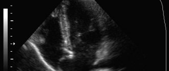
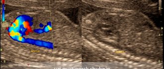
Ultrasound examination (ultrasound) of the aorta and heart. Ultrasound allows us to identify the presence and size in diameter and along the length of aneurysms of the ascending, descending aorta, aortic arch, abdominal aorta, the condition of the vessels extending from the aorta, as well as the presence of aortic valve defect, the nature of changes in the aortic wall.
Computed tomography (CT). When the lumen of the aorta is more than 4 cm, its expansion is considered to be aneurysmal. When performing computed tomography, it is possible to determine the involvement of large arteries in the process and identify signs of dissection (dissection) of the walls (in case of dissecting aortic aneurysm). Angiographic examination (aortography). It is used, as a rule, before surgery when planning its nature and volume.
Causes of aneurysm
- Atherosclerosis, characterized by the appearance of fatty plaques on the vascular walls of the aorta and its branches. The influence of atherosclerosis on the course of an aneurysm has not been fully studied, but the relationship between atherosclerosis and circulatory disorders and, as a consequence, cessation of the delivery of nutrients to the abdominal organs has been proven.
- Diabetes mellitus affecting the blood arteries. There are often cases when it is accompanied by nephropathy and retinopathy.
- Genetics. There are several congenital syndromes, for example, Ehlers-Danlos syndrome, Marfan syndrome, etc. They affect the abdominal aorta. In most cases, there is a relationship between the aneurysm and hereditary diseases.
- Infectious diseases. These are ailments that affect the heart, syphilis, salmonellosis and others.
- Abdominal injuries. So, with a strong blow to the chest or stomach, the aorta may be damaged.
- Inflammatory processes that can cause thinning of the walls of the aorta.
How is an abdominal aortic aneurysm treated?
The complexity of the situation is that often the first signs of an abdominal aortic aneurysm appear at the moment of its rupture with the occurrence of severe bleeding; in the conditions of modern Russian healthcare, the fatal outcome in such cases reaches 90-100%.
Considering the suddenness of the onset of complications of an aneurysm, the only way to treat it is to remove the dilated vessel and replace it with an artificial vessel or prosthesis when it is first detected. Based on the examination, we recommend surgery for you.