Etiology:
- Inflammatory lesions: rheumatism, systemic scleroderma, aortoarteritis.
- Degenerative processes: myxomatous degeneration.
- Atherosclerosis.
- Infectious processes: infective endocarditis.
- Congenital pathology: bicuspid aortic valve.
There is a tendency to increase the frequency of involutional (degenerative-atherosclerotic) causes of aortic stenosis, which has led to an increase in age patients over 60-65 years of age requiring surgical correction of aortic disease.
With congenital or rheumatic valve damage, a long latent period without clinical manifestations is observed. Mortality and the risk of complications increase significantly with the onset of symptoms of the disease. With angina pectoris, fainting and manifestations of heart failure caused by systolic dysfunction of the left ventricle, the average life expectancy is 5, 3 and 2 years, respectively. In asymptomatic patients, the risk of sudden death is low (even with severe aortic stenosis), while in the presence of symptoms, 15–20% of patients die suddenly.
Causes of coronary artery stenosis
The main cause of stenosis is atherosclerosis, in which cholesterol deposits accumulate on the walls of blood vessels, gradually forming plaques that block the lumen.
Also among the provoking factors include:
- anomalies in the development of coronary vessels (tortuosity, incorrect location);
- cardiomyopathy;
- endarteritis;
- systemic diseases (vasculitis, systemic lupus erythematosus, rheumatoid arthritis);
- benign and malignant tumors;
- heart transplantation.
Increase the risk of vasoconstriction:
- hereditary predisposition;
- hypertension;
- diabetes;
- pathologies of the thyroid gland;
- excess weight;
- sedentary lifestyle;
- smoking;
- frequent stress;
- elderly age.
Rate of progression of aortic valve damage:
- mild aortic stenosis
(aortic valve opening area -1.2 - 2 cm2) becomes severe, requiring aortic valve replacement, over 10 years - in 10% of patients, over 25 years - in 38%; - with moderate asymptomatic aortic stenosis
(the area of the aortic valve opening is 0.75 - 1.2 cm2), aortic valve replacement is required after 10 years in 25% of cases; - asymptomatic severe aortic stenosis
(aortic valve opening area <0.75 cm2) usually progresses more quickly; 30–40% of patients develop symptoms within 2 years and there is a need for aortic valve replacement.
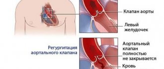
Atrial fibrillation, aortic regurgitation (spontaneous or caused by infective endocarditis), mitral regurgitation, and acute myocardial infarction accelerate decompensation.
In uncomplicated aortic stenosis, the characteristic auscultatory picture is: systolic murmur at Botkin’s point and at the base of the heart on the right, carried out to the vessels of the neck, weakening of the 2nd sound due to the aortic component. The intensity of the systolic murmur may decrease with the development of left ventricular systolic dysfunction and is not a criterion for the severity of the defect. Often the course of aortic stenosis is complicated by pathology of the mitral valve (“mitralization”).
Symptoms of coronary artery stenosis
The main sign of narrowing of the coronary arteries is discomfort, burning and pain in the heart area that occurs during physical and emotional stress. There may be burning pain lasting from 30 seconds to 10 minutes, radiating to the shoulder blade and left arm.
Clinical manifestations may include:
- attacks of dizziness;
- increased heart rate;
- heart rhythm disturbance;
- increased blood pressure;
- shortness of breath;
- weakness.
In some cases, the disease is asymptomatic for a long time and manifests itself only when the vessel is completely blocked by a thrombus or atherosclerotic plaque, which causes ischemia and myocardial infarction. Heart attack is the leading cause of death (90%) from heart disease.
Usually the heart experiences oxygen starvation when the lumen is blocked by 51%. If the artery narrows by 75%, then any physical activity can cause a heart attack, accompanied by necrosis of cardiac tissue. But due to the individual characteristics of the body, some people feel a deterioration in their condition already at 44%, while others do not notice changes even with a narrowing of 75%.
Diagnosis of aortic stenosis
- ECG;
- Chest X-ray;
- Coronary angiography - performed when there are indications for surgical treatment or suspected coronary atherosclerosis;
- MSCT of the ascending aorta with contrast – indicated for poststenotic dilatation of the aortic root;
- EchoCG. Echocardiography allows you to determine: the structure of the valve (bicuspid valve, thickening of the leaflets, fibrosis, calcification, vegetation), the nature of its movement (leaf mobility, degree of opening) and the area of the opening; changes in the aortic root (poststenotic dilatation), left ventricular volume, severity of left ventricular hypertrophy, disturbances of local contractility of the left ventricle (indicating coronary artery disease), EF, left atrium volume, condition of other valves. Doppler studies can accurately determine the pressure gradient between the aorta and left ventricle.
Echocardiographic criteria for the severity of aortic stenosis, taking into account the area of the aortic valve (normal area is 3–4 cm2):
- moderate stenosis 1 – 1.4cm2
- severe stenosis < 1 cm2
- critical stenosis < 0.75 cm2
The average systolic gradient between the aorta and LV at the level of the aortic valve is less than 50 mm Hg. Art. – hemodynamically insignificant aortic stenosis, 50 – 80 mm Hg. – moderate aortic stenosis, more than 80 mm Hg. – severe aortic stenosis.
Indications for surgical treatment (prosthetics) of aortic stenosis
(American Heart Association, American College of Cardiology)
Classification of recommendations and levels of evidence in ACC/AAC format:
- Class I: Conditions for which there is evidence and/or general agreement that a procedure or treatment is beneficial, useful and effective.
- Class II: Conditions for which there is conflicting evidence and/or differences of opinion about the usefulness/efficacy of a procedure or treatment.
- Class IIA: Weight credibility/opinion in favor of usefulness/effectiveness.
- Class IIB: Benefit/effectiveness less well established by evidence/opinion.
- Class III: Conditions for which there is evidence and/or general consensus that the procedure/treatment is not beneficial, not effective, and in some cases may be harmful.
In addition, the levels of evidence to support the recommendations are outlined as follows:
- Level of Evidence A: Data are from multiple randomized clinical trials.
- Level of Evidence B: Data are from single randomized trials or non-randomized trials.
- Level of Evidence C: Expert consensus only, individual case studies and standards of care.
CLASS I
- 1. Aortic valve replacement is indicated in symptomatic patients with severe aortic stenosis (Level of Evidence: B).
- 2. Aortic valve replacement is indicated for patients with severe aortic stenosis during coronary artery bypass grafting (CABG). (Confidence level: C).
- 3. Aortic valve replacement is indicated for patients with severe aortic stenosis, during surgery on the aorta or other heart valves. (Confidence level: C).
- 4. Aortic valve replacement is indicated for patients with severe aortic stenosis and LV systolic dysfunction (EF less than 50%). (Confidence level: C).
CLASS IIA
Aortic valve replacement is indicated for patients with moderate aortic stenosis, undergoing CABG surgery, surgery on the aorta or other heart valves. (Confidence level: B).
CLASS IIB
- 1. Aortic valve replacement is indicated for asymptomatic patients with severe aortic stenosis and an abnormal response to exercise (eg, the development of symptoms of hypotension). (Confidence level: C).
- 2. Aortic valve replacement is indicated for older patients with severe asymptomatic aortic stenosis if there is a high likelihood of rapid progression (age, calcification). (Confidence level: C).
- 3. Aortic valve replacement is indicated for patients with mild aortic stenosis during CABG surgery, when there are signs of moderate to severe valve calcification, which can lead to rapid progression. (Confidence level: C).
- 4. Aortic valve replacement is indicated for asymptomatic patients with extremely severe aortic stenosis (aortic valve area less than 0.6 cm2, mean gradient more than 60 mmHg and flow velocity more than 5.0 m/s), when the expected operative mortality of patients is less than 1.0%. (Confidence level: C).
CLASS III
- 1. Aortic valve replacement is not indicated for the prevention of sudden death in asymptomatic patients with aortic stenosis who do not have any of the indicators from the list of Class IIA/IIB recommendations. (Confidence level: B).
- 2. In older patients with severe, symptomatic, calcific aortic stenosis, aortic valve replacement is the only effective treatment.
- 3. Young patients with congenital or rheumatic aortic stenosis may be candidates for valvotomy. Although there is no consensus regarding the optimal timing of surgery in asymptomatic patients, reasonable recommendations can be made for most patients.
Aortic valve replacement
In recent years, significant progress has been made in the surgical treatment of heart valves. Improvements in technology (including artificial blood circulation machines), the development of uniform standards and protocols for both preoperative examination and the course of the operation have made it possible to reduce the risks of perioperative complications, making the operation on the heart valve apparatus safer than refusing surgery and trying to live with valve dysfunction.
Types of surgical treatment: heart valve replacement surgery
In principle, there are two types of heart valve surgery: prosthetics with an artificial or biological prosthesis and repair of your own valve. It is quite natural that a person’s own valve, after successful reconstruction, functions better than an artificial prosthesis. But if it is impossible to preserve your own valve, the only way out is to replace it with a prosthesis.
Types of valves used in prosthetics
- Biological valves.
They can be made from animal or human tissues (heterografts, homografts, autografts). Biological valves may contain some artificial components to provide valve support and placement. The main advantage of such a valve is the absence of the need for lifelong anticoagulant therapy (constant strict use of drugs that significantly thin the blood and require constant blood tests), and the main disadvantage is its limited service life (15 - 20 years). - Mechanical valves
. They consist entirely of mechanical elements (titanium and pyrolytic carbon) and are designed to replace the patient's own valve functions. The mechanical valve is very reliable and durable, designed for many years of full-fledged operation, which is the main advantage, but requires the patient to constantly take anticoagulants.
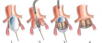
When replacing the aortic valve, access to the valve is achieved by dissecting the aorta in the ascending section (transverse aortotomy). After excision of the aortic valve leaflets and thorough decalcification of the fibrous ring, the latter is sutured with U-shaped sutures. In order to prevent the annulus fibrosus from cutting through, the seams are reinforced with Teflon gaskets. To select the size of the implanted prosthesis, the diameter of the fibrous ring is measured with special calibrators. The selected prosthesis is implanted in the aortic position by stitching its braid. After fixing the prosthesis, the mobility of the locking elements of the mechanical prosthesis or the coaptation of the biological valve leaflets is assessed. If there are no restrictions on their mobility, the dissected aorta is sealed with two rows of sutures.
Treatment of coronary artery stenosis
The doctor selects the method of therapy depending on the degree of development of pathological changes, the cause of stenosis, the presence of concomitant diseases, the age and general health of the patient.
Conservative treatment includes:
- drug therapy (the doctor selects medications individually);
- diet therapy;
- lifestyle changes (giving up bad habits, moderate physical activity).
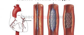
In severe cases, surgery is indicated. Typically, stenting is performed (the vessel is expanded with a balloon and secured with a stent) or bypass surgery (a bypass is created for blood flow).
Primary appointment (examination, consultation) with a cardiovascular surgeon
1850 rub.
Sign up
About technology
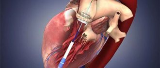
This technology is the implantation of an aortic valve using a small (or no) incision on the thigh, which means without opening the chest and without cardiac surgery!
Through a puncture (or small incision) in the artery, an artificial valve is inserted in a compressed state, which expands when it is installed in place of the damaged aortic valve. After opening, the valve begins to function and restores the full and normal functioning of the aorta.
For the first time in Russia, the private multidisciplinary clinic “Center for Endosurgery and Lithotripsy,” relying only on its own resources and without spending budgetary funds on healthcare, performed this unique and high-tech operation on two patients aged 74 and 82 years.
Moreover, in one of the patients, multiple atherosclerotic lesions of the heart vessels were previously eliminated using a “non-surgical” method without anesthesia. Thus, the patient’s complex lesion of the heart vessels and aortic valve was completely cured using two high-tech minimally invasive techniques without cardiac surgery requiring anesthesia, opening the chest and artificial circulation.