Diagnosis of the disease
STD aneurysm (type R)
As mentioned earlier, STD aneurysm is a congenital developmental anomaly. Therefore, it can be detected without difficulty even in childhood. Another situation is observed if its progressive stretching has occurred, then for the first time it can be noticed at a more mature age.
The main methods for diagnosing an aneurysm are:
- Echocardiographic study. Ultrasound of the heart can reveal protrusion of the interatrial septum.
When an aneurysm is accompanied by the existence of a patent foramen ovale, the following methods may be informative:
- ECG is the simplest method. An electrophysiological study of the heart helps to see overload of the right side of the heart. Heart rhythm disturbances are also detected. In this case, signs of atrial fibrillation can be detected.
- X-ray of the chest organs. An aneurysm with a patent foramen ovale is characterized by the presence of a specific symptom on an x-ray - “pulsation of the roots of the lungs.”
- Echocardiographic study. As in the case of non-communicating aneurysm, this method is very important in diagnosing a patent foramen ovale. Thanks to this method, you can see the turbulence of the blood flow in the area of the hole. Valvular abnormalities may also be detected.
Instrumental diagnostics can also be supplemented by the following methods:
- Transesophageal ultrasound.
- Transthoracic ultrasound.
- CT scan.
- Catheterization of the heart chambers.
Atrial septal defect
Atrial septal defect (ASD) is the second most common congenital heart defect.
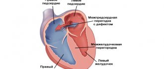
With this defect, there is a hole in the septum that separates the right and left atria into two separate chambers. The fetus, as we said above, not only has this hole (open oval window), but is also necessary for normal blood circulation. Immediately after birth it closes in the vast majority of people. In some cases, however, it remains open without people being aware of it. The discharge through it is so insignificant that a person not only does not feel that “there is something wrong with the heart,” but he can calmly live to a ripe old age. (It is interesting that, thanks to the capabilities of ultrasound, this defect in the interatrial septum is clearly visible, and in recent years, articles have appeared that show that among such adults and healthy people who cannot be classified as patients with congenital heart disease, there is a significantly higher number of people suffering from migraines - severe headaches. These data, however, have yet to be proven).
Unlike patent foramen ovale, true atrial septal defects can be very large. They are located in different parts of the septum itself, and then they speak of a “central defect” or “a defect without an upper or lower edge,” “primary” or “secondary.” (We mention this because the type and location of the hole may depend on choice of treatment type).
If there is a hole in the septum, a shunt occurs with blood discharge from left to right. With an ASD, blood from the left atrium partially flows into the right atrium with each contraction. Accordingly, the right chambers of the heart and lungs become overfilled, because they have to pass through themselves a larger, extra volume of blood, and one more time that has already passed through the lungs. Therefore, the pulmonary vessels are filled with blood. Hence the tendency to pneumonia. The pressure in the atria, however, is low, and the right atrium is the most “distensible” chamber of the heart. Therefore, while increasing in size, it copes with the load for the time being (usually up to 12-15 years, and sometimes more) quite easily. High pulmonary hypertension, which causes irreversible changes in the pulmonary vessels, never occurs in patients with ASD.
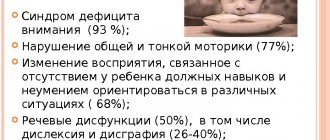
The vast majority of newborns, infants, and young children grow and develop absolutely normally. Parents may notice their tendency to frequent colds, sometimes resulting in pneumonia, which should be alarming. Often these children, in 2/3 of cases girls, grow up pale, thin and somewhat different from their healthy peers. They try to avoid physical activity as much as possible, which in the family can be explained by their natural laziness and reluctance to tire themselves out.
Heart complaints can and, as a rule, appear in adolescence, and often after 20 years. Usually these are complaints about “interruptions” in the heart rhythm that a person feels. Over time, they become more frequent, and sometimes lead to the patient becoming incapable of normal, ordinary physical activity. This does not always happen: G.E. Falkowski once had to operate on a patient aged 60, a professional driver, with a huge atrial septal defect, but this is an exception to the rule.
To avoid such a “natural” course of the defect, it is recommended to close the hole surgically. Unlike a VSD, an interatrial defect will never heal on its own.
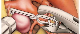
Surgery for ASD is performed under artificial circulation, on an open heart, and consists of suturing the hole or closing it with a patch. This patch is cut out from the heart sac - the pericardium - the sac surrounding the heart. The size of the patch depends on the size of the hole. It must be said that the closure of an ASD was the first open-heart surgery, and it was performed more than half a century ago.
Sometimes an atrial septal defect can be combined with an irregular, anomalous entry of one or two pulmonary veins into the right atrium instead of the left. Clinically, this does not manifest itself in any way, and is a finding when examining a child with a large defect. This does not complicate the operation: the patch is simply larger and is made in the form of a tunnel in the cavity of the right atrium, directing blood oxidized in the lungs to the left parts of the heart.
Today, in addition to surgery, in some cases it is possible to safely close the defect using X-ray surgery
technology. Instead of suturing the defect or sewing in a patch, it is closed with a special umbrella-shaped device - an occluder, which is passed along the catheter in a folded form and opened, passing through the defect.
This is done in the X-ray surgery room, and we described everything related to such a procedure above when we touched on probing and angiography. Closing a defect with such a “non-surgical” method is not always possible and requires certain conditions: anatomical location of the hole, sufficient age of the child, etc. Of course, if they are present, this method is less traumatic than open-heart surgery. The patient is discharged after 2-3 days. However, it is not always feasible: for example, in the presence of abnormal vein drainage.
Today, both methods are widely used, and the results are excellent. In any case, the intervention is elective and not urgent. But it needs to be done in early childhood, although it can be done earlier if the frequency of colds and, especially, pneumonia becomes frightening and threatens bronchial asthma, and the size of the heart increases. In general, the sooner the operation is performed, the sooner the child and you will forget about it, but this does not mean that you should be in a particular hurry with this defect.
How to get treatment at the Scientific Center named after. A.N. Bakuleva?
Online consultations
Mechanisms of aneurysm formation
Patent foramen ovale
During the prenatal period in the fetus, the oval window is located in the septum between the atria. Through this window, blood is discharged from the left atrium to the right. Thus, the blood flow does not involve the pulmonary circulation, since during this period there is no need for the lungs to work.
After birth, the baby's lungs begin to function and the oval window closes (overgrows). If the window does not close completely, thin connective tissue forms in this place, or a communication remains between the atria.
- Aortic aneurysm: symptoms and treatment
How is atrial aneurysm treated?
Aneurysm of the membranous part of the interventricular septum is treated with the same methods as other types of similar pathologies. Once the disease is identified, drug treatment is usually prescribed, during which the doctor monitors the growth of the aneurysm. The following drugs are used:
- Medicines that stimulate the production of collagen are needed to strengthen the walls of blood vessels;
- B vitamins;
- Microelements including zinc and copper;
- Medicines to relieve blood pressure if the patient is prone to hypertension;
- Drugs that resolve blood clots;
- Medicines to regulate heart rate.
The medicinal method is used for small pathologies, but if accelerated growth is observed, surgical intervention cannot be avoided. During the operation, the doctor’s task is to strengthen the walls of the aneurysm. The following methods are used:
- A synthetic patch is installed;
- Sutures are placed through a small hole;
- The oval window is closed using an endoscope.
Surgical treatment does not exclude the use of medications during the rehabilitation period. You will also have to take medications that strengthen blood vessels, vitamins and minerals. It is required to comply with all recommendations and prescriptions of the doctor for the treatment to be successful.
4.Treatment
With an uncomplicated aneurysm, there is no need for either conservative or cardiac surgery (for obvious reasons, any surgical intervention on the heart is in itself risky and is used only as a last resort measure with no alternatives). However, observation and regular follow-up examinations by a cardiologist are strictly necessary: the condition and size of the aneurysm must be monitored. In cases of threatening development of the clinical situation, the patient is immediately hospitalized, and in the cardiac surgery hospital the defect or plastic suturing of the MPP is performed using bioinert materials.
What is an aneurysm of the bladder?
Atrial aneurysm is a small anomaly of the heart, which is a protrusion of the vessel wall. In this case, blood circulation is disrupted, and the neoplasm puts pressure on the walls of the atrium. The disease is less common in adults than in children. There are several different forms:
- Protrusion from the left to the right atrium;
- Bulging of the septum of the heart to the left;
- Fusiform pathology, when the upper part protrudes in one direction, the right part in the other.
An aneurysm of the bladder in a newborn and in adults is dangerous not only because of a possible rupture - this happens infrequently. The danger is that if blood flow is disrupted in the acute form of the disease, deviations in the oxygen supply to the brain are likely. Blood clots, clots, and anomalies associated with defects in blood vessels and arteries appear, and a stroke is likely.
Heart aneurysm
According to the time of occurrence, acute, subacute and chronic cardiac aneurysm are distinguished. Acute cardiac aneurysm forms within 1 to 2 weeks from myocardial infarction, subacute - within 3-8 weeks, chronic - over 8 weeks.
Acute aneurysm
In the acute period, the wall of the aneurysm is represented by a necrotic area of the myocardium, which, under the influence of intraventricular pressure, bulges outward or into the ventricular cavity (if the aneurysm is localized in the area of the interventricular septum).
Subacute aneurysm
The wall of a subacute cardiac aneurysm is formed by a thickened endocardium with an accumulation of fibroblasts and histiocytes, newly formed reticular, collagen and elastic fibers; in place of destroyed myocardial fibers, connecting elements of varying degrees of maturity are found.
Chronic aneurysm
A chronic cardiac aneurysm is a fibrous sac microscopically consisting of three layers: endocardial, intramural and epicardial. In the endocardium of the wall of a chronic cardiac aneurysm there are proliferations of fibrous and hyalinized tissue. The wall of a chronic cardiac aneurysm is thinned, sometimes its thickness does not exceed 2 mm. In the cavity of a chronic cardiac aneurysm, a parietal thrombus of various sizes is often found, which can line only the inner surface of the aneurysmal sac or occupy almost its entire volume. Loose mural thrombi are easily fragmented and are a potential source of risk of thromboembolic complications.
There are three types of cardiac aneurysms: muscular, fibrous and fibromuscular. Typically, a cardiac aneurysm is single, although 2-3 aneurysms may be detected at the same time. Cardiac aneurysms can be true (represented by three layers), false (formed as a result of a rupture of the myocardial wall and limited to pericardial adhesions) and functional (formed by a section of viable myocardium with low contractility, bulging during ventricular systole).
Taking into account the depth and extent of the lesion, a true cardiac aneurysm can be flat (diffuse), saccular, mushroom-shaped, and in the form of an “aneurysm within an aneurysm.” In a diffuse aneurysm, the contour of the external protrusion is flat, gentle, and on the side of the heart cavity there is a cup-shaped depression. A saccular cardiac aneurysm has a rounded, convex wall and a wide base. A mushroom aneurysm is characterized by the presence of a large protrusion with a relatively narrow neck. The concept of “aneurysm within an aneurysm” refers to a defect consisting of several protrusions enclosed within one another: such cardiac aneurysms have sharply thinned walls and are most prone to rupture. During examination, diffuse cardiac aneurysms are more often detected, less often - saccular aneurysms, and even less often - mushroom-shaped and “aneurysms within an aneurysm”.