Ways to get rid of bruises
Most often, the appearance of a bruise is preceded by a strong blow, which cannot be ignored. This injures small vessels, a small amount of blood flows out of them and permeates the subcutaneous tissue and lower layers of the skin. In response to injury, local swelling occurs - a protective reaction of the body. At this stage, the bruise is not yet visible, but you already need to take active action. The goal is to reduce tissue permeation with blood.
In order to quickly reduce swelling at the site of a bruise, it is necessary to use agents that cause vasospasm. Cooling with ice works well for this, but a frozen piece of meat wrapped in film and a thin towel will also work. It must be applied to the site of injury for 20 minutes. After cooling, the ice is removed, but the procedure can be repeated after 15-20 minutes. Constantly keeping the cold near the bruise is dangerous and can cause even more damage.
Home remedies can help relieve swelling. For this you can use:
- a compress made from a solution of vinegar or vodka;
- onion gruel with salt;
- applying a cut aloe leaf;
- half a raw potato.
Folk remedies against bruises can be used at an early stage, immediately after a bruise. A mixture of garlic and vinegar pulp and lavender essential oil, which should be applied to the injury site, are considered effective. But you need to be careful; minor bruises can be treated at home without the help of a doctor.
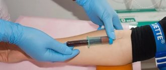
Orthopedics and traumatology services at CELT
The administration of CELT JSC regularly updates the price list posted on the clinic’s website. However, in order to avoid possible misunderstandings, we ask you to clarify the cost of services by phone: +7
| Service name | Price in rubles |
| Appointment with a surgical doctor (primary, for complex programs) | 3 000 |
| X-ray of the chest organs (survey) | 2 500 |
| Ultrasound of soft tissues, lymph nodes (one anatomical zone) | 2 300 |
All services
Make an appointment through the application or by calling +7 +7 We work every day:
- Monday—Friday: 8.00—20.00
- Saturday: 8.00–18.00
- Sunday is a day off
The nearest metro and MCC stations to the clinic:
- Highway of Enthusiasts or Perovo
- Partisan
- Enthusiast Highway
Driving directions
What to put on a bruise
In order for treatment for a bruise to be effective, it is necessary to take into account the time that has passed since the injury. The formation of a hematoma goes through several stages. Immediately after the injury, agents are used that relieve swelling. At home, at an early stage, you can use folk remedies, but after that, purchase pharmaceutical drugs.
On the first day, you can apply ointment or gel containing heparin to the bruise. It helps reduce swelling by increasing the speed of blood flow. Heparin is well absorbed through the skin, thins the blood and prevents the formation of a hematoma. Therefore, treatment of the bruise will proceed quickly.
But when using heparin, you need to remember that it is not recommended to use it in the first hour or two after a bruise. It can increase bleeding at the injury site and cause an enlarged hematoma.
If a soft tissue injury is accompanied by pain, it can be reduced with the help of non-steroidal anti-inflammatory drugs. Quickly relieve discomfort with a gel or ointment that contains diclofenac, ibuprofen or piroxicam. They can be applied to the impact site after a few hours.
After a day, these remedies for treating bruises are no longer effective. Blood permeates the tissues, coagulates, and its cells are destroyed. Therefore, the bruise turns blue due to the protein hemoglobin. From this moment, its disintegration and hematoma begins to bloom. She herself will not be able to pass in a short time until all hemoglobin is destroyed into bilirubin.
We need products that resolve bruises by heating the tissues and increasing blood flow in them. You can use dimethyl sulfoxide creams or ointments at home without a doctor's prescription. This substance is obtained from hot peppers, so it has a warming effect. Minor bruises after using it disappear within 2-3 days.
WHEN YOU SHOULD SOUND THE ALARM, THE FIRST SYMPTOMS OF HEMATOMA
A hematoma gives its symptoms and signs almost immediately after injury.
- Firstly, the skin at the site of the hematoma is sharply painful.
- After a short period of time, the site of injury begins to swell, the tumor can spread significantly and interfere with movement (for example, with a hematoma on the ankle, the swelling may be such that it is impossible to move independently or step on the affected leg).
- After swelling, the site of hemorrhage quickly turns red. Patients feel internal tension in the area of the hematoma; it is hard to the touch.
The color of the hematoma can be different - from bright red to purple, most often it is heterogeneous - its edges are darker, bluish in color, and the inside of the hematoma is red.
How to quickly remove a bruise on your face
A black eye appears much faster than in other parts of the body. If measures are not taken quickly, the resorption of the bruise will take a long time. The reason is the location of the vessels and the loose subcutaneous tissue that surrounds the eyes. It easily becomes saturated with blood and swells.
First aid after injury is to apply ice to the face. Of the folk methods of struggle, it is necessary to use safe ones that will not irritate the mucous membrane of the eyes. Therefore, a paste of onion, vinegar or garlic will not work. But you can use:
- raw potatoes;
- parsley paste;
- a piece of raw pineapple (not canned);
- essential oils.
They are also treated with pharmaceuticals containing heparin, diclofenac and ibuprofen. Bruises on the face can be smeared with gel with troxevasin. It reduces swelling, pain, relieves inflammation, increases capillary tone and reduces their fragility. Troxevasin helps to quickly remove bruises and strengthen small blood vessels. It is recommended to use it 2-3 times a day.
If the bruise is large and does not want to dissolve, you can try to hide it with the help of cosmetics. Women apply a special color corrector to the defect on the face, which covers the blue tint, after which the skin is smoothed with foundation and powdered.
Difference
What is the difference between a hematoma and a bruise? From the above it is clear that both are accumulations in the formed cavities of a large or small amount of blood that has flowed out from ruptured blood vessels. There are several differences between these two concepts.
- The word “bruise” is used colloquially, and “hematoma” in official medicine, criminology, jurisprudence, and scientific literature.
- The medical term hematoma comes from the fusion of two Greek words, ema, meaning blood, and oma, meaning tumor. People called bruises the spots on the skin that appear after bruises or other injuries, because of their characteristic blue with various shades of color.
- A bruise is always visible to the naked eye because it forms in the subcutaneous fat layer. A hematoma can appear not only in the subcutaneous tissue, but also in the internal organs, so sometimes instrumental examination is required to detect it.
- Bruises are commonly called spots that go away without treatment. Hematomas can be so complex that they require mandatory medical intervention.
How to quickly remove bruises on legs
First aid for a bruise on the leg is a cold compress. This can be done by soaking a cloth in water mixed with vinegar or alcohol. Due to rapid evaporation from the surface of the skin, they remove the tumor or prevent it from forming.
To avoid having to wait for the hematoma to heal, you need to lie down and raise your leg above the level of your heart. This will speed up the flow of blood from the limb and prevent it from swelling. Ointment with heparin, painkillers and troxevasin also effectively resolve the bruise.
There are hardware methods that help heal bruises. In cosmetology clinics they are treated with a laser. Irradiation of a tumor causes tissue heating and accelerates the breakdown of hemoglobin. You can remove a small hematoma at one time, but a large one will only change color. The disadvantage of this treatment is its high cost.
Hematoma. general information
Hematoma is a condition characterized by the accumulation of liquid or coagulated blood inside the body, which occurs as a result of rupture of blood vessels and is localized in soft tissues. Hematomas can be small in size, or they can compress soft tissues and nearby organs. Hematomas form under the skin, mucous membranes, deep within the muscles, in the walls of internal organs, and in the brain.
When treating Hematoma, doctors at the BIOSS clinic use both time-tested and the latest developments and proprietary techniques.
Our clinic employs the best doctors in Moscow who have extensive experience in treating Hematoma
There are several classifications of hematomas:
Medicines for bruises
Major injuries are accompanied by severe pain, which is difficult to relieve with local remedies. Therefore, soon after injury, you can take painkillers orally based on:
- nimesulide;
- diclofenac;
- indomethacin;
- ibuprofen;
- ketorolac.
They help relieve pain, reduce swelling and inflammation.
Sometimes hematomas appear due to increased fragility of blood vessels, so it is necessary to use agents to strengthen the capillaries against them. Riboxin and complexes containing vitamin C and ascorutin help with this.
It is difficult to remove blueness under the eyes that is not associated with injury. It appears due to kidney pathologies or due to the proximity of blood vessels under the skin. In this case, it is necessary to eliminate the cause, apply cooling compresses before bed or apply special patches.
TYPES OF HEMATOMA AND THEIR TREATMENT
There are several approaches to the classification of hematomas.
Hematomas are distinguished depending on:
- the nature of bleeding - they can be arterial, venous and mixed;
- localizations – subcutaneous, intramuscular, intracranial, etc.;
- clinical signs - encysted, pulsating, simple.
In addition, situational hematomas are distinguished in the treatment, which require a special approach, for example, hematomas during childbirth, hematomas during pregnancy, etc.
An arterial hematoma is a hematoma that contains arterial blood in the cavity. As a rule, such hematomas are bright red, they are often diffuse - with a wide distribution on the surface. Venous hematoma occurs when there is compression and disruption of the integrity of the vein. These hematomas are bluish-violet in color, they are inactive and hard to the touch. The most common hematomas are mixed, when both arterial and venous blood enters the cavity.
A subcutaneous hematoma forms under the layer of skin and looks more like a bruise. They can form both due to injuries and due to various diseases - tuberculosis, syphilis, scarlet fever, lupus erythematosus. Often such hematomas form in people suffering from hemophilia. At the slightest damage to the vessel, spots appear on the skin. Subcutaneous hematomas can be of three degrees.
With a mild hematoma, its symptoms appear prolonged - approximately a day after the injury, while it absolutely does not interfere with the functioning of the organ on which it appeared. Painful sensations are weak and sometimes do not occur at all. If the hematoma is not complicated by anything, then it goes away on its own without any treatment. A moderate hematoma forms within three to four hours. In this case, the hematoma can partially disrupt the functioning of the organ on which it originated. A slight swelling and swelling of the soft tissues forms around such a hematoma. Apply ice and a pressure bandage to the site of the hematoma and contact a medical facility. A severe hematoma can occur with serious injury. In this case, the presence of a hematoma disrupts the functioning of organs. Hemorrhage forms quickly - literally within an hour you can notice a blue spot at the site of damage. Most often this is a subcutaneous hematoma, which is visible to the naked eye. Over time, the hematoma intensifies and can become intramuscular. In this case, the patient will feel numbness and soreness in the muscles. Such a hematoma requires a mandatory examination by a doctor and further treatment. If a hematoma is not treated, it can cause serious harm to the human body.
Intramuscular hematoma is characterized by the accumulation of blood in the muscles. In this case, the patient feels significant pain in the area of damage. Muscle function is impaired. In order to cure such a hematoma, it is necessary to consult a doctor. You may need to surgically open the hematoma and drain the cavity.
of intracranial hematomas - epidural, intracerebral, subdural, intraventricular.
Epidural hematomas are a collection of blood between the meninges and the skull bone. Most often, such hematomas occur near the temple; their development is associated with a traumatic moment (a blow to a stone, a blow to the head with a blunt object). With an epidural hematoma, the artery most often suffers, so such a hematoma is also arterial. At the site of the rupture, up to one hundred and fifty milliliters of blood quickly accumulates. The appearance of such a hematoma leads to compression of the brain. In this case, the patient loses consciousness for a short time, and then regains consciousness, but feels a headache, weakness, and vomiting. After several hours of improvement, a sharp deterioration occurs. Depending on the size of the hematoma, a state of coma can quickly occur. Heart contractions slow down, blood pressure drops, and the eyes stop responding to stimuli (except for the pupils). When such a hematoma is diagnosed, emergency surgery is prescribed to eliminate it.
Subdural hematomas are hemorrhages between the arachnoid and dura mater of the brain.
Venous blood collects in such hematomas, so they are also venous. Quite often, such hematomas are bilateral - the first occurs at the site of the impact, and the second - at the counter-impact. These hematomas have a larger area than epidural ones and can sometimes contain up to three hundred milliliters of blood. In the presence of such a hematoma, crisis phenomena in the patient may increase over the course of two days - hemiparesis, breathing problems, epilepsy, and bradycardia occur. The surgical treatment of such a hematoma consists of excision of the hematoma itself, restoration of the integrity of the bone and revision of the brain. In some cases, drainage is applied.
Intracerebral hematoma is very difficult to diagnose. Hemorrhage may occur slowly, increasing in volume day by day. Symptoms of intracerebral hematoma gradually appear. In some cases, immediate bruising may appear some time after the injury. The symptoms of such a hemorrhage depend on where it occurred - there may be hearing, speech, vision impairment, loss of consciousness, memory impairment, loss of sensitivity, or vice versa, hypersensitivity. Most often, such a hematoma is treated with special drugs that help it resolve. They are used if the volume of spilled blood is less than thirty milliliters. Otherwise, surgery may be necessary.
Intraventricular hematoma occurs when hemorrhage occurs in the ventricles of the brain. Treatment of such a hematoma depends on its size. If it is impossible to treat such a hematoma conservatively, then they resort to surgical intervention.
Color change
Probably everyone has noticed that bruises on the body tend to change their color over time.
Basically, in the first hours after its occurrence, the hematoma visible to the eye has a red-purple color, which is caused by the content of oxyhemoglobin in the spilled blood. It soon turns into hemoglobin, causing the hematoma to turn blue-violet. In about 5-6 days, hemoglobin breaks down into methemoglobin and verdochromogen, the color of which is green. Therefore, the hematoma acquires greenish tints. The transformation of blood particles continues, and after about a week, verdochromogen breaks down into bilirubin and biliverdin, which color the hematoma yellow. After this, it gradually becomes discolored and is compared with the overall skin tone.
The rate at which the hematoma changes color depends on its size, thickness, location and individual characteristics of the patient’s body. In hematomas of large area and thickness, several colors can be observed simultaneously.