Home Articles How to measure blood pressure correctly
Adapting medical technologies to the needs of patients makes it possible to monitor blood pressure (BP) values independently. To do this, you need to buy a mechanical or electronic tonometer at the pharmacy. Knowing how to correctly measure blood pressure, you can assess your condition or provide assistance to a loved one. Regular monitoring of blood pressure is necessary for patients with cardiovascular pathologies, diabetics, elderly people, and bedridden patients.
About pressure
Blood pressure indicators are a digital assessment of the force with which circulating blood presses on the inner wall of the vessel. The upper value is recorded at the moment of systole (contraction of the heart muscle), the lower value at the moment of diastole (relaxation of the myocardium). The systolic indicator reflects the strength of muscle tension at the moment of blood ejection, the diastolic indicator reflects the tone of the blood vessels. A decrease, and more often an increase in blood pressure, occurs:
- due to physical and nervous overload;
- against the background of chronic diseases;
- due to age-related changes in the body.
In a healthy person, a compensatory mechanism can cope with the “jump” in blood pressure in half an hour. People with heart and vascular diseases require medication assistance, so they need to measure their blood pressure twice a day. Elderly relatives living separately should be taught self-control skills. If a person cannot use a blood pressure monitor for health reasons, a specially trained caregiver can be hired.
Nuances of blood pressure control
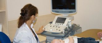
There are two types of tonometers used in everyday life: mechanical and electronic. A mechanical device is considered more accurate. It consists of a dial, an inflation bulb, a valve and a cuff. But an electronic tonometer, equipped with a processor, display and cuff, is more convenient to use.
Despite the simplicity of self-diagnosis, not everyone can measure blood pressure correctly. The procedure requires compliance with certain conditions, and evaluation of results requires knowledge of reference values.
Preliminary preparation includes avoiding nicotine, energy drinks and coffee an hour before measuring blood pressure. Before taking measurements, you need to empty your bladder and rest for 5-10 minutes.
Field of this:
- sit comfortably, leaning your back against the back of a chair (armchair), relax your limbs;
- position your hand so that your elbow rests on the table;
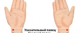
- put the device cuff on 20 mm above the elbow (the tube should be on the vein in the elbow);
- turn on the tonometer;
- wait for stable values on the screen (the numbers should not “jump”).
It would be correct to measure blood pressure 1.5 hours after physical activity, 20-30 minutes after eating. While readings are being taken, you must not move, talk, or chew. To obtain objective results, it is recommended to repeat the procedure at intervals of 3-5 minutes.
When caring for a bedridden patient, do not disturb him. You need to measure your blood pressure in a supine position, but at the same time, take into account an adjustment of 5-7 mm Hg. Art.
The problem of modern pathogenetic intensive therapy for intracranial hemorrhages of non-traumatic origin is the most important in clinical neuroreanimatology, which is associated with a high prevalence, mortality rate, disability and social maladaptation of patients who have suffered hemorrhagic stroke [1-3].
Changing the position of the angle of inclination of the head end of the bed in neurosurgical patients is a generally accepted and proven method for correcting increased intracranial pressure (ICP) [2, 4, 5]. In intensive care practice, positions from 15 to 60° are used, but preference is given to 30° [2, 5, 6]. To date, there is no consensus on which position most effectively and significantly reduces ICP without reducing cerebral blood flow. It is generally accepted that for most patients with cerebral pathology, regardless of the etiological factor, a position of 15-30° is preferable. Early work carried out on patients with various brain pathologies confirmed the effective reduction of ICP at various positions of the head end in the range from 15 to 60° [5-7]. This position is justified from a physiological point of view, since even a slight elevation of the head end of the bed improves venous and liquor outflow from the cranial cavity and leads to a decrease in ICP [4, 5, 8]. However, the choice of the position of the angle of inclination of the head end in case of intracranial hemorrhage after surgery remains a controversial issue [6].
The purpose of the study was to study the nature of the relationship between hemodynamic parameters (ICP, cerebral perfusion pressure (CPP), mean arterial pressure (MAP) at different positions of the head end of the bed in patients with intracranial hemorrhage.
Material and methods
The study included 36 patients (11 men and 25 women) with intracranial hemorrhages of non-traumatic origin aged from 16 to 65 years (average - 52.9±10.5 years). Upon admission and over time, all patients underwent multislice computed tomography (MSCT) of the brain. Of these, 27 were diagnosed with subarachnoid hemorrhage using MSCT angiography due to rupture of an arterial aneurysm of cerebral vessels, and 8 were diagnosed with hemorrhagic stroke with the formation of an intracerebral hematoma of hemispheric localization. All patients were operated on on the 1st day from the onset of the disease. Treatment of patients was carried out taking into account the indicators of multimodal neurophysiological monitoring of ICP, blood pressure, CPP, cerebral blood flow (CBF) using transcranial Dopplerography (TCDG). ICP was measured using parenchymal sensors (Codman Microsensor “Johnson & Johnson”, USA). The sensor was installed on the opposite side of the lesion (on the intact side). The parameters of ICP, BP, and CPP were recorded using Philips MP 40 and MP 60 bedside monitors. Data were collected through an analog output on a personal computer and analyzed using the ICM Plus program. Radial artery catheterization was performed to measure invasive blood pressure. The invasive blood pressure sensor was placed at the level of the external auditory canal (in the projection of the foramen of Monroe) in order to correctly measure CPP. Doppler examination was performed according to standard methods using the Sono Scape S8 apparatus. When conducting a test with a change in the position of the head end of the bed, three consecutive measurements were taken, each lasting 5 minutes in positions 30-0-60°; in all cases, the initial ICP did not exceed 25 mm Hg. The study began in the standard position of the patient, when the angle of inclination was 30°. Subsequently, following the research algorithm, the patient was given a horizontal position with subsequent elevation of the head end to 60°. After stabilization of the condition at each point, the parameters of average ICP, blood pressure, and central pressure were recorded for 5 minutes. The study was carried out on days 1, 2, 3 and 5 after surgery. The criterion for inclusion in the study was survival of the operated patients for at least 1 week from the onset of the disease.
Statistical processing of the obtained data was carried out using the Statistica 6.0 software package in accordance with the main objectives of the study. The normality of the distribution of variables was checked using the Shapiro-Wilks method. Parametric and nonparametric statistical methods were used for analysis. Data are presented as medians and quartiles.
Results and discussion
Starting from the 1st day from the onset of the disease, a gradual increase in intracranial pressure figures was observed (Table 1)
.
The recorded ICP values in the standard position of the head end of the bed of 30° increased sharply and by the 3rd day of monitoring reached 27 (22-33) mm Hg, increasing by 22% compared to the initial parameters.
The greatest differences in ICP threshold values were recorded on the 5th day after surgery - on average 27% compared to the 1st day (from 21.5 (19-24) to 27 (20.5-36) mm Hg by the 5th day). At the same time, according to invasive monitoring data, the average ICP numbers from the 1st to the 5th day increased insignificantly. A sharp decrease in CPP was observed already on the 2nd day from the onset of the disease. Its values decreased by 18% compared to the initial ones - from 89.5 (84-101) to 73.5 (63.5-84) mm Hg. Further, the CPP indicators gradually increased, but by the 5th day they did not reach the initial level, equaling 83 (69.5-88.5) mm Hg, and on average became 7% lower than on the 1st day.
Also on the 2nd day there was a decrease in mean arterial pressure: from 105 (97-120.5) to 91 (81.5-100) mmHg, which was on average 13% lower than the initial values. Subsequently, the blood pressure readings began to gradually increase, but never reached the initial level, amounting to 97.5 (89.5-105) mm Hg. on the 5th day. Thus, the difference in the recorded blood pressure values between days 1 and 5 was 7%.
When performing a sequential change in the angle of inclination of the head end of the bed (30-0-60°) on the 1st day, the maximum ICP values (18 (16-22) mm Hg) were recorded in a horizontal position (Table 2)
.
ICP figures in the horizontal position were 33% higher than at 30 and 60°.
There were no significant differences between the 30 and 60° positions, and the average ICP values were 12 mmHg. The minimum CPP values on the 1st day from the onset of the disease were recorded in the 30° position, but did not differ significantly from the CPP figures in the horizontal position - 86 (78-99) and 87 (79.5-99.5) mm Hg . respectively. At the 60° position, the CPP was on average 5.5% higher than at 30° - 91 (81.5-99) mm Hg.
It was also noted that in a horizontal position in patients with non-traumatic hemorrhages, blood pressure increases by 5%, approaching 103.5 (98-117.5) mm Hg. At the same time, at the 30 and 60° positions the differences were insignificant: 98 (90.5-111) and 101 (94.5-113.5) mmHg. respectively. Thus, on the 1st day from the onset of the disease, the position of the head end of the bed 0° was the least preferable, since in this case a pronounced increase in ICP values was observed in combination with minimal perfusion of the brain parenchyma. The 60° position should be considered the most favorable on the 1st day after surgery, since it was in it that optimal CPP values were observed, ICP values remained relatively stable and the blood pressure level increased slightly (on average by 3% compared to the 30° position).
On the 2nd day from the onset of the disease, a similar pattern was observed: when the position of the head end of the bed changed from 30 to 0°, the maximum ICP and critically low CPP were recorded in the horizontal position: 20 (16-25) and 71.5 (60-83) mm Hg respectively. However, at the 60° position (compared to 30°), ICP was increased by an average of 3%, and CPP decreased by 5.5%. Blood pressure values were maximum in the horizontal position - 91.5 (83.5-105.5) mm Hg; When the head end of the bed was raised, the blood pressure readings remained stable. Thus, on the 2nd day from the onset of the disease, preference should have been given to an angle of inclination of the head end of the bed of 30°. It was in this position that optimal cerebral perfusion was ensured and intracranial hypertension did not increase.
On the 3rd day after surgery, there is a critical increase in ICP (an average increase of 7-14 mm Hg) in combination with low cerebral perfusion (a decrease of 5-7 mm Hg) and high systemic blood pressure ( an increase of 4-8 mm Hg) were still recorded in a horizontal position (see Table 2)
. However, by the 3rd day the difference in the values of hemodynamic parameters in the 30 and 60° positions disappeared. Therefore, both of these positions can be considered optimal.
On the 5th day, the horizontal position of the patient was again the least preferable. At the same time, the increase in ICP in the 0° position became less significant (6-8 mm Hg compared to 8-11 mm Hg on the 1st day), however, a longer period of time was required to restore ICP values to the original numbers When recording CPP, the minimum values were recorded at 60° - 77.5 (69-89.5) mm Hg. At the same time, a significant decrease in blood pressure in the 60° position was observed: from 99 (90.5-107) to 94.5 (88-102) mm Hg. Thus, changing the angle of inclination of the head end of the bed upward to 60° was just as undesirable as keeping the patient in a horizontal position. The optimal angle of inclination again became 30°.
It should be noted that an increase in the ICP level in a horizontal position leads to the formation of a Doppler pattern of difficult perfusion, characterized by a relative decrease in the average linear blood flow velocity and an increase in peripheral resistance indices. According to M. Rosner et al. [6], an elevated position of the head end can effectively reduce ICP in only 1/2 of patients with neurosurgical pathology. In our study, on the 1st day from the onset of the disease, a significant decrease in ICP figures when moving from the horizontal to the 60° position occurred in 78% of patients. However, by the 5th day, an effective decrease in intracranial pressure when changing the angle of inclination of the head end of the bed towards its increase was registered only in 62% of patients.
Despite the different etiological factors, in patients with hemorrhagic stroke with the formation of intracerebral hematomas and patients with subarachnoid hemorrhages due to rupture of cerebral arterial aneurysms, no significant differences were found in the studied parameters.
The conducted research gave grounds to formulate the following conclusions.
In patients with non-traumatic intracranial hemorrhage, the maximum increase in ICP was observed starting from the 3rd day from the onset of the disease in combination with reduced cerebral perfusion. Starting from the 2nd day after surgery, continuous monitoring of hemodynamic parameters is advisable to avoid the development of uncontrolled intracranial hypertension and ischemic brain damage.
On the 1st day from the onset of the disease, an angle of 60° should be considered optimal, since it was in this position of the head end of the bed that optimal cerebral blood flow was maintained and intracranial hypertension syndrome did not increase. On the 2nd day, the angle of 30° became the most favorable, and by the 3rd day, the differences in the values of hemodynamic parameters in the 30 and 60° positions disappeared, and both of these positions were optimal. On the 5th day after surgery, an inclination angle of 30° again became preferred.
Considering the critical increase in intracranial hypertension and a significant decrease in cerebral perfusion when the head end of the bed is positioned at 0°, regardless of the patient’s length of stay in the intensive care unit, it is necessary to ensure the short duration and maximum painlessness of invasive manipulations, which are possible only when the patient is in a horizontal position (tracheostomy, catheterization of central veins, changing the endotracheal tube, etc.).