Cyanosis is a symptom characterized by a blue tint of the skin and mucous membranes, which occurs due to an increased level of carbhemoglobin in the blood. Cyanosis, which is associated with the entry of chemical compounds into the body or the deposition of any substances in the skin, is considered to be false cyanosis.
Cyanosis resulting from hypoxemia is considered true. Capillary blood in the presence of true cyanosis contains more than fifty g/l of reduced hemoglobin.
Cyanosis is a fairly common symptom of heart disease. The further a part of the body is from the heart muscle, the more intense its cyanosis.
Cyanosis of the fingers and toes, nasolabial triangle, and ears is called acrocyanosis. Acrocyanosis occurs when the level of reduced hemoglobin increases in the venous blood. Acrocyanosis occurs due to excessive absorption of oxygen by tissues when blood flow slows down. Central cyanosis is a widespread cyanosis. The cause of diffuse cyanosis is a lack of oxygen in the pulmonary circulation.
Patients with both peripheral and central cyanosis often come to the Yusupov Hospital. Cyanosis of the face brings aesthetic discomfort to patients, while cyanosis of the extremities may remain unnoticeable for a long time. By signing up for a consultation by phone or online, during an individual conversation you can find out the differences between cyanosis and acrocyanosis, learn about the possible causes of cyanosis, view photos of patients with cyanosis of the skin, photos of patients with cyanosis of the nasolabial triangle and calculate the financial side of the issue.
The Yusupov Hospital is engaged in scientific activities, diagnostics, treatment, rehabilitation and prevention. This is the only medical institution in Moscow of such a high level.
There are total and regional cyanosis
There are total and regional cyanosis (perioral - around the mouth, cyanosis of the nasolabial triangle, cyanosis of the distal parts of the body (acrocyanosis) - the tip of the nose, earlobes, lips, tip of the tongue, hands, feet). More often, cyanosis is observed in diseases of the respiratory system and cardiovascular system. In lung diseases, cyanosis occurs as a result of blood passing through poorly ventilated areas of the lungs, while the amount of unsaturated hemoglobin increases as a result of a mismatch between ventilation and perfusion. In congenital heart defects, cyanosis is caused by intracardiac mixing of venous and arterial blood. Peripheral cyanosis can occur as a result of a decrease in peripheral blood flow; the amount of unsaturated hemoglobin in the capillary bed increases due to increased extraction of oxygen by tissues. Cyanosis in a healthy child can develop at high altitudes, where the partial pressure of oxygen in the inspired air is reduced.
Types of cyanosis
The pathological condition can be divided on several grounds. A generally accepted way of typing a disorder is by origin.
Accordingly, they are called:
- Acrocyanosis is a blue discoloration of the tip of the nose, lips, fingers, that is, the end parts of the human body. Occurs in cardiovascular disorders and diseases associated with insufficient oxygen supply (respiratory disorders).
- Central or diffuse cyanosis. accompanied by a change in the shade of all tissues of the body as a whole, without individual localizations. It is especially common in circulatory pathologies and respiratory failure. Treatment must be carried out quickly, because the process is generalized and affects all systems.
- Peripheral variety. When your fingers, nails, arms, legs, face turn blue. In a word - limbs and their individual segments or the head area. A pathological change occurs with intense ischemia against the background of problems with the blood, heart, defects or other changes in cardiac structures.
- Other local varieties. When the process affects only certain parts of the body. For example, the mucous membranes of the oral cavity and genital structures acquire a bluish tint.
It is impossible to say exactly why. The development factor can be anything: from a local inflammatory, degenerative process to a generalized blood flow disorder.
Cyanosis can be divided into several more varieties:
- By location: total or local, when the change in shade covers individual areas of the body.
- By exact location: perioral cyanosis (around the nasolabial fold) or distal (affects the limbs, individual segments of the arms or legs).
- By duration: permanent (which does not end at all and characterizes the patient’s condition over a long time) or transient (develops in a paroxysmal manner: it begins and resolves itself, without outside intervention from doctors).
These classifications are actively used in medical practice to describe the pathological process. Determining the vector of diagnosis, developing a strategy for therapeutic measures.
Central (warm, arterial) cyanosis
Central (warm, arterial) cyanosis is a condition in which the oxygen content in the circulating blood is less than 85%, warming the limbs and other parts of the body does not lead to their redness, and when pressure is applied to the skin, a bluish spot appears. Central cyanosis is divided into hemoglobin and methemoglobin.
With hemoglobin cyanosis, the amount of reduced hemoglobin in the peripheral blood is increased. This form of cyanosis is more common with a right-to-left shunt of blood (tetrad, pentad and triad of Fallot, transposition of the great vessels, tricuspid valve atresia, truncus arteriosus; late cyanosis with reverse shunt: ventricular and atrial septal defects, Ebstein's syndrome, patent ductus arteriosus), with severe right ventricular failure.
Cyanosis, more pronounced on the arms than on the legs, indicates transposition of the large arteries with the presence of high coarctation or stenosis of the aorta; the resulting pulmonary hypertension reduces the degree of discharge through the patent ductus arteriosus, resulting in more oxygenated blood flowing to the extremities. Drumstick fingers and cyanosis more pronounced on the legs than on the left arm, while the right arm is relatively normal in color, supports the diagnosis of pulmonary hypertension with backflow of blood through the patent ductus arteriosus, resulting in less delivery to the lower extremities oxygenated blood.
Skin cyanosis appears when the respiratory system is damaged (dyspnea due to narrowing of the respiratory tract, restriction of the respiratory surface of the lungs, impaired respiratory movements, damage to the respiratory center) and depends on the severity of respiratory failure. More often it is observed in the syndrome of respiratory disorders in newborns, with pneumonia, atelectasis, pneumothorax, croup, etc.
Treatment
Therapy depends on the specific pathological process. Let's look at the problems in order with possible solutions:
- Heart defects. The operation is indicated. Plastic or prosthetics. For example, replacing a valve with an artificial one. There are no other options.
- Myocardial inflammation. Antibiotics and beta blockers are prescribed to relieve rhythm disturbances. If necessary, use cardioprotectors (for example, Mildronate).
- Functional deviations from cardiac structures. Protective agents (Riboxin and analogues), antiarrhythmics, and beta blockers are indicated as support measures.
- Coronary insufficiency. The same drugs are used. Plus, you need to find the root cause and eliminate it.
- Heart attack. The list is the same. Additionally, organic nitrates (not always), vitamins are used, and intravenous infusions of solutions are performed.
- Asthma. Bronchodilators (Berodual and similar), inhaled corticosteroids, oral hormones (Prednisolone and others), first and third generation antihistamines (Cetrin, Suprastin, Tavegil).
- Inflammation of the respiratory tract. Antibiotics, antivirals, hormonal drugs and similar.
- Respiratory failure. Glucocorticoids, bronchodilators. They look for the primary cause and carry out correction. In extreme cases, the patient is connected to a ventilator.
- Anemia. Iron supplements, B vitamins, also need to eliminate the cause of insufficient absorption of nutrients.
- Poisoning. Detoxification measures, introduction of antidotes. The correction is carried out in the hospital.
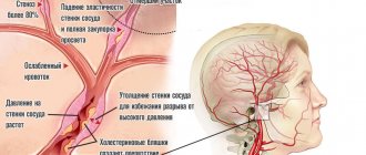
- Atherosclerosis. Statins, fibrates, nicotinic acid, special diet, giving up bad habits.
- Thrombosis. Antiplatelet agents, anticoagulants, also thrombolytics.
- Drug addiction and alcoholism. Detoxification measures, coding, rehabilitation.
- Infections. Antibiotics, anti-inflammatory drugs, analgesics, antipyretics, as well as antiviral and immunomodulators are prescribed.
There are a lot of options. The strategy needs to be developed individually.
Peripheral (cold, venous) cyanosis
Peripheral (cold, venous) cyanosis is a condition in which the blood in the arterioles of the cutaneous vascular plexuses has a normal oxygen content, warming the skin is accompanied by redness, and after pressing, a pink spot first appears, which later acquires a bluish tint. It is observed with a decrease in minute blood volume (heart failure, pericarditis, armored heart), local stasis in the final sections of the bloodstream (cooling, collapse and shock of various origins, arterial embolism, polycythemia, etc.).
In case of carbon monoxide poisoning (carbon monoxide, CO), carbon monoxide, entering the blood, combines with the iron of hemoglobin, converting it into carboxyhemoglobin, whose concentration in the blood is more than 35 g/l, hemic hypoxia, respiratory failure and neurological disorders occur (children's neurologist - polyclinic "Markushka"). Typically, patients have a bright red (cherry-red) face, acrocyanosis, and in a later stage severe cyanosis develops.
Development mechanism
The pathogenesis of the pathological process is based on a group of factors. If we talk about them in a generalized format:
Breathing problems
They are especially common. In this case, the oxygen concentration itself decreases. During biochemical processes, carbon dioxide is released into the riverbed. This is a harmful, waste product. It needs to be brought out faster.
And this is where the problem arises. Not only are the lungs unable to deliver enough oxygen, but they are also unable to eliminate harmful gases that are toxic to the body.
The concentration grows, poisoning of all tissues and systems of the body begins. Need urgent medical attention.
Cardiovascular disorders
Another common factor in the development of the pathological process. There are a lot of diseases that cause cyanosis. Often we are talking about heart defects and functional disorders.
It makes sense to check this part with a cardiologist. The mechanism is based on a violation of blood transfer, its stagnation in the large and small circles.
Hence dysfunctional disorders. The heavier they are, the worse the situation is overall.
Blood abnormalities
In some cases, red blood cells themselves are not able to combine normally with oxygen and form full-fledged hemoglobin. This leads to the accumulation of carbon dioxide, which begins to poison the patient's entire body.
As a rule, the problem is acquired, but there are congenital types of the disorder.
It is necessary to approach the diagnosis carefully. A hematologist works with patients of this profile.
Endocrine disorders
Deviations from hormonal levels. They are extremely common. In this case, the tissues themselves are unable to normally receive and absorb oxygen. They produce carbon dioxide as usual.
The concentration of carboxyhemoglobin increases several times. Cyanosis is pronounced and clearly visible. Treatment is very difficult.
Intoxication processes
In case of poisoning with salts of heavy metals, vapors of acids and volatile compounds, cellular respiration is disrupted. Rapid ischemic processes occur that pose a danger to life and health. Urgent inpatient correction is needed.
These are the basic mechanisms of the problem. Typically they occur individually and not as a system. There are exceptions to what has been said.
Methemoglobinemia
A large number of drugs and chemical compounds, including nitrates, cause methemoglobinemia . In this case, hemoglobin iron is oxidized into trivalent iron, which is not able to firmly bind oxygen. The cyanosis that develops gives the skin a brownish rather than a bluish tint. The blood is dark in color and has a chocolate tint.
The severity of symptoms is determined by the level of methemoglobin in the blood: with a methemoglobin level of 15-20%, there is no cyanosis or there is mild cyanosis; at 24-45% methemoglobin, severe cyanosis, lethargy, dizziness, respiratory distress, and cardiac arrhythmias occur; at 45-55% methemoglobin, the effects of central nervous system depression increase; at 55-70% coma, convulsions, shock, arrhythmias develop; if the methemoglobin level exceeds 70%, death occurs.
Hereditary methemoglobinemia is associated with a hereditary defect in methemoglobin reductase, the latter ensures the reduction of ferric iron to ferrous iron. Blood test for children - medical.
Diagnostics
The examination is carried out under the supervision of cardiologists, pulmonologists and therapists. Subspecialty doctors are hired as needed.
A sample list of events would be:
- Questioning the patient about complaints and symptoms of the pathological process.
- Taking a history to better understand the origin of the disorder.
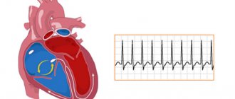
- ECG, ECHO-Kg. In order to investigate the condition of the heart. Anatomical (structural) and functional, detect defects and other disorders.
- Spirometry. Function of external respiration. To assess the condition of the pulmonary structures.
- General blood tests, biochemical, hormone tests. An expanded picture of lipids (lipid profile) is also needed.
- Allergy tests.
- Serological studies, bacteriological cultures, PCR, ELISA and other methods.
The question is put to rest by a specialized specialist or a group of doctors.
“Gray” syndrome is a form of cyanosis in children in the first months of life associated with the use of chloramphenicol
“Gray” syndrome is a form of cyanosis in children in the first months of life associated with the use of chloramphenicol. In children, regardless of age (especially with glucose-6-phosphate dehydrogenase deficiency), sulfonamides in high doses can cause sulfhemoglobinemia, and for clinical signs of cyanosis to appear, a blood level of 5 g/l of sulfhemoglobin is sufficient. Ingestion of 100-300 mg of methylene blue per day restores the oxygen transport function of the blood in all forms of methemoglobinemia, while ascorbic acid in a dose of 100 to 500 mg is effective in congenital methemoglobinemia. Sulfhemoglobin is a strong compound, and methylene blue has no effect on the restoration of the oxygen-containing space of the blood.
The light blue coloration of the skin in argyria may resemble cyanosis, but abnormal skin pigmentation due to skin color can occur in Addison's disease or hemochromatosis.
Therapy
During neurorehabilitation
Doctors are trying to normalize hemoglobin and increase oxygen levels; after using oxygen therapy, cyanosis should completely disappear in infants. If cyanosis becomes chronic, then it will not be possible to quickly eliminate the blue color of the skin; it may reappear even after complete disappearance.
During the treatment of facial cyanosis in a newborn, medications belonging to the following groups are often used:
- cardiac glycosides;
- bronchodilators;
- antihypoxants;
- analeptics.
Only a specialist can determine the reasons for the appearance of a bluish color on the skin of newborns, and he will also prescribe the most effective treatment in a particular case. Most often, the disease is stopped in the early stages, but if a child was born with a heart defect, cyanosis can only be removed surgically.
How to deal with cyanosis?
This pathology is not treated as a separate disease, since the symptoms indicate the presence of a more serious anomaly. Restorative measures are aimed at diagnosing the root cause and getting rid of the main pathological process. If the patient feels a constant lack of air, oxygen therapy is used. With regular use of the mask, the color of the fabrics is gradually restored, which indicates the effectiveness of the measure.
At risk are people who regularly consume ethanol-containing drinks, abuse inhalation of nicotine and combustion products, do not follow a diet, abuse fatty foods, are regularly in stressful situations, have an old post-traumatic condition of the lower extremities, diabetic syndrome, frostbite, varicose veins, maintain a regime of activity and proper rest.