Heart valve surgery
Heart valve surgery ¦ Closed and open mitral commissurotomy
Closed mitral commissurotomy consists of surgical separation of the adhesions of the left atrioventricular orifice in case of its stenosis. This operation is performed on a closed heart without the use of artificial (extracorporeal) circulation from a left- or right-sided thoracotomy approach.
After transesophageal echocardiography has excluded the presence of a thrombus in the left atrial appendage, a purse-string suture is placed on it, the heart cavity is penetrated and a digital inspection is performed, thus assessing the condition of the mitral valve, determining the degree of its stenosis (narrowing) and the mobility of the leaflets.
Loose adhesions can be separated manually, dense adhesions - only with the help of special instruments (dilators, commissurotomes), which expand the narrowed mitral orifice to a diameter of 3.5-4 centimeters. In order to reduce the risk of embolism of the arterial vessels of the brain, it is recommended to apply pressure on the carotid arteries at the time of performing this manipulation. The inability to perform an adequate closed commissurotomy is considered a reason to switch to extracorporeal circulation and perform open surgery.
By the way, since the 1950s, since the introduction of the artificial blood circulation method into practice, closed mitral commissurotomy began to be replaced by prosthetics. Its use is now considered justified only when there is no possibility for one reason or another to carry out artificial blood circulation. Also, closed mitral commissurotomy is sometimes performed in pregnant women with severe mitral stenosis.
Open mitral commissurotomy is used when there is mild damage to the patient’s valve apparatus in the absence of massive calcification. In this case, the operation involves performing a median sternotomy and switching to extracorporeal circulation with the so-called bicaval cannulation. The surgeon enters the left atrium from its posterior surface in the area of the interventricular groove. After the mitral valve is exposed, they begin the commissurotomy itself, first of all identifying the area of fusion of the leaflets, and then cutting this area in the direction from its free end to the fibrous ring. Then the chordae tendineae are carefully separated and the incision continues longitudinally to the papillary muscles.
In this case, small calcium deposits can be locally removed, while being careful not to damage the valve apparatus in any way. Finally, the left atrium incision is sutured and artificial circulation is stopped. Unlike closed commissurotomy, the open technique allows for more reliable removal of blood clots from the atrium and dissection of existing adhesions. Open mitral commissurotomy gives excellent long-term results, but is associated with the risk of developing restenosis or valve insufficiency. Only a small proportion (no more than 7%) of all patients subsequently require mitral valve replacement. With closed commissurotomy, there is a high probability of mitral regurgitation and embolism, especially in the case of atrial thrombosis and the presence of valve calcification.
(495) 506-61-01 — Where is the best place to have heart valve surgery?
REQUEST TO THE CLINIC
Coronary angiography
MITRAL VALVE SURGERY – MITRAL COMISSUROTOMY
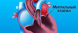
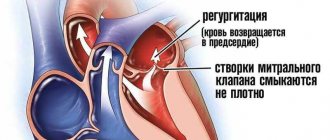
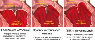
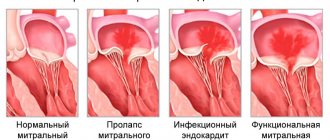
The mitral valve (bicuspid valve) of the heart is the valve between the left atrium and the left ventricle of the heart. It is represented by two connective tissue plates that prevent, during left ventricular systole, regurgitation (backflow) of blood into the left atrium.
Various pathological processes, both acquired and congenital, can cause valve dysfunction.
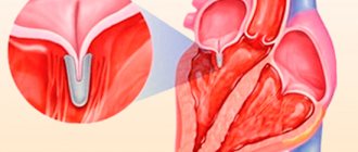
Mitral commissurotomy is a method of surgical treatment of congenital or acquired pathology of the mitral valve. MITRAL COMISSUROTOMY is a surgical operation whose purpose is to dissect the adhesions of the petals of the mitral heart valve (commissures).
Mitral commissurotomy is performed to treat mitral stenosis, a pathological narrowing of the mitral valve of the heart (between the left atrium and ventricle). The operation is performed on all patients with mitral stenosis, with the exception of the first stage of the disease, when the body’s own compensatory mechanisms allow it to easily cope with the resulting disorders.
There are two types of mitral commissurotomy:
Closed
Open
CLOSED MITRAL COMISSUROTOMY
Closed mitral commissurotomy is a surgical separation of the fusions of the left atrioventricular orifice in case of its stenosis.
This operation is performed on a closed heart without the use of artificial (extracorporeal) circulation from a left- or right-sided thoracotomy approach.
After transesophageal echocardiography has excluded the presence of a thrombus in the left atrial appendage, a purse-string suture is placed on it, the heart cavity is penetrated and a digital inspection is performed, thus assessing the condition of the mitral valve, determining the degree of its stenosis (narrowing) and the mobility of the leaflets.
Loose adhesions can be separated manually, dense adhesions - only with the help of special instruments (dilators, commissurotomes), which expand the narrowed mitral orifice to a diameter of 3.5-4 centimeters.
In order to reduce the risk of embolism of the arterial vessels of the brain, it is recommended to apply pressure on the carotid arteries at the time of performing this manipulation.
The inability to perform an adequate closed commissurotomy is considered a reason to switch to extracorporeal circulation and perform open surgery.
Since the 1950s, since the introduction of artificial blood circulation into practice, closed mitral commissurotomy has been replaced by prosthetics.
The use of closed commissurotomy is now considered justified only when it is not possible for one reason or another to perform artificial circulation. Closed mitral commissurotomy is sometimes performed in pregnant women with severe mitral stenosis.
With closed commissurotomy, there is a high probability of mitral regurgitation and embolism, especially in the case of atrial thrombosis and the presence of valve calcification.
OPEN MITRAL COMISSUROTOMY
Open mitral commissurotomy is an operation that involves performing a median sternotomy and switching to extracorporeal circulation with so-called bicaval cannulation.
Open mitral commissurotomy is used when there is mild damage to the patient’s valve apparatus in the absence of massive calcification.
The surgeon enters the left atrium from its posterior surface in the area of the interventricular groove. After the mitral valve is exposed, they begin the commissurotomy itself, first of all identifying the area of fusion of the leaflets, and then cutting this area in the direction from its free end to the fibrous ring. Then the chordae tendineae are carefully separated and the incision continues longitudinally to the papillary muscles.
In this case, small calcium deposits can be locally removed, while being careful not to damage the valve apparatus in any way.
Finally, the left atrium incision is sutured and artificial circulation is stopped.
Unlike closed commissurotomy, the open technique allows for more reliable removal of blood clots from the atrium and dissection of existing adhesions.
Open mitral commissurotomy gives excellent long-term results, but is associated with the risk of developing restenosis or valve insufficiency. Only a small proportion (no more than 7%) of all patients subsequently require mitral valve replacement.
Mitral valve stenosis - symptoms and treatment
Treatment tactics for mitral stenosis depend on its stage.
In stage I stenosis, patients are not indicated for surgical treatment, since compensatory mechanisms develop in the body. Compensation is supported by primary and secondary prevention of rheumatic fever.
The main task of primary prevention is to prevent the development of rheumatism. It involves the complete and timely elimination of streptococcal infections in sinusitis, rhinitis, sore throat and other diseases of the upper respiratory tract.
The goal of secondary prevention is to reduce the risk of re-exacerbation of rheumatism. It involves courses of bicillin prophylaxis (intramuscular injections of bicillin-5), observation by a rheumatologist, and the use of non-steroidal anti-inflammatory drugs in the event of colds and during various operations.
For primary and secondary prevention of rheumatism, it is important to follow the regimen. It consists of a healthy lifestyle, proper nutrition, exercise, absence of bad habits, good living conditions, timely visits to the dentist, medical supervision and treatment for colds.
In stage II stenosis, the indications for surgery are relative. Treatment options considered:
- closed mitral commissurotomy - an operation to widen the mitral valve, which is performed using a special dilator;
- reconstructive surgeries are valve-sparing surgeries in which valve function is restored using various suture technologies while preserving the valve leaflets and structures of the valve apparatus.
These operations make it possible to save patients' lives without leading to specific complications.
For stages III and IV of stenosis, surgical treatment is mandatory. Conservative therapy (taking cardiac glycosides and diuretics) brings only a short-term effect, since with an increase in physical activity, circulatory disorders still occur.
Closed mitral commissurotomy is indicated for patients with isolated mitral stenosis or re-fusion of the valve leaflets (restenosis) without gross changes in the valve structures, as well as with concomitant mitral regurgitation or grade I calcification. Mortality with this type of treatment is 0.5-1%. Repeated operations are performed in 35% of cases 10-15 years after the first surgical intervention [12].
In patients at high surgical risk, transcatheter balloon valvuloplasty is performed. It also aims to dilate the mitral valve. To do this, a percutaneous puncture of the common femoral vein is first made. Through it, the doctor inserts conductors and catheters into the right atrium. There, a puncture of the interatrial septum is performed, after which a balloon is inserted into the mitral valve ring and inflated [13].
Indications for reconstructive operations on the mitral valve are defects with predominant insufficiency without gross changes in the leaflets and structures of the valve apparatus, as well as the absence of calcification. Mortality after reconstructive surgery is 1.5-4%. Long-term survival rate is 90% [14].
In all other cases: valvular infective endocarditis, post-infarction defects, grade II or III calcification, the most effective operation is valve replacement with a mechanical or biological prosthesis. Mortality after such treatment ranges from 2-8%. The five-year survival rate of patients using modern types of artificial and biological prostheses is 75-90% [15].
Before any operation, the doctor must explain to the patient the course of the operation, anesthesia, possible risks and complications. After this, the patient signs consent for the operation. A few days before the planned intervention, drug therapy is carried out to stabilize the patient’s condition and prevent complications that may arise during or after surgical treatment. Hygienic treatment of the patient’s skin is also necessary: taking a shower, shaving surgical access sites. The patient should not eat food on the day before surgery.
After the operation, the patient is under observation in the intensive care unit. After stabilizing his condition and restoring the vital functions of the body, he is transferred to a specialized department under the supervision of the attending physician. At this time, the patient follows all recommendations for taking medications, activity and nutrition. With the help of a physical therapy doctor, the patient performs restorative exercises (breathing, motor) daily. The attending physician monitors the increase in the patient’s physical activity (metered walking), performs the necessary dressings and helps to recover psychologically.
After discharge, the patient must strictly follow all the doctor’s recommendations. It is necessary to be observed on an outpatient basis by a cardiologist and perform ECHO-CG annually. For patients with a mechanical heart valve, it is important to monitor such laboratory blood indicators as the INR (International Normalized Ratio) level. This is due to the use of warfarin, a blood thinning drug. Its target values should be in the range of 2-3.
SURGERY
J. Bender
AND
Infectious complications after surgery pose a threat to patients, cause morbidity, affect the workload of hospital staff, and their development leads to increased costs for the hospital and health insurance companies for treatment. Antibiotic prophylaxis has been beneficial for the past 20 years, and there has recently been some agreement that effective prophylaxis can be achieved with a single dose of an appropriate drug (or combination of antibiotics) at the start of surgery. Administration of a single dose reduces the likelihood of the emergence of resistant strains of microorganisms and is cheaper than repeated doses of antibiotics. G. Anderson et al., from two university hospitals in Melbourne, Australia, conducted a prospective study to evaluate the effectiveness and costs of three antimicrobial regimens used to prevent infectious complications after abdominal surgery. 1070 patients were randomly assigned to receive one of three treatment regimens consisting of ceftriaxone (1 g IV) or cefotaxime (1 g IV)—both third-generation cephalosporins—or ticarcillin, a moderately broad-spectrum penicillin, in combination with clavulanic acid (3.1 g of this combination intravenously). Interventions included appendectomy, elective and emergency colorectal surgery, esophagogastroduodenal surgery, small bowel resection, cholecystectomy, and common bile duct examination. Patients undergoing appendectomy or colorectal surgery were given an additional 500 mg intravenous metronidazole, which is effective against anaerobic organisms and protozoa (Trichomonas), if they were randomized to receive one of the cephalosporins. Metronidazole was not added if the patient received ticarcillin plus clavulanic acid after randomization. Wound infections were defined as dangerous when hospital stay was prolonged or reoperation was required; A wound infection was considered minor if it did not affect the length of hospital stay. The following results were obtained: the rate of wound infections was 4.3%. Twenty-one (2%) patients developed a serious wound infection, as expected primarily after colorectal surgery, whereas 25 (2.3%) patients had minor wound infections. There was no difference in the severity of wound infections between the three groups receiving different treatment regimens; The fewest minor complications (virtually none) were observed in the ceftriaxone group; infections were caused mainly by Staphylococcus aureus and colon inhabitants such as Enterobacteriaceae and E. coli. No differences were found in the incidence of postoperative infections of other sites (lungs, urinary tract) between the three groups. The side effects of the antibiotics used were minimal: in the ceftriaxone group there was 1 case of mild thrombocytopenia, which lasted only 1 day; another patient receiving cefotaxime experienced urticaria for a short period of time. Given that there was no difference in the incidence of infectious complications between the three groups, the researchers analyzed treatment costs. They accurately took into account not only the cost of purchasing drugs, but also the cost of necessary treatment for infectious complications that developed in any of the three groups. The cost of purchasing ceftriaxone was twice that of the other two regimens, which cost the same. Costs for treatment of infectious complications were highest in the ticarcillin plus clavulanic acid group, whereas they were similar in the other two groups. The authors concluded that both cephalosporins are equally effective against infectious complications after abdominal surgery. Since the acquisition costs of cefotaxime are lower, it may be the drug of choice for antimicrobial prophylaxis.
Literature:
Anderson G, Boldiston C, Woods S, O`Brien P. A Cost-effectiveness evaluation of 3 antimicrobial regimens for the prevention of infectious complications after abdominal surgery. Arch Surg 1996;131:744-8.
PERCUTANEOUS MITRAL COMISSUROTOMY
N. Mazur N. Mazur
D
This treatment method has been used for about 15 years, which has made it possible to obtain immediate and medium-term results of the intervention, as well as to determine indications for it. Percutaneous commissurotomy (PCT), performed by experienced specialists, is characterized by a low risk of complications. In general, the need for this surgical intervention does not exceed 1%; the proportion of patients in whom this intervention is impossible ranges from 1 to 17%. In case of successful commissurotomy, the area of the atrioventricular opening increases more than 2 times (on average from 0.9 - 1 to 1.9 - 2.4 cm2). The risk of hemopericardium is 0.5 - 7%, embolism - 0.5 - 5%, severe mitral regurgitation - 2 - 19%, hemodynamically insignificant atrial septal defect - 10 - 20%. Other complications (vascular, arrhythmias) are very rare. Immediate improvement is observed when the orifice area increases to 1.5 cm2 or by at least 25% and is manifested by a decrease in pressure in the left atrium, pulmonary artery and a decrease in pulmonary vascular resistance. Long-term 5-year observations showed that the harbingers of an unfavorable outcome are age, an initially high functional class of circulatory failure, the value of vascular resistance of the pulmonary bed, and the area of the atrioventricular orifice after commissurotomy. Indications for PCI are determined by the clinical condition, anatomical changes of the valves and the experience of specialists. Intervention is usually not indicated for patients who do not complain, with the exception of those who require major surgery on other organs, want to give birth to a child, have a high risk of thromboembolism or recurrent supraventricular arrhythmia. The absolute indication for PCI is the impossibility of surgical intervention. It should be preferred in the following cases: with restenosis after surgical commissurotomy; in patients with an increased risk of poor outcome during surgery; in patients with high-grade pulmonary hypertension; in patients with severely impaired left ventricular function; in patients with aortic disease in order to improve their condition before subsequent surgery on both valves; in elderly patients. Anatomical factors that are contraindications for PCI are the following: the presence of a thrombus in the atrium, severe mitral regurgitation, severe aortic heart disease, an atrioventricular orifice area greater than 1.5 cm2. The results are significantly worse if there is valvular calcification. In cases of massive calcification of both leaflets, surgical treatment is preferable. The significant impact of specialist experience on the results of PCCT is demonstrated by data on the frequency of complications depending on the number of procedures performed. For example, if experience is limited to approximately 100 interventions, the need for emergency surgery occurs 4 times more often than if PCI was performed by a specialist who has already performed 1000 procedures. Thus, PCI should be considered as an additional treatment method for patients with mitral stenosis.
Literature:
Vahanian A. Percutaneous mitral comissurotomy. Eur Heart J 1996;17:1465-9.
PERCUTANEOUS TRANSLUMINAL ANGIOPLASTY FOR KAWASAKI DISEASE
S. Berreklouw
A
Orthocoronary artery bypass grafting is the standard therapy for severe coronary artery stenosis associated with Kawasaki disease lesions. Long-term patency of the bypass graft in elderly patients is satisfactory, which cannot be said about young children suffering from this disease. According to previous studies, the short- and long-term patency of saphenous vein grafts after 1 year is 67% and decreases to 55% after 5 to 9 years. Moreover, these shunts have a number of disadvantages: the lumen diameter of native saphenous vein grafts is too small to achieve satisfactory long-term patency in young children, and these shunts have limited ability to expand as the child grows. Currently, shunts from the anterior thoracic artery have found use in coronary artery bypass surgery due to their growth potential. However, the long-term patency of these shunts has not been determined. On the other hand, percutaneous transluminal coronary angioplasty (PTCA) is rarely performed as an alternative treatment for severe coronary stenosis associated with Kawasaki disease lesions. Since 1982, at the institute where T. Ino et al. work, all patients in whom two-dimensional echocardiography reveals a coronary aneurysm or significant dilatation in the acute stage have undergone coronary angiography within 6 months of the onset of Kawasaki disease. As a result, angiographic abnormalities in the coronary arteries were detected in 220 (79%) of 278 patients. The remaining 58 (21%) patients had no visible abnormalities suggestive of coronary aneurysm recurrence or expansion. Of the 220 patients with angiographic abnormalities, 46 had an aneurysm with or without significant stenosis and 174 had dilatation. Ultimately, 5 patients (1 female) aged 2 to 18 years (median 8 years) underwent PTCA for severe coronary artery stenosis. In the acute phase, these patients were diagnosed with Kawasaki disease and were prescribed aspirin and prednisolone (2 patients), aspirin alone (1), aspirin in combination with high doses of gamma globulin (1). There is no information about the treatment prescribed for the fifth patient. All patients received an anticoagulant (aspirin 5–10 mg per day with or without dipyridamole 3–5 mg per day orally) from the time the lesion was first detected until PTCA was performed. The time from the onset of Kawasaki disease to PTCA ranged from 2 to 16 years (median 6 years). Fluoroscopy revealed coronary artery calcification in 2 patients. One patient had three ring-shaped calcifications in the area of three coronary arteries. In another patient, annular calcification was consistent with an aneurysm of the left anterior descending artery. PTCA was considered successful if the degree of stenosis was less than 50% of that before dilatation. Control angiography was performed 6–12 months after the initial one. PTCA was found effective in 4 out of 5 patients. The lesion that was the “target” of PTCA was localized in the middle right coronary artery in 3 patients, and in the proximal left anterior descending artery in 2 patients. In all patients, the stenosis was localized proximal to the aneurysm. PTCA was performed successfully in two right-sided and two left-sided coronary stenoses. In the 4 patients in whom angioplasty was effective, the degree of stenosis decreased from 84 to 33% (p < 0.005). In the fifth patient, who had annular calcification, the percentage of stenosis at PTCA remained unchanged even when the filling pressure was increased to 10 atm. Two-dimensional echocardiography revealed no significant differences in left ventricular wall motion before and after PTCA. 4 patients in whom PTCA was performed successfully underwent control angiography after 6–12 months; the study did not reveal significant restenosis or progression of stenosis. There were no significant complications that could be attributed to the PTCA performed. PTCA was performed in these patients because the stenosis could progress and acute myocardial ischemia could develop after a few years. Criteria for performing PTCA in children with Kawasaki disease, different from those for adults, have yet to be determined. If a pronounced initial thickening is detected, then it is more advisable to use not a conventional angioplasty balloon, but a rotational destructor (rotablator) or an arterectomy catheter. Thus, PTCA can be used as an alternative to coronary artery bypass grafting in patients with coronary artery stenosis due to Kawasaki disease. Conventional PTCA should be performed in patients younger than 8 years of age due to the specific histopathological features of this disease.
Literature:
Ino T, Akimoto K, Ohkubo M, Nishimoto K, Yabuta K, Takaya J, Yamaguchi H. Application of percutaneous transluminal coronary angioplasty to coronary arterial stenosis in Kawasaki disease. Circulation 1996;93:1709-15.