Heart valve prolapse is a disease accompanied by a malfunction of the valve located between the ventricle and the left atrium and is a common and relatively harmless anomaly, during which an unnatural protrusion of the valve leaflets appears when the heart contracts.
A heart valve is a movable valve consisting of individual elements that blocks the openings for blood circulation from one part of the heart to another. The valves' job is to control blood flow. There are four valves in the heart - mitral, tricuspid, aortic and pulmonary, which allow blood to flow in one direction and prevent it from returning back. When the heart muscle contracts, pressure is created, and blood is ejected from the heart, and the valves that regulate the movement of blood in this direction at the time of muscle contraction open. After the muscle contracts, the pressure in the heart drops, the valve closes, and blood cannot return to the heart. Among other types of prolapse, the most common is mitral valve prolapse, which is caused by congenital weakness of the connective tissue that makes up the heart valves.
The heart of an expectant mother: what are the dangers of mitral valve prolapse?
The human heart is a tireless worker. On average, this muscular organ weighs only about 300 grams, but daily pumps 2 thousand liters of blood through 90 thousand kilometers of blood vessels.
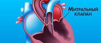
The heart consists of four chambers - the right and left atria and ventricles. Its right and left halves perform different functions. Normally, they are isolated from each other, but the atria and ventricles of the same name are separated from each other by special valves. The right atrium and right ventricle have a tricuspid valve, and the left-sided sections have a bicuspid, or mitral, valve. The purpose of these valves is to ensure proper blood flow. Namely: when the atria contract, blood should freely enter the ventricles, and when the ventricles contract, it should be sent further into the blood vessels. That is, the valves prevent reverse blood flow (regurgitation). Venous blood (saturated with carbon dioxide) enters the right atrium, and then into the right ventricle. Then it goes to the lungs, where it is enriched with oxygen and becomes arterial, acquiring a bright scarlet color. This is the so-called pulmonary circulation. Arterial blood is pumped into the left atrium and, bypassing the mitral valve, ends up in the left ventricle. From here, thanks to powerful muscle contractions, blood flows into the aorta, the largest artery in the body. Further, through the system of blood vessels, oxygen-rich blood is sent to all tissues and organs. This is a large circle of blood circulation. But if there is damage to the blood vessels, heart or valves, then malfunctions may occur in this ideal mechanism.
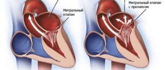
Mitral valve prolapse: “flapping” leaflet
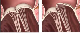
The mitral valve consists of two leaflets - anterior and posterior, which separate the cavities of the left ventricle and left atrium. This ensures proper blood flow - when the heart muscle contracts, blood should only move in one direction. With mitral valve prolapse (other names for this pathology are “flapping” or “sailing” valve, Barlow’s syndrome, late systolic murmur syndrome), its leaflets do not close completely, as if “sagging”. In this case, the left ventricle and left atrium experience a constant increased load caused by excess blood volume. In severe cases, such long-term effects on the heart can lead to serious problems.
Looking for the causes of mitral valve prolapse
What is the cause of this anomaly? Very often - heredity, namely, a violation of the development of connective tissue (connective tissue dysplasia). The fact is that connective tissue makes up half of the body’s weight and performs many functions: protective, supporting, mechanical, etc. In other words, it forms the skeleton, joints, tendons, ligaments, outer integument, forms the walls of blood vessels, and is included in all organs and systems of the body. The proper functioning of connective tissue largely depends on how the child developed in the womb. Violation of the formation of connective tissue in the fetus is the cause of many congenital diseases, including mitral valve prolapse. And very often, prolapse is accompanied by other “companion diagnoses” caused by insufficiency of connective tissue, varicose veins, prolapse of internal organs, myopia, and disorders of the nervous system.
What causes connective tissue dysplasia? The answer from doctors is unanimous: one of the main reasons is a deficiency of microelements, in particular potassium and magnesium. How can you enrich your diet with essential microelements? Include fresh meat dishes, milk, buckwheat, dried apricots, baked potatoes, spinach, bananas, and nuts in your menu. Completely eliminate simple carbohydrates - sweet pastries, sugar, as well as tea and coffee. If connective tissue dysplasia is suspected, the pregnant woman is prescribed additional potassium and magnesium supplements along with a special diet.
Other causes include previous inflammatory diseases, injuries, age-related changes, and disorders of the elasticity of connective tissue.
Types of mitral valve prolapse
Mitral valve prolapse (MVP) is divided into two groups:
anatomical (congenital);
as a syndrome associated with disruption of the nervous and hormonal systems , as well as arising against the background of various diseases - rheumatism, coronary heart disease or chest trauma.
The more significant the non-closure of its valves, the more the load on the heart increases, which has to pump a larger volume of blood. The left atrium, under constant pressure, enlarges. And during pregnancy and childbirth, when the heart is already working “for two,” additional loads can pose a threat to the health of the expectant mother and fetus.
There are three degrees of mitral valve prolapse:
- I degree – non-closure of the valves by 3–6 mm;
- II degree – 6–9 mm;
- III degree – over 9 mm;
It is also customary to distinguish the severity of reverse blood flow (regurgitation):
- I degree – regurgitation at the level of the leaflets;
- II degree – regurgitation to the middle of the atrium;
- III degree – regurgitation to the opposite side of the atrium.
Let's evaluate the risks of mitral valve prolapse during pregnancy
During pregnancy, a third circulation is added to the systemic and pulmonary circulation - the uteroplacental one. If a woman is expecting one baby, then the blood volume increases on average by 30–50%. If there are twins or triplets, then even more. Wise nature has prepared a woman’s body for such stress - during pregnancy, the size of the heart increases slightly, its muscle walls thicken. From about the 20th week, the heart rate increases - by 10-12 beats per minute. Thanks to these changes, the developing fetus is provided with all the necessary substances supplied by the blood.
As a rule, antenatal clinics require visits to a cardiologist during pregnancy, even if the expectant mother has absolutely no complaints. It is important to promptly inform your gynecologist about all existing (and past) heart problems.
In young women, minor mitral valve prolapse, as a rule, does not manifest itself in any way and is detected only by echocardiography.
With mild prolapse and slight regurgitation (grades I–II), the expectant mother usually feels normal. The pregnancy proceeds well, and the woman gives birth on her own. In this case, prolapse is considered not as a disease, but as an individual anatomical feature. Such pregnant women do not need specialized observation or treatment by a cardiologist.
If the degree of regurgitation and sagging of the mitral valve is somewhat greater, it is enough for a woman to undergo a follow-up examination with a cardiologist every 3 months.
With age, with deep prolapse, specific symptoms appear: rhythm disturbances, shortness of breath, pain in the heart. And if the expectant mother has grade III prolapse with symptoms of heart failure (such as swelling, shortness of breath, heart rhythm disturbances), then both a gynecologist and a cardiologist should monitor her condition during pregnancy.
To reduce all possible risks, it is very important for such expectant mothers to follow a strict sleep, nutrition and physical activity regime. They will have to eliminate salt from the menu and limit fluid intake to reduce the risk of swelling.
Is hospitalization necessary for mitral valve prolapse during pregnancy?
As already mentioned, for mild prolapse, only observation by a cardiologist is sufficient.
Indications for hospitalization in pregnant women with MVP may be the development of gestosis (a complication of the second half of pregnancy, in which pressure increases, swelling and protein appear in the urine), an increase in prolapse, an acute increase in pressure in the left atrium, accompanied by shortness of breath, fainting and dull pain in the left atrium. breasts
In this case, the expectant mother is hospitalized and prescribed treatment aimed at eliminating the main manifestations of mitral valve prolapse, heart rhythm disturbances, and also preventing damage to the heart muscle.
Treatment of mitral valve prolapse
No special treatment is required for this diagnosis. Intervention is necessary only in cases where arrhythmia (any disturbance in the regularity or frequency of normal heart rhythm) or pain in the heart is observed. For these conditions, sedatives (for example, products containing motherwort and valerian) help well. If necessary, the doctor will prescribe medications to prevent arrhythmia, as well as medications containing magnesium.
Perhaps the most frequently asked question when diagnosing mitral valve prolapse is – is it possible to give birth on your own? Will the heart survive? Doctors are quick to reassure: this diagnosis is not a contraindication for pregnancy and childbirth. It is only important to monitor your well-being and remember that you are responsible not only for your health, but also for the health of your child.
Is mitral valve prolapse hereditary? Alas, yes. The presence of this anomaly in a pregnant woman is one of the most common risk factors, and in the vast majority of cases, prolapse is a congenital defect of the heart valve.
Most people with this diagnosis have close relatives with a similar pathology. Children with prolapse have a thin build, they often have disorders of the musculoskeletal system (scoliosis, flat feet, weak ligaments). They are restless and get tired quickly.
But there is also good news. Mitral valve prolapse, as a rule, does not have any significant symptoms and does not require treatment (if we are talking about degrees I and II). Often the condition of the mitral valve remains unchanged throughout life. In some cases, as the child grows, the prolapse may decrease or disappear altogether. And to a large extent this will depend on the proper nutrition of the expectant mother and baby.
With minor prolapse (grades I–II), childbirth takes place in regular maternity hospitals; special observation in this case, as a rule, is not required.
In more serious cases, you will have to give birth in a specialized maternity hospital, which delivers births to women with serious cardiovascular diseases. The team of doctors, in addition to the obstetrician-gynecologist, neonatologist and anesthesiologist, also includes a cardiologist who closely monitors the work of the mother’s heart. If there are indications, the expectant mother undergoes a caesarean section.
Publications in the media
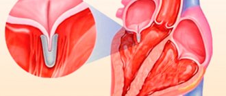
Mitral valve prolapse (MVP) is a pathological sagging (bending) of one or both mitral valve leaflets into the left atrium during left ventricular systole. Statistical data. MVP is found in 3–8% of people in the general population (apparently, the data is overestimated). Manifestations of MVP are first recorded at the age of 10–16 years; after 10 years, girls are observed 2 times more often.
Etiology. MVP can be primary or secondary • Primary MVP •• A disease inherited in an autosomal dominant manner with myxomatous deformation of the mitral valve leaflets •• MVP is also observed in patients with Marfan syndrome and other congenital connective tissue diseases, such as Ehlers-Danlos syndrome, elastic pseudoxanthoma, osteogenesis imperfecta •• In the occurrence of MVP, exposure to toxic agents on the fetus on the 35–42nd day of pregnancy may also be important • Secondary MVP can occur with: •• IHD (ischemia of the papillary muscles) •• rheumatism (post-infectious sclerotic changes) • • hypertrophic cardiomyopathy (disproportionally small left ventricle, change in the location of the papillary muscles).
Pathogenesis • Primary MVP •• Myxomatous degeneration of collagen leads to excessive accumulation of mucopolysaccharides in the middle spongy part of the mitral valve leaflets and its hyperplasia, which causes the appearance of break areas in the fibrous part of the valve. Local replacement of the elastic fibrous tissue of the valve leaflet with a weak and inelastic spongy structure leads to the fact that during systole, under the influence of blood pressure from the left ventricle, the leaflet bulges towards the left atrium (prolapses) •• In the occurrence of primary MVP in Marfan syndrome, it is also important dilatation of the annulus fibrosus of the mitral valve - it does not decrease by 30% in systole, as is normal, which leads to protrusion of one or both leaflets into the cavity of the left atrium • Secondary MVP occurs as a result of thinning and elongation of the tendon threads or their separation or dilatation of the fibrous ring. Lengthening of the tendon threads, separation of part of them lead to the fact that the leaflet is not held in place and begins to prolapse into the left atrium • If the mitral valve leaflet is excessively bowed, mitral regurgitation may occur with dilatation of the left atrium and left ventricle. It should be noted that MVP can be combined with prolapse of other valves: tricuspid valve prolapse in 40% of cases, pulmonary valve prolapse in 10%, aortic valve prolapse in 2%. In this case, in addition to mitral valve insufficiency, manifestations of insufficiency of the corresponding valve will occur. There is often a combination of MVP with other congenital anomalies of the heart - ASD, additional conduction pathways (usually left-sided).
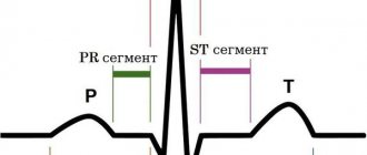
Clinical manifestations • Complaints •• In most cases, MVP is asymptomatic and is detected by chance during a preventive examination •• With more severe prolapse, patients complain of palpitations (ventricular extrasystole, paroxysmal supraventricular tachycardia, less often ventricular tachycardia) •• A common complaint is pain in the chest. It can be either atypical or typical anginal due to spasm of the coronary arteries or ischemia as a result of tension of the papillary muscles •• Shortness of breath during physical exertion, fatigue are also noted by patients with MVP •• Extremely rare manifestations are visual impairment as a result of thromboembolism of the retinal vessels, and also transient ischemic attacks as a result of cerebral thromboembolism. In the occurrence of embolic complications, importance is attached to the separation of fibrin threads located on the atrial side of the mitral valve •• Often the above-described complaints are accompanied by psycho-emotional lability • Upon examination, concomitant congenital abnormalities of the chest shape can be identified - kyphoscoliosis, funnel-shaped chest, pathologically straightened back, reduced anteroposterior chest size cells or signs of Marfan syndrome • Auscultation of the heart (a “silent” form of MVP is possible) •• The main auscultatory sign of MVP is a short mid-systolic high-frequency click (pathognomonic). It appears as a result of sagging of the mitral valve leaflets in systole into the cavity of the left atrium and their sharp tension •• The systolic click may be followed by a mid- or high-frequency late systolic murmur, better heard at the apex of the heart •• To clarify the manifestations of MVP, dynamic auscultation of the heart is used •• • Changes in left ventricular end-diastolic volume result in changes in the timing of the click and murmur. All maneuvers that contribute to a decrease in end-diastolic volume, an increase in heart rate, or a decrease in resistance to ejection from the left ventricle lead to MVP appearing earlier (the click noise approaches the first sound). All maneuvers that increase blood volume in the left ventricle, reduce myocardial contractility or increase afterload increase the time from the beginning of systole to the appearance of a click noise (moves back to the second sound) ••• In the supine position, the click occurs later, the noise is short ••• In the position standing, the click occurs earlier, and the noise is longer ••• In the squatting position, the click occurs later, and the noise is shorter (may even disappear). Instrumental data • Usually no changes are detected on the ECG in patients with MVP. Of the detected deviations, depression of the ST segment or negative T waves in leads III, aVF are most often noted. These changes may reflect ischemia of the inferior wall of the left ventricle as a result of tension in the posterior papillary muscle due to leaflet prolapse. In patients with ECG changes, cardiac arrhythmias also occur. Some patients experience prolongation of the QT interval. Recording an ECG after taking beta-blockers increases the specificity of this method • EchoCG •• In one-dimensional mode, the “hammock” symptom is detected - sagging of one or both leaflets in systole by more than 3 mm •• In two-dimensional mode, sagging of the mitral valve leaflets into the cavity of the left atrium in systole is detected left ventricle, thickening of the leaflets more than 5 mm in diastole, lengthening of the tendon filaments, lengthening of the leaflets, dilatation of the fibrous ring •• There are three degrees of MVP, determined in a four-chamber section ••• I degree (minor) - sagging of the leaflets into the cavity of the left atrium up to 5 mm ••• II degree (moderate) - sagging of the leaflets into the cavity of the left atrium 5-10 mm ••• III degree (severe) - sagging of the leaflets into the cavity of the left atrium more than 10 mm •• Doppler examination can reveal a regurgitation jet in the left atrium. With severe MVP, dilatation of the left atrium and left ventricle occurs, detected in one- and two-dimensional modes. It should be remembered that in the presence of typical auscultatory signs of MVP, its EchoCG signs may be absent in 10% of patients. When conducting the study, you should remember about other congenital heart defects (in particular, ASD). Differential diagnosis • Mitral valve insufficiency of rheumatic origin • Isolated aneurysm of the interatrial septum • Isolated tricuspid valve prolapse • VSD.
TREATMENT Management tactics • Treatment of the underlying disease in secondary MVP • Risk groups for the development of complications (patients with severe systolic murmur, thickened prolapsed mitral valve leaflets, left ventricular hypertrophy, rhythm disturbances, fainting) undergo regular ECG, echocardiography • Prevention of endocarditis is indicated for persons with severe systolic murmur. Treatment of various options • For asymptomatic MVP without signs of mitral valve insufficiency, there is no need for treatment •• The patient should be given recommendations for normalizing lifestyle, optimizing physical activity (a decrease in the tone of the sympathetic nervous system can lead to a decrease in valvular dysfunction) •• EchoCG is recommended -control once every 1–2 years •• It is necessary to avoid drinking strong tea, coffee, alcohol, as well as smoking • For severe MVP •• If there are symptoms of MVP such as tachycardia, palpitations, chest pain, is prescribed -adrenergic blockers in small doses (for example, propranolol at a dose of 30–60 mg/day) •• In case of dilatation of the left atrium and left ventricle, prolongation of the QT interval, a history of fainting, dilatation of the initial part of the aorta, physical activity is prohibited •• Prevention is recommended infective endocarditis using amoxicillin •• For symptoms of embolization, acetylsalicylic acid is prescribed at a dose of 80–325 mg/day •• With significant changes in hemodynamics, increasing symptoms of mitral valve insufficiency, mitral valve replacement or annuloplasty is performed.
Prognosis and complications . Typically, MVP is benign. Complications of MVP most often occur in patients with systolic murmur, thickened, elongated mitral leaflets, or enlarged left ventricular cavity or left atrium. Complications include: • separation of tendon threads • severe mitral valve insufficiency (0.06%) • fibrin deposition on the mitral valve leaflets • cardiac arrhythmias • cerebrovascular pathology (0.02%) • infectious myocarditis (0.02%) • sudden cardiac arrest death (0.06% of cases with severe mitral valve insufficiency). Synonyms • Systolic click-murmur syndrome • Barlow's syndrome • Inflated mitral valve syndrome. Reduction. MVP - mitral valve prolapse.
ICD-10 • I34.1 Prolapse [prolapse] of the mitral valve
The effect of mitral valve prolapse on a child
Unless complications develop during pregnancy or childbirth, MVP and mitral valve regurgitation usually do not pose any threat to the fetus. In some cases, heart valve problems can affect the fetus, and some studies have suggested that women with MVP are at risk for preterm birth. They are also more likely to have low birth weight babies.
Additionally, it is important to point out that women with mitral valve prolapse are more likely to give birth to children with a similar disease.
Is mitral valve prolapse a hereditary disease? Yes, most often MVP has a strong hereditary tendency. The good news is that most babies born with prolapse do not experience symptoms or complications of the condition.
Mitral valve prolapse in children rarely causes health problems. Most often, many people live completely unaware that they have a mitral valve disorder. However, patients with MVP must monitor and monitor their condition. Parents should monitor symptoms of mitral prolapse and ensure that their affected child has regular medical checkups, as mitral valve disease can progress over time.
The effect of mitral valve prolapse on pregnancy
Most women with mitral valve prolapse tolerate pregnancy well. Sometimes, women with MVP often find that symptoms of the disease decrease in frequency and severity during pregnancy. Even women with severe mitral regurgitation can have a safe, successful pregnancy if there are no other cardiovascular problems.
During pregnancy, various physiological changes occur in the body:
- Blood volume increases (up to 50%).
- Blood vessels dilate.
- Blood pressure drops.
- There is a decrease in systemic vascular resistance.
- Cardiac output increases.
These cardiovascular changes can significantly improve the symptoms of MVP and cause a decrease in the severity of mitral valve regurgitation.
However, not all women experience a problem-free pregnancy with mitral valve prolapse and regurgitation. MVP can negatively affect pregnancy by causing or worsening symptoms. In particular, the following may occur:
- Heartbeat
- Fatigue.
- Fainting.
Such signs are often possible symptoms of pregnancy against the background of MVP. With mitral regurgitation, complications such as heart failure, arrhythmia and endocarditis can develop.