Classification
Diagnosed cases of the disease in adolescents and adults are divided into two types:
- MVP with a high risk of developing regurgitation;
- moderate prolapse with a low risk of developing minor regurgitation.
Doctors can also divide the disease into stages:
- moderate prolapse 0-1 degree: also known as primary (even during pregnancy, no negative symptoms are felt; the body independently regulates the additional load on the heart);
- MVP stage 2 (transitional): signs - intracardiac pressure increases, in adolescents and during pregnancy there is constant shortness of breath and fatigue;
- 3rd (decompensated stage): significant damage to the heart muscle can be diagnosed.
Departments of the heart
The heart consists of four sections: two atria and two ventricles. They are connected using valves. And they also ensure the movement of blood in the right direction.
The structure of the human heart
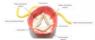
The following types of heart valves are distinguished:
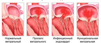
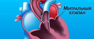
- Mitral valve , which is located on the left side of the heart between the atrium and ventricle. It consists of two doors. This area is the first to be exposed to various pressure changes, so pathologies develop more often here.
- The tricuspid heart valve is located on the right side, connecting the atrium and ventricles. Consists of three doors. During complications at the third stage, this area suffers.
- Arterial and aortic heart valves connect the corresponding vessels to the heart muscle. Each has 3 doors.
Normally, the valves close very tightly when blood enters the compartment, but in some cases their operation may be disrupted, and blood leaks through them.
It happens that the disease does not reveal itself and is noticed accidentally during a routine examination or during the treatment of other diseases. Valves that do not close completely create some turbulence, which results in a reverse flow of liquid tissue through the vessel, but it is so insignificant that it does not affect the body as a whole. According to statistics, this is observed in seventy percent of the healthy population.
The root causes may be violations of the walls of the heart, valves, and papillary muscles.
Causes
At the moment, the exact reasons why moderate mitral valve prolapse of the 1st degree occurs are not known. But the relationship between the occurrence of mitral valve prolapse with 1st degree regurgitation and some concomitant diseases has been determined:
- if adolescents have already been diagnosed with signs of connective tissue diseases, especially Marfan syndrome, then the risk that the primary stage of the disease will develop increases several times;
- signs of disturbances in the functioning of the autonomic nervous system during pregnancy also lead to the occurrence of primary MVP.
- disproportionately small left ventricle;
- genetic factors are especially dangerous during pregnancy.
Degrees
There are four degrees of reverse blood transfusion:
- With grade 1 valve regurgitation, there are no symptoms for several years. A large amount of returning blood enlarges the heart, which can cause, if proper treatment is not followed upon detection, a persistent increase in blood pressure. When examining the patient, a heart murmur is detected, ultrasound shows a slight discrepancy in the valve and a slight disturbance in blood flow.
- Stage 2 regurgitation of heart valves is characterized by greater severity of the returning flow. There is stagnation in the small circle.
- Stage 3 valve regurgitation is characterized by a large backflow, the flow of which reaches the posterior wall of the atrium. Here an increase in blood pressure develops in the pulmonary artery, due to which there is an overload on the right side of the heart muscle. As a result of this disorder, insufficiency occurs in the systemic circulation.
In the last stage, shortness of breath, heart rhythm disturbances, asthma, and pulmonary edema appear. If you do not consult a doctor for help, swelling, blueness of the skin (skin acrocyanosis), weakness, fatigue, and pain in the chest area appear.
The severity of the stages is determined by the power of the blood stream that returns to the ventricle or atrium:
- does not extend beyond the anterior leaflet of the valve connecting the left ventricle to the atrium;
- reaches or crosses the gate;
- the magnitude of the flow approaches half the length of the ventricle;
- the stream touches its top.
There is also prolapse of the bicuspid heart valve, due to which there is a backflow of body fluid of varying degrees. Previously, this diagnosis was not made often. This is due to newer ways of detecting the disease. The use of the Doppler method helped to determine the exact amount of the returning jet.
Heart valve prolapse is found in thin, tall people, and teenagers. In most cases, the disease does not cause any ailments in the patient and is detected in young people by chance, undergoing various medical examinations, for example, when entering college, or before being drafted into the army.
If the degree is first or even zero, then there is no need for treatment. The main thing is not to miss the transition to the formation of complications; for this you need to be examined by a doctor.
Diagnostics
For the initial diagnosis of mitral valve prolapse with grade 1 regurgitation, routine listening to the chest using a stethoscope is sufficient. With grade 0-1 MVP, a click will be heard immediately after ventricular contraction. If the patient is suffering from MVP with stage 2 regurgitation, then when listening with a stethoscope the blood will be heard as a “whooshing” sound immediately after the click.
But the most accurate diagnostic test is echocardiography. Only with the help of echocardiography can a cardiologist determine the degree of mitral regurgitation.
What happens in the heart during the development of regurgitation
Probably many people remember from school that a person consists of 2 atria and 2 ventricles, the work of which can be schematically described as follows:
- Blood enters the atria from the vessels (venous to the right, arterial to the left).
- Filled with blood, the atria contract, pushing their contents into the ventricles, and the valves open (tricuspid on the right, mitral on the left).
- After all the fluid has flowed into the ventricles, the valves close tightly, preventing reverse blood flow.
- After the valves close, a powerful contraction of the ventricles occurs, and blood rushes through the arteries.
The left side of the heart works with a higher load, because it supplies blood to all organs and tissues of the body. This happens normally.
The following happens:
- To accommodate the blood flowing from the ventricle and entering through the veins, the atrium chamber gradually increases. Moreover, due to the fact that the atrial cavity is partially filled at the moment of relaxation, blood flow in the pulmonary veins worsens, leading to stagnation in the pulmonary circulation.
- The flow of increased blood flow from the atrium into the ventricular cavity gradually leads to its increase.
The degree of health risk associated with this pathology depends on the size of the regurgitation and how it develops.
Treatment
Treatment of MVP depends on the patient's health status and associated factors. The goal of treatment is to improve heart function, minimize unpleasant symptoms, and avoid future complications.
- Drug treatment.
Of course, pills and injections will not be able to restore a damaged mitral valve. But the right medications will help prevent the formation of blood clots and fluid accumulation.
- Surgery.
Surgery is the optimal treatment method. But this method may be contraindicated during pregnancy.
- Waiting tactics.
Patients diagnosed with primary moderate prolapse do not require surgical treatment. But regular examinations by a cardiologist will be necessary in order to begin the necessary treatment on time.
Forecast
The prognosis for first-degree heart valve regurgitation is favorable. With constant monitoring by the attending physician, complications are identified immediately and, if necessary, treatment is prescribed.
With the second degree the situation is different. Once diagnosed, only sixty percent remain on their feet, and then only for fifteen years. Death occurs due to heart attack, heart failure, embolism, pulmonary pneumonia.
Preventive measures are aimed at reducing the risk of reverse blood flow in the heart.
Thus, heart valve regurgitation is a serious disease. Which can be either acquired or congenital. It is localized between different parts of the heart (in its right or left part). It has various degrees of development, the first of which is the simplest, has no symptoms, so the disease is difficult to calculate.
If pathology is detected, treatment is carried out using surgical methods or medication. The main thing is not to be late, so a systematic examination of the body by a specialist is recommended.
sostavkrovi.ru
The size of the divergence of the valves when closing the valve
Based on the size of the valve gap remaining after the closure of the valves, the following degrees of development of the pathology are distinguished:
- I degree. The gap after the valves are closed is insignificant, and patients do not show any complaints. The presence of regurgitation of the first degree is revealed only during a medical examination, when upon auscultation a heart murmur is heard in the apical region and a weakening of the first tone. Some people experience physiological regurgitation, where the valve leaflets are healthy but allow little blood to pass through when the ventricle contracts.
- II degree. With it, the return of blood volume can reach the middle of the atrial chamber. If the returned volume is a quarter or more, then the patient, in addition to the appearance of characteristic noises, may experience signs of stagnation in the pulmonary circulation.
- III degree. The leaking fluid reaches almost the posterior wall of the atrium, causing its overload and provoking congestion in the pulmonary veins. Gradually, overload occurs in the right half of the heart. Patients with stage III regurgitation complain of fatigue, shortness of breath, and in some cases, attacks of suffocation are possible.
- IV degree. Congestion becomes more pronounced, patients complain of shortness of breath, heart pain, and severe fatigue. Due to stagnation in the pulmonary circulation, a person experiences hypertrophy of the right heart. In severe cases, cardiac asthma and pulmonary edema may occur.
With stage I regurgitant reflux, treatment is not required; such patients are only under medical supervision. Stage II is treated conservatively, medications are prescribed to relieve the symptoms that have arisen and to treat the underlying disease that caused the deformation or prolapse of the valve.
Indications for surgical intervention are non-closure of the leaflets up to 45 mm and return of more than 60% of the volume at the time of ventricular contraction.
Diagnostic methods
In order to identify the presence of pathology and the degree of its development, the following methods are used:
- X-ray. An X-ray will show enlargement of the left chambers of the heart. With advanced pathologies, enlargement of all parts of the organ is possible.
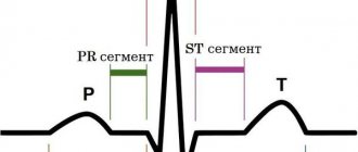
- ECG. This method is not very informative. Only in the case when the posterior papillary muscle of the myocardium is affected, negative T waves are recorded on the electrocardiogram in leads II and III, and moderate tachycardia is noted.
- Echocardiography. The study makes it possible to determine the size of the valve gap at the moment of closure of the valves, the amount of backflow and the cause of the disorder (prolapse, rupture of the valve muscles or destructive changes).
- Through esophageal echocardiography. It is used infrequently, mainly in severe cases. Allows you to determine whether there is myocardial ischemia, the presence of fibrosis or calcification in the valve tissue.
- Dopplerography. Study of the magnitude and speed of blood flow. Gives a complete picture of blood exchange in the entire vascular bed.
Mitral valve prolapse 1st degree
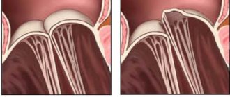
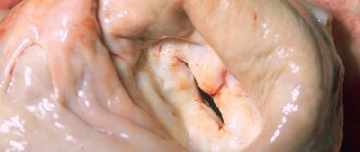
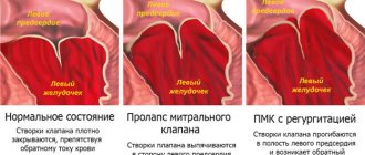
The mitral valve is located on the border between the atrium and the ventricle. Thanks to it, blood flows in one direction. If there are no abnormalities, the valve fits tightly, preventing backflow of blood. Mitral valve prolapse is a condition in which there is sagging of the valve. In this case, sagging of any leaflet may be observed, but grade 1 prolapse of the anterior leaflet of the mitral valve is usually detected.
What are the risk factors?
First degree mitral valve prolapse develops due to structural disorders of the connective tissue. As a result of these changes, the valves cannot resist the reverse flow of blood to the required extent and bend into the atrium cavity.
There are 3 degrees of pathology depending on the amount by which the valves diverge. Mitral valve prolapse with regurgitation of the 1st degree is a condition when blood moves in the opposite direction in a small volume, since the deviation of the valves is 3-6 mm. As a rule, due to a slight deviation, grade 1 mitral valve prolapse without regurgitation is diagnosed. This condition is not dangerous and can be considered as a variant of the norm.
Symptoms are more pronounced in grade 2 pathology. In this case, the doors diverge by 6 to 9 mm.
8
24/7
The most dangerous is grade 3 mitral valve prolapse, in which the protrusion of the leaflets exceeds 9 mm. This option is dangerous, as it leads to hypertrophy of the left ventricle and atrium, resulting in impaired heart function.
Causes
The reasons why grade 1 mitral valve prolapse develops are divided into congenital and acquired.
Congenital may be caused by a genetic mutation during the formation of connective tissue. We are talking about such cases with Marfan and Ehlers-Danlos syndrome. Also, this anatomical change can be inherited.
To acquired ones, i.e. Factors that had a negative impact after birth include the following:
- valve defects of a rheumatic nature;
- arterial hypertension and left ventricular hypertrophy;
- infective endocarditis;
- detachment of the chord due to trauma to the chest;
- chord avulsion due to myocardial infarction.
The most common reason for the development of grade 1 mitral valve prolapse is rheumatic disease. This is an autoimmune reaction that occurs as a result of exposure to certain types of bacteria. Simultaneously with damage to the mitral valve, other valves and joints are also negatively affected.
Symptoms
Mitral valve prolapse of the 1st degree does not manifest itself with severe symptoms. Abnormalities may be indicated by pain in the left side of the chest. The duration of pain varies in each case. It can last a few minutes or several days.
There is no dependence on physical activity, but pain can be triggered by emotional stress.
Other signs that may occur include:
- feeling of lack of oxygen, desire to take a deeper breath;
- arrhythmias;
- headaches and dizziness;
- temperature rise to low-grade levels.
It happens that grade 1 mitral valve prolapse is detected during pregnancy during an ECG. In the absence of regurgitation, this condition is not dangerous for either the mother or the fetus. However, in any case, a pregnant woman should regularly visit a cardiologist to monitor her condition.
Mitral valve prolapse increases the risk of developing gestosis in pregnant women. This condition is dangerous because the fetus does not receive enough oxygen, which leads to growth retardation and the risk of premature onset of labor. In such cases, birth is performed by caesarean section, which reduces the likelihood of complications.
8
24/7
Diagnostics
If you complain of chest pain, you should contact a cardiologist. If regurgitation is severe, a specialist will be able to detect the disorder by listening with a stethoscope.
To obtain more informative data, the following methods are used:
- Ultrasound;
- ECG;
- Doppler.
The main diagnostic method is echocardiography. It allows you to assess the degree of sagging of the valves and the volume of reverse outflow of blood. In addition, secondary changes occurring in the myocardial tissue are visible: chamber dilatation, leaflet hypertrophy, pathology of the annulus fibrosus.
An electrocardiogram is an additional research method. The ECG reflects changes in rhythm, ischemia, and myocardial hypertrophy.
In the process of making a diagnosis, it is necessary to carry out differential diagnosis with the following conditions:
- heart defects – acquired and non-acquired;
- myocarditis of an infectious nature;
- MPP aneurysm;
- prolapse of the 3-leaf valve;
- mitral insufficiency;
- pathology of the interventricular septum.
Treatment
Grade 1 mitral valve prolapse is a condition that does not require treatment if there are no symptoms. In other cases, treatment is prescribed taking into account the patient’s age, gender, and existing symptoms.
As a rule, the following groups of drugs are used:
- sedatives - since the symptoms are caused by disorders of the functioning of the autonomic nervous system;
- beta blockers - aimed at restoring heart rhythm;
- anticoagulants - preventing the formation of blood clots.
The main place in the treatment of early-stage prolapse is adjusting the patient’s lifestyle. It is necessary to choose the optimal level of both physical and mental stress. Protecting the body from emotional overstrain and stress is carried out with the help of sedatives in combination with consultations with a psychotherapist. For physical activity, visiting the pool is recommended. You should also contact a specialist who can create an individual set of physical therapy exercises.
Load regulation is of particular importance when grade 1 mitral valve prolapse is diagnosed in a child. It is important to ensure that the child does not experience any mental or physical stress, as this will lead to an increase in symptoms and the transition of the pathology to the next stage.
Both adults and children are recommended to relax from time to time in specialized sanatoriums, where they will have the opportunity to combine relaxation with undergoing the necessary procedures: acupuncture, massages, etc. Visiting sanatoriums is recommended at least once a year.
At the initial stage of the pathological process, folk remedies can be used. The use of decoctions of motherwort, sage, and St. John's wort is indicated as sedatives. Ginseng infusions will also have a general strengthening effect on the body.
Drugs that may be prescribed:
- Cinnarizine – intended to improve blood microcirculation;
- Cardiometabolites – improve metabolic processes;
- Beta blockers - normalize heartbeat;
- Vitamin and mineral complexes.
Surgical intervention is not used at the initial stage of prolapse.
If there are no symptoms, the patient does not need treatment. The only limitation that exists is control over physical activity. However, it is necessary to regularly visit a cardiologist to monitor the condition and exclude progression of the disease.
Complications
Mitral valve prolapse rarely causes complications - in only 2% of cases.
Possible complications include:
- mitral regurgitation of acute or chronic form - caused by reverse blood flow;
- endocarditis, which can lead to rupture of chords or separation of valve fragments;
- thromboembolism;
- arrhythmias.
Stage 1 mitral valve prolapse is not dangerous due to these complications, but we must not forget that this pathology is prone to progression.
Forecast
Mitral valve prolapse with regurgitation of the 1st degree usually does not lead to circulatory disorders, so the prognosis is favorable if the pathology is detected in time and adequate treatment is prescribed. With an adequate work and rest schedule, changes do not negatively affect the patient’s quality of life.
8
24/7
Prevention
Since mitral valve prolapse is often a congenital anomaly, prevention is discussed only in terms of preventing the development of complications.
In order to reduce the impact of negative factors, the following measures are used:
- regularly visiting a cardiologist and undergoing the necessary examinations;
- complete rest;
- transition to a healthy diet;
- physical therapy exercises to strengthen the heart muscle;
- timely and complete treatment of infectious diseases;
- control of the level of physical and mental stress;
- undergoing sanatorium-resort treatment;
- giving up bad habits - alcohol and nicotine addiction;
- taking medications must be agreed with a doctor;
- protection from stressful situations.
Patients can engage in the following sports:
- sport shooting;
- curling;
- golf;
- bowling;
- karate;
- gymnastics;
- volleyball;
- basketball;
- billiards.
In fact, with the first degree of mitral prolapse there are no strict restrictions. You shouldn't just do weightlifting. Excessively high loads increase the pressure exerted by the blood flow, which can result in increased sagging of the valve leaflets.
Regular visits to a cardiologist will eliminate the further development of pathology. Worsening of the situation requires the prescription of drug treatment.
Treatment methods
Prolapse of both the anterior and posterior leaflets of the mitral valve is eliminated surgically. The conservative way is ineffective.
However, the use of medications is indicated at the stage of planning the operation and after the intervention to maintain the condition of the cardiac structures in a working position.
The main method of treatment is mitral valve replacement. Plastic surgery does not make much sense and gives a worse prognosis even with identical or even greater complexity of the intervention.
At the same time, there is no point in immediately going under the knife. And not a single doctor will prescribe radical therapy at first glance at a person. At an early stage, the process may spontaneously slow down. Surgery is indicated for stable progression within 3-6 months.
What medications are used:
- Cardioprotectors. To improve metabolic processes in the heart muscle. Mildronate will do.
- Antiarrhythmics as needed. Amiodarone. But in a minimal dosage, for a short course.
- Medicines to lower blood pressure. ACE inhibitors, beta blockers, centrally acting agents, calcium antagonists. With great caution and strictly according to indications.
Lifestyle changes are mandatory. No stress, smoking, alcohol, minimum physical activity.
A new diet is also required, but it is not necessary to prescribe and follow a strict diet. If possible, you should contact a specialized nutritionist. It is independently recommended to adhere to treatment table No. 10.