The formed blood cells known as white blood cells are heterogeneous in nature. Moreover, they make up a huge number of the total mass of such structures. They perform an important function of protecting the body from harmful external influences.
They are the defensive force that does not allow viruses, bacteria, or fungi to pass through. Sometimes malfunctions also occur and immune cells begin to attack healthy tissue. This is how allergies develop, for example. But this is a slightly different topic.
As for leukocytes, they can be divided into two large groups:
- Granulocytes. They have a clearly defined core. Perform the task of the primary immune reaction. That is, as soon as pathogenic structures penetrate the body, they are the ones responsible.
- Agranulocytes. Lymphocytes and some others. They enter the “battle” second. They are responsible for the formation of stable and long-term, lifelong immunity to a particular disease.
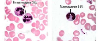
Segmented neutrophils are granulocytes that are most abundant in the blood. According to research, the number of such formed cells varies from 65 to 75% of the total mass of leukocytes, white blood cells in general.
The indicators of segmented cells are very sensitive to any deviation in the functioning of the body. Whether it is infectious or autoimmune inflammation, the analysis provides informative information that can be used as a basis for further diagnosis.
As a rule, it is not necessary to adjust the indicators of segmented neutrophils. This is a consequence, not a cause.
It is necessary to fight the primary provoking factor. This will be the basis of high-quality, competent therapy.
Position of neutrophils in blood tests
Blood consists of plasma and formed elements, which are counted and studied during a general clinical analysis (CCA) - the most common, informative blood test. Based on the results of the analysis, disturbances in microbiological processes in the body are determined, indicating the development of a particular disease.
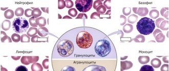
Blood cells are divided into three main groups according to their external characteristics and functional purpose:
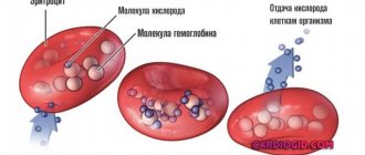
- Erythrocytes or red blood cells. They have the shape of a sphere, are responsible for transporting iron-containing protein - hemoglobin, and ensure gas exchange in the body (delivery of oxygen from the lungs to the body tissues and carbon dioxide molecules in the opposite direction).
- Platelets. Blood cells in the form of platelets. The main purpose is to ensure the preservation of the integrity of blood vessels and proper coagulation (blood clotting).
- Leukocytes or white (colorless) cells. A special group of blood cells includes two subgroups: granulocytes (granular) and agranulocytes (non-granular), which, in turn, are divided into several varieties. Agranulocytes are monocytes and lymphocytes, granulocytes are eosinophils, basophils and neutrophils (band and segmented). The totality of leukocyte cells in the OKA is represented by a leukogram, otherwise the leukocyte formula.
Colorless blood cells protect the body from the invasion of foreign agents (antigens) - viruses, bacteria, fungi, parasites. When antigens penetrate, leukocytes are mobilized and strive to eliminate them.
The biochemical process of capture and digestion of “strangers” by leukocytes is called phagocytosis. This protective reaction explains blood leukocytosis (increase in the number of white cells) during inflammatory and infectious processes. Neutrophils are pronounced phagocytes with the function of capturing and destroying pathogenic microorganisms.
Designation of the main blood elements in OKA
| WBC | leukocytes | R.B.C. | red blood cells | |
| Leukogram | ESR | erythrocyte sedimentation rate (ESR) | ||
| NEU or NEUT | neutrophils | PLT | platelets | |
| LYM | lymphocytes | RET | reticulocytes (immature red blood cells) | |
| MON | monocytes | |||
| BAS | basophils | |||
| EOS | eosinophils | |||
Neutrophil norms and their functions
If we do not specify individual levels in men and women by age (they are described here), the average number of formed cells will be from 51 to 60% of the total mass of white blood cells.
Another approximately 7-10% is accounted for by immature forms of structures. The so-called band neutrophils, which are yet to enter “adult”, functionally active life.
The main task of formed cells, white blood cells of this type, is to bind antigens and fight pathological structures, be they bacteria or other offenders. These bodies play a major role in eliminating helminthic infestations and in the development of an allergic reaction.
The laboratory, artificial function is to mark inflammatory processes, oncological phenomena and other pathological conditions, which will be discussed further.
Segmented neutrophils in OKA
The typing of neutrophils is determined by their maturation. Having formed in the bone marrow, neutrophil granulocytes enter the plasma and are divided into segments:
Causes of increased neutrophils in the blood
- Rods. They have the shape of rods without a segmented nucleus - these are immature formed elements of blood. Increasing their concentration means cell rejuvenation. In laboratory medicine it is called “shift of the leukocyte formula to the left.” A chronic shift of the leukogram to the left is observed in patients with oncological tumors and in diabetics during a diabetic crisis.
- Segmented. Mature cells with a formed nucleus and a clear structure. An increase in their number means a cellular structure or a shift of the leukogram to the right, which indicates the weakness of the bone marrow reserves and the inability to produce young cells.
Non-pathological cellular aging is caused by blood transfusion (blood transfusion). When there is a threat to the body (penetration of antigens), segmented ones are aimed at capturing and digesting bacteria and fungi. By changing the shape of the shell, the cells move towards pathogenic microorganisms, capture and digest them.
A single segmented granulocyte is capable of destroying from 20 to 30 bacterial agents. Mature neutrophils take part in the regulation of body temperature, the severity of the inflammatory process, and the level of immunity. Partially influence the coagulation process.
The lifespan of a segmented granulocyte is from 3 to 4 days. After completed phagocytosis, the blood cells die and become part of the purulent discharge along with other products of biochemical breakdown.
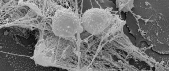
Neutrophil cells on blood microscopy
The neutrophil response (increased NEU concentration in the blood) is the primary reaction in acute processes. With a large-scale bacterial invasion or attack of certain types of bacteria that neutrophils cannot resist, the infection existing in the body becomes chronic, for which high values of neutrophilic leukocytes are not characteristic.
Reference! Low levels of neutrophils (segmented and band in aggregate) are designated as neutropenia, high values – neutrophilia (neutrophilic leukocytosis, neutrophilia).
Pelger's anomaly of neutrophils
Pelger's anomaly refers to a hereditary morphological change in the structure of neutrophil cells, namely, in the process of segmentation of their nucleus.
The disease has no clinical manifestations and is detected accidentally during a general blood test with a leukocyte count.
Due to segmentation anomalies, old segmented neutrophils in the analysis look like young band neutrophils. That is, such neutrophils have one unsegmented nucleus.
Pelger's anomaly does not lead to changes in cell granularity and does not impair their functionality. Neutrophils with pelgerized nuclei are not defective and are able to adequately perform their functions.
However, such cells often cause misinterpretation of the CBC due to the presence of a false left shift.
To confirm the Pelger anomaly, it is necessary to examine the blood of the parents (since the pathology is hereditary). In the future, before taking tests, it is necessary to notify the doctor and laboratory staff about the presence of this anomaly.
For reference. Patients with pelgerized neutrophils also do not require special treatment.
Normal values
Laboratory calculation of NEU in a clinical analysis is the number of cells per 1 ml of blood or 1,000,000,000 cells per liter (in the analysis form it is indicated as 10^9/l) and is called the absolute number (abs.) of neutrophil granulocytes. The norm for the absolute number of segmented (mature) cells in adolescents over 16 years of age and in adults is 1.8-6.5 * 10^9/l.
Norm abs. stab (immature) cells – 0.04–0.30 * 10^9/l. A simpler calculation is to measure the relative number of formed neutrophilic elements as a percentage of the total number of leukocytes. Starting from the age of 16, reference values for mature neutrophils are 45-70%, immature neutrophils are 1-6%.
In the blood of children, the number of mature and immature neutrophil granulocytes depends on age.
Normal relative indicators by age (in%)
| Child's age | After birth | Baby up to one year old | 2-4 years | 4-8 years | 8-12 years | 12-14 years old | 14-16 years old |
| Mature cells | 48-72 | 15-46 | 33-56 | 33-59 | 43-59 | 44-60 | 44-65 |
| Immature cells | 4-11 | 1-6 | 1-5 | 1-5 | 0-5 | 0-4 | 1-5 |
Minor neutrophilia is allowed in women carrying a child. During pregnancy, hematopoiesis is activated, immunity decreases, and biochemical processes are completely rearranged.
In the case when segmented neutrophils are significantly increased (1.5-2 times), this means that an infection is actively progressing in the body, threatening premature birth or miscarriage.
General meanings for pregnant women
Deviations from normal values indicate violations. High rates most often indicate acute infectious and inflammatory processes associated with bacterial or fungal activity.
A low level of mature neutrophil leukocytes requires dynamic monitoring. The deciphering of the blood OCA results obtained from the laboratory is carried out by the doctor who referred for the study.
Functions
Band and segmented neutrophils protect the body from infections, primarily bacterial and fungal. They are able to migrate outside the bloodstream to the source of inflammation, absorb microorganisms, and destroy them with the help of enzymes.
This ability of leukocytes is called phagocytosis, which happens:
- completed;
- unfinished.
When phagocytosis is completed, the microbe is completely digested inside the neutrophil, and its remains are thrown out of the cell. If not completed, the leukocyte dies along with fragments of microorganisms and is excreted in the form of purulent discharge. One segmented neutrophil can digest up to 30 bacteria.
- Band neutrophils: normal in the blood, reasons for increase and decrease
The membranes of segmented neutrophils can easily change their shape, forming protrusions, with the help of which the leukocyte moves to the site of introduction of the microorganism. The neutrophil chooses the direction of movement based on chemicals released by damaged tissues. This method of movement is called chemotaxis.
In addition to phagocytosis, segmented granulocytes perform the following functions:
- bactericidal - release substances that kill bacteria;
- influence the blood coagulation system;
- affect body temperature;
- play an important role in regulating the intensity of inflammation and activation of the immune system.
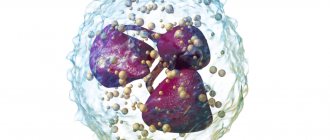
What does a segmented neutrophil look like?
Increased level of segmented neutrophils in the blood
Segmented neutrophilia (indicator more than 75%) can have physiological and pathological causes of development. Elevated levels of neutrophil leukocytes cause:
- prolonged neuropsychological tension (distress), psycho-emotional shock;
- PMS in women;
- excessive physical activity (intense sports training, other stress).
The number of mature leukocyte neutrophils is increased in chronic alcoholics and with single alcohol intoxication. A pathological shift of the leukogram to the right is a clinical sign of acute infectious and necrotic processes.
Generalized and local infections caused by the activity of pathogenic bacteria (Escherichia coli, staphylococci, enterococci, streptococci, anaerobes, Koch's bacillus). The most common ones include:
- inflammation of the appendix (appendicitis) and purulent inflammation of the abdominal cavity (peritonitis);
- acute pneumonia and pulmonary tuberculosis;
- development of acute or relapse of chronic pyelonephritis;
- sepsis (blood poisoning);
- suppuration (abscesses) of different locations (in muscles, subcutaneous tissue, bones);
- intestinal infectious diseases (dysentery, cholera, salmonellosis, etc.);
- parasitic infestations (in the acute period);
- streptococcal and streptococcal lesions of the oropharynx.
Neutrophils higher than normal in a blood test are recorded when infected with sexually transmitted diseases (in the acute period of symptoms). Necrosis:
- gangrenous lesions of the limbs;
- infarction (myocardium, spleen, kidney, etc.);
- pancreatic necrosis (death of pancreatic cells).
Chronic segmented leukocytosis accompanies non-healing trophic ulcers, ketoacidosis crisis in diabetes mellitus, and oncohematological pathologies (cancer of the circulatory and lymphatic systems). The total level of neutrophil cells may be increased:
- in the first days after surgery;
- with cyanocobalamin vitamin deficiency (chronic deficiency of vitamin B12);
- after a course of treatment with hormone-containing medications.
Neutrophilia is sometimes observed after routine vaccination.
Stages of neutrophilia
According to the degree of shift of the leukogram to the right, three stages of neutrophilia are distinguished. The first stage is moderate (7-10 absolute units * 10^9/l) and the second severe stage (10-20 absolute units * 10^9/l) are characteristic of local (local) inflammation. The third severe stage (20-60 ab. units * 10^9/l) is a sign of a generalized infectious-inflammatory process. An increase in NEU values can indicate the extent of the infectious lesion.
Education mechanism
After the initial myeloblast cell has developed, the neutrophil gradually matures into a band cell.
It then penetrates the blood plasma. After a period of time, the core of the element changes.
It is divided into segmented shares. This is how mature neutrophils appear.
Full-fledged cells, ready for protection, penetrate the capillary walls in 2-5 hours.
Decreased cell concentration
A decrease in the levels of segmented neutrophils (less than 45%) is characteristic of chronic low-grade inflammatory processes, viral diseases, blood diseases, and depletion of bone marrow reserves.
Neutropenia accompanies:
- chronic toxoplasmosis;
- zoonotic infections (brucellosis and tularemia);
- ARVI and influenza,
- viral hepatitis A, B, C;
- hemolytic and hypoplastic types of anemia;
- dysfunction of the bone marrow to produce blood cells caused by radiation or chemotherapy.
“Childhood” viral diseases in which NEU values are reduced in a child: measles, rubella, chickenpox. Neutropenia can occur due to incorrect therapy with medications: cytostatics (antitumor drugs), sulfonamides, analgesics.
The physiological reasons for the lack of segmented blood cells are due to the characteristics of dietary habits (when the diet does not contain enough foods containing B vitamins). Nutritional neutropenia is associated with a psychopathological eating disorder (anorexia) and wasting of the body (cachexia).
Stages of neutropenia
Decrease in neutrophil values by stages:
- light (1-1.5 absolute units * 10^9/l);
- moderate (0.5-1.0 absolute units * 10^9/l);
- heavy (0-0.5 absolute units * 10^9/l).
A low level of neutrophil leukocytes in a blood test without significant disturbances in the functioning of the body may be associated with genetic Kostmann syndrome - the inability of stab cells to mature.
Important! When assessing a general blood test, the doctor never considers the indicators of certain parameters separately. Conclusions about disorders and diseases are made based on a comparison of all values and the presence of somatic symptoms.
Causes in children
In younger patients, the list of diseases will be approximately the same. There are some additions. A number of conditions occur only in young patients and appear immediately.
Pediatric infectious diseases
Scarlet fever, rubella, chicken pox, whooping cough, mumps (mumps) and much more.
They are not fundamentally different from other infectious pathologies. All white blood cells rush to fight foreign agents.
Pediatricians are involved in recovery. Antibiotics are prescribed in strictly controlled dosages. Anti-inflammatory drugs, also drugs for viral infections, antipyretics. An individual regimen is prescribed.
Regular monitoring of the condition is indicated.
Congenital bone marrow disorders
Segmented neutrophils in a child can be increased as a result of hyperfunction of the bone marrow, when too many formed cells are produced: including leukocytes in different variations.
This is usually a genetically determined disorder. It is difficult to correct. All that remains is to deal with the symptoms.
Corticosteroids and immunosuppressants are prescribed. The disease often occurs in periods. The violation is replaced by remission and so on in a circle. You need to be constantly monitored by a hematologist.
Results
Segmented neutrophils are mature neutrophils (a type of leukocyte) responsible for eliminating infections, primarily the activity of pathogenic bacteria. With bacterial infection, the number of segmented cells increases.
In clinical practice, this is an indicator of an acute infectious process. Sluggish chronic diseases of bacterial etiology and viral diseases are not accompanied by an increase in the values of segmented cells. Chronic neutrophilia may indicate the development of oncological processes.
Classification
The cells are produced in the bone marrow.
Depending on their maturity, neutrophils are divided into 6 groups:
- Myeloblasts. It is formed from stem cells in the bone marrow. This cell is the basic one. Subsequently, it is transformed and modified.
- Promyelocytes. The cell is of considerable size and round in shape. The key to the structure of promyelocytes is the nucleus, which fills almost the entire volume of the cell.
- Myelocytes. The element decreases in size and acquires the correct shape. The color of the kernel is bright purple.
- Metamyelocytes. They are small in size and round in shape. The core resembles the outline of a horseshoe.
- Band neutrophils. The main part of the cell is occupied by the cytoplasm - semi-liquid content. The core narrows.
- Segmented neutrophils. The cell differs from the rod cell in the structure of the nucleus, which is divided into segments. A segment is a part in the form of a lobule. Other characteristics: size, cytoplasmic volume, granular structure do not change. A segmented neutrophil is a mature cell. The remaining groups are immature elements and are much smaller in number.