01 Aug 2021 at 10:37 MRI of the heart in Tushino 7916
“Aneurysm of the aorta of the heart - what is it?” Many patients in whom a specialist has just discovered this disease ask a similar question. An aortic aneurysm is a widespread or limited dilation of the largest vessel in the human body. With this disease, the diameter of the aortic lumen exceeds the norm by two or more times. Aortic aneurysm is one of the most dangerous heart diseases.
Which method of diagnosing aortic anomalies to choose: MRI, CT, ECHO-CG, angiography
Selection Methods
- ECHO CG in young children,
- MRI examination in adults.
What will a chest x-ray show?
Double aortic arch:
- Soft tissue compaction on both sides of the trachea
- Bilateral narrowing of the trachea.
Retroesophageal right aortic arch:
- Soft tissue compaction to the right of the trachea
- Narrowing of the trachea or displacement of the trachea on the right side.
What will fluoroscopy with barium show in case of anomaly of the aortic arch?
- Anterior view demonstrates bilateral esophageal narrowing at various levels
- The lateral view demonstrates marked posterior narrowing of the esophagus.
Why is ultrasound of the heart and aorta performed for arch abnormalities?
- Two separate aortic arches, each giving rise to the carotid and subclavian arteries.
What MRI and MSCT images of the heart and aorta will show
- May demonstrate double or right aortic arch
- The right arch is often larger (75%)
- The right duta is often located posteriorly and superiorly and often passes behind the esophagus.
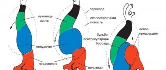
Diagrammatic representation of a double aortic arch with a predominance of the right arch.
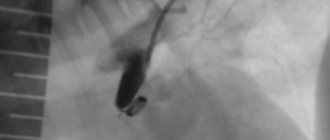
The right aortic arch (arrow) in a 36-year-old man. A chest x-ray demonstrates displacement of the trachea to the left side.
Structure and departments
The anatomical structure of the aorta differs slightly from other arteries of the elastic type. Its shape is linear almost throughout its entire length, with a gradual decrease in diameter from the center to the periphery.
The walls of the tube consist of three layers, differing in histological characteristics:
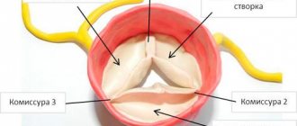
- The intima, located on the inner surface, is the endothelial layer from which the aortic valve is formed. Normally it is tricuspid, but sometimes there is a genetic mutation when it is bicuspid.
- The media, located under the intimal part, consists of elastic and smooth muscle fibers that can stretch and contract quite strongly, maintaining a normal blood flow rate.
- The adventitia, located on the outer surface, consists of connective tissue fibers, collagen and elastin cells, which together create a fairly rigid frame.
Up to 5 liters of blood are transported through the vessel every minute. This volume creates constant pressure. Its normal values are considered to be 120-140 mm. rt. Art. (systolic indicator, that is, at the moment of cardiac output) and 70-90 mm. rt. Art. (diastolic indicator, that is, at the moment of filling of the left ventricle before the next ejection).
Depending on the distance from the heart, the diameter of the aorta gradually decreases from 40 at the root to 18 mm at the place where the bifurcation of the aorta into the iliac arteries is located. The exception is the isthmus of the aorta, which is located near the heart on the arcuate part of the vessel. It has a smaller diameter than the areas located above and below. Topographically, the bloodstream is divided into three parts: the ascending section, the arch and the descending section.
Rising department
The ascending aorta of the heart is located on the left side of the sternum at the level of the third rib. At the point where the heart tissue enters the vessel, the tricuspid semilunar valve is located. This system is designed to prevent the backflow of arterial blood from the vessels into the left ventricle. Distal to the valve, close to them, are the sinuses - small bulges on the wall in which the coronary arteries originate, delivering oxygenated blood to the myocardium.
At the exit from the heart, the aorta forms a bulb - a small extension, the walls of which are thicker than those of further sections. It is more durable and has increased elasticity to dampen sudden fluctuations in blood during cardiac output. At the level of the junction of the second rib with the sternum bone, the bulb narrows and becomes an arch.
Aortic arch
The aortic arch is a continuation of the ascending section and, as its name suggests, is a curved section that deviates to the left. Its end is located at the level of the fourth thoracic vertebra, where the arch passes into the descending section.
Sometimes, in the presence of a genetic abnormality of the vascular system, the right aortic arch is found instead of the left one. Its isthmus has the shape of a ring, inside which the esophagus and trachea can be located. With a right-sided aortic arch, there is no threat to the patient’s life, but the pulsation of the vessel provokes difficulties with breathing and swallowing.
Three branches branch off from the left and right aortic arches in the isthmus region, supplying the upper part of the body: brachiocephalic, left common carotid and left subclavian.
Descending department
The descending section has the greatest extent in the systemic circulation and is located in two sections:
- the thoracic aorta passes through the chest cavity;
- The abdominal aorta passes through the abdominal cavity.
Both the thoracic and abdominal aortas have a linear shape, from which arteries of smaller diameter periodically arise. The beginning of the section is located at the level of the fourth thoracic vertebra, and the end is at the level of the fourth lumbar vertebra.
The thoracic part of the vessel extends to the 12th thoracic vertebra, where it passes through an opening in the diaphragm and continues with the abdominal aorta. Along its entire length, unpaired and paired branches extend from it, supplying blood to the mediastinum, lungs, ribs, chest muscles, pleura, etc.
Arteries also branch from the abdominal part of the aorta, which are involved in the blood supply to the abdominal organs: the muscles of the anterior abdominal wall, liver, stomach and intestines, etc. It is also responsible for the blood circulation of the pelvic organs. The end of the abdominal aorta is the bifurcation of the vessel into the iliac arteries.
Forecast
{banner_banstat9}
In itself, aortic thickening is not a diagnosis, but just an instrumental finding. You can say anything concrete after assessing the root cause.
The following factors play a major role:
- Dynamics of deviation. Progression is associated with more severe general condition and outcome.
- The age of the person. Elderly people have a relatively unfavorable prognosis.
- Family history. If there was at least one representative in the family with the condition in question, it is necessary to carefully check every six months. In such individuals, the anomaly progresses rapidly. Therefore, delay reduces the chances of successful treatment.
- The presence of concomitant somatic pathologies. Among the especially dangerous ones are hypertension, diabetes mellitus, metabolic syndromes, and atherosclerosis.
- Possibility and necessity of radical treatment. If surgery is required, the prognosis is initially worse. Further, everything depends on the success of the therapy. The absence of such a need gives a better outcome, although it does not guarantee it.
In general, even an aneurysm, as one of the most severe complications, is associated with a survival rate of 85-90%.
Attention:
Heart failure and circulatory disorders in the brain reduce the patient's chances by 30-40%.
Pathologies
The load to which the abdominal aorta and the thoracic section of the vessel located above are subjected often leads to the appearance of serious pathologies. Such diseases of the aorta are called acquired, that is, arising during the functioning of the body or the influence of external factors on it. These include:
- atherosclerosis - deposition of cholesterol on the walls of the vessel with subsequent calcification of plaques, as a result of which they lose their elasticity and worse regulate blood pressure;
- aneurysm - a pathological protrusion (pocket) on the wall, resulting from increased blood pressure on a vessel in which dissection or rupture may occur;
- aortic dissection - splitting of the vessel wall with blood entering the cavities, which often accompanies an aneurysm;
- inflammation of the vessel wall due to intoxication or an autoimmune process.
The list of congenital anomalies is more diverse, since it includes pathologies of an anatomical and physiological nature. Such diseases include:
- valve insufficiency, in which blood is thrown back into the ventricle, causing hypertrophy of the heart muscle and other pathologies;
- valve stenosis - fusion of the semilunar valves, due to which blood is not able to penetrate from the ventricle into the circulatory system at a normal speed;
- stenosis is a limited narrowing of a vessel, as a result of which the blood pressure in the upper area remains too high, and in the lower area – too low;
- Marfan syndrome - insufficiency of the connective tissue layer of the wall, in which aneurysms occur on the vessel and the valves become dysfunctional;
- bifurcation of the arch is a pathology in which the vessel has an additional bifurcation in the area of the esophagus and trachea, the latter are compressed in a ring, which makes swallowing and breathing difficult;
- right-sided arch - an abnormal position of the bend of the vessel, which sometimes leads to difficulties with breathing and swallowing.
The higher the affected part of the vessel is located, the greater the impact on health the pathology has. For example, the abdominal aorta affected by atherosclerosis provokes changes in the functioning of individual organs of the gastrointestinal tract, sometimes the pelvis and lower extremities, and with changes in the thoracic region, the heart, all organs located below the pathological focus, and even the brain will suffer.
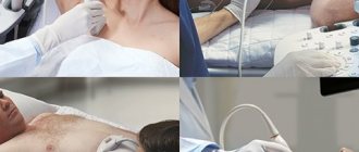
Diagnostic methods
When signs of disease appear, a comprehensive diagnosis is carried out. It consists of instrumental studies:
- computed or magnetic resonance imaging;
- X-rays of the abdominal or chest cavity;
- EchoCG.
The listed procedures help the doctor detect pathological changes, determine their scale and exact location. To clarify changes in the functionality of the aorta of the abdominal cavity or thoracic region, Dopplerography and ultrasound of the vessel are used.
Laboratory blood tests for pathologies of the circulatory system are not very informative. They are used as auxiliary tools to confirm the diagnosis. For example, after visualizing a foreign body attached to the wall, the doctor uses tests to determine what it could be: if cholesterol is high, atherosclerosis is diagnosed, and if platelet levels are high, thrombosis is diagnosed.
Causes
There are both fundamental factors and risk factors that do not directly determine the development of the pathological process. First group:
Current or previous tuberculosis
This is a dangerous infectious disease. It is provoked by mycobacterium, also called Koch's bacillus. Can exist for a long time without symptoms. The clinic is already formed at pronounced stages.
Damage to the aorta is a complication, the prevalence is up to 25% of the total number of cases. Timing does not matter; involvement is possible at the very first stages, which affects the overall forecast.
The walls of the vessel are destroyed and scarred, the lumen narrows, and the process progresses independently. Recovery is possible only with comprehensive treatment of tuberculosis in a hospital setting.
The total duration of supervision is 6-12 months. Hospital stage - no more than 3. The rest of the period is spent on outpatient care. Throughout the rest of life, the patient should be monitored for relapses.
Syphilis
Danger does not come immediately. This sexually transmitted infection can be transmitted non-sexually, but the likelihood of this option is extremely low.
Urgent treatment of the condition is required. Because aortic damage occurs in approximately 30% of situations.
Thickening of the aortic walls does not occur immediately; it is a later complication. There are known cases of the development of a delayed phenomenon after 10-20 years from the conditional cure of syphilis.
Therefore, such patients are recommended to undergo at least echocardiography every 6 months and closely monitor their well-being.
Aortitis
Inflammatory process in the corresponding vessel. Relatively rare. Again, as a consequence of an infectious disease.
It is a complication of herpes, damage by pyogenic flora, and septic myocarditis. In rare cases, it develops as a result of autoimmune pathologies. For example, rheumatism or vasculitis.
Inflammatory diseases need to be treated in a timely manner, because the likelihood of complications is significant.
Aortitis can be treated with antibiotics or immunosuppressants, depending on the etiology of the process. In any case, hospitalization in a cardiac hospital is indicated.
Hypertension
Stable increase in blood pressure. It occurs for many reasons, which themselves require identification and qualified assessment by doctors.
At the first stage of deviation, when the disease is still unstable in its manifestations, there is a chance for normalization. As the condition progresses, the risks of impairment increase. The third degree of hypertension cannot be treated.
As mentioned, hardening of the aorta of the heart is the result of degeneration, excessive stress on the walls, or a combination of these factors.
In the absence of therapy, there is a high probability of vessel rupture. Sooner or later this will happen. Specific medications are prescribed to lower blood pressure (ACE inhibitors and others, as indicated).
Age-related changes
Violation of the functional activity, anatomical characteristics of the connective tissue that makes up the aorta and its walls. Mainly appears to be the result of age-related changes.
Variations are possible. The same phenomena are observed in diabetes mellitus, genetic pathologies, anomalies of intrauterine development with spontaneous features.
Atherosclerosis
There are two types.
- The first is a narrowing of the vessel, stenosis. Occurs in smokers, alcoholics, drug addicts, and people with arterial hypertension. It is a complication and requires urgent help. If the process is stable, surgery may be required if conservative methods do not have an effect.
- The second option is occlusion or blockage of the vessel with a cholesterol plaque. The result of a disorder of lipid metabolism in the body. It is less dangerous because the condition develops over months or even years.
Gradually, the formation becomes calcified and becomes covered with mineral salts. Such an atherosclerotic plaque cannot be dissolved with statins. Surgical treatment is required.
Functions
The aorta of the heart has a huge role to play. Being the beginning and main highway of the systemic circulation, it delivers blood to all organs of the chest, abdominal cavity and small pelvis. From its arch originate the vessels that supply blood to the head and upper limbs, and from the abdominal part of the aorta blood enters the lower limbs. From this we can conclude that almost all organs and tissues of the human body are supplied with blood through the aorta of the heart.
To the point where the aortic bifurcation begins, arteries branch off from it to all vital organs:
- in the chest cavity - to the heart and surrounding tissues;
- in the abdominal cavity from the infrarenal abdominal aorta - to the gastrointestinal tract and urinary organs;
- in the abdominal cavity from the intrarenal part of the vessel to the organs of the reproductive system.
The abdominal part of the aorta is completed by a bifurcation, that is, the bifurcation of the aorta, into the left and right iliac arteries, through which blood flows to the lower extremities.
In addition to nourishing tissues, the vessel takes part in the regulation of blood pressure. The mechanism of stretching and relaxing the walls prevents the formation of a large gap between blood pressure in systole and diastole.
Complications
{banner_banstat10}
Possible consequences:
- Thrombosis. The formation of a clot in one of the arteries, sometimes in the aorta itself. Causes local ischemia. When torn off and moved along the riverbed, it often blocks the pulmonary vessel and coronary structures, which immediately leads to death.
- Aneurysm. Wall protrusion. It is observed against the background of stable degeneration and dystrophy of aortic tissue.
- Rupture, delamination. Leads to death within a matter of seconds. There were cases to the contrary, but they were casuistic. There are hardly more than 0.5%.
Thickening of the aortic walls is not an independent disease. It is not considered a diagnosis and is not represented in the ICD-10 classifier. This is a consequence, an accidental find.
After eliminating the root cause, there is every chance to stop the progression and achieve results without affecting the quality and length of life.