Pulmonologist
Prokhina
Maria Egorovna
Experience 38 years
Pulmonologist
Make an appointment
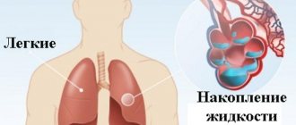
Pulmonary edema is a pathological, very serious condition, which is characterized by the release of transudate into the lung tissue. As a result, gas exchange is disrupted, which leads to serious consequences, including death.
Emergency care for pulmonary edema is the only thing that can increase the patient’s risks of survival and recovery. A person in such a situation requires immediate medical attention.
Pulmonary edema itself is most often a complication that accompanies serious problems of organs and body systems, for example, the cardiovascular system, gastrointestinal tract, etc.
The main symptoms of pulmonary edema in humans
Pulmonary edema is a life-threatening condition for the patient that requires urgent hospitalization and emergency care.
The pathology is characterized by the accumulation of intercellular fluid inside the alveoli and in the lung tissue. The main symptoms of pulmonary edema in humans in the initial stage are: increasing weakness, tachycardia, increased sweating, dry paroxysmal cough, worsening in a lying position. Depending on the course, the following types are distinguished: acute, subacute, fulminant and prolonged edema. The main reasons for the development of pathology:
- acute intoxication of the body;
- acute left ventricular failure;
- chronic lung diseases;
- TELA;
- severe traumatic brain injury with convulsive activity;
- long-term artificial ventilation;
- diseases accompanied by increased intracranial pressure.
The main causes of toxic pulmonary edema: inhalation of irritating or suffocating gases.
Causes and pathogenesis
Pulmonary edema is not considered an independent nosological form. This is rather a consequence, a complication of diseases. Common reasons:
- diseases that release toxins (sepsis)
- pneumonia
- taking too high doses of certain medications
- drugs
- pulmonary embolism
- effects of radiation on the lungs
- cardiac pathologies in which LV failure develops, blood stagnates in the pulmonary circle
- diseases in which there is too little protein in the blood
- lung diseases with congestion in the right circle
- infusion of solutions in large quantities without forced diuresis after this
As for pulmonary embolism, a blood clot can form in the body, which then breaks off, traveling through the bloodstream, it enters the pulmonary artery, blocking it. The pressure in it increases, which causes pulmonary edema.
Classification of pathology
There are cardiogenic, non-cardiogenic and mixed pulmonary edema. Non-cardiogenic edema is a condition not associated with heart disease. This term combines toxic, nephrogenic, allergic and other forms of pulmonary edema. Cardiogenic is a consequence of heart disease. First aid for pulmonary edema due to heart failure will be discussed below.
Classification according to the development of pathology:
- fulminant edema (develops very quickly and, as a rule, ends in the death of the patient);
- acute (develops within 4-6 hours and is a complication of heart attack, severe traumatic brain injury and anaphylaxis);
- subacute (characterized by a wave-like increase in symptoms, being a complication of uremia and liver failure);
- prolonged edema (develops over the course of a day and is characteristic of chronic lung diseases).
Clinical manifestations in adults
Pathology does not always develop rapidly. In some cases, it is preceded by weakness, dizziness, chest tightness, dry cough, headache and tachypnea. These symptoms can appear a few minutes or several hours before swelling develops.
Primary manifestations: compressive chest pain, increasing tachycardia, dry wheezing turning into wet wheezing, increased breathing.
Signs of progression of pathology:
- shallow breathing;
- bubbling wet rales;
- earthy skin color;
- increased suffocation;
- cold sweat;
- the appearance of foamy sputum with a pink tint from the mouth.
On our website Dobrobut.com you can learn more about pathology and, if necessary, make an appointment with a specialist. The doctor will answer questions and tell you the difference between interstitial and alveolar pulmonary edema.
Symptoms
The attack most often begins at night. The patient wakes up with a feeling of suffocation, takes a forced semi-sitting or sitting position with his hands resting on the bed. This position helps to connect the auxiliary muscles and makes breathing somewhat easier. There is a cough, a feeling of lack of air, shortness of breath more than 25 breaths per minute. In the lungs, dry whistling rales can be heard, visible at a distance, and breathing is harsh. Tachycardia reaches 100-150 beats/min. Upon examination, acrocyanosis is revealed.
The transition of interstitial cardiogenic pulmonary edema to alveolar edema is characterized by a sharp deterioration in the patient’s condition. The wheezing becomes moist, large bubbles, and the breathing is bubbling. When you cough, pinkish or white foam is produced. The skin is bluish or marbled, covered with a lot of cold, sticky sweat. There is anxiety, psychomotor agitation, fear of death, confusion, and dizziness. The pulse gap between systolic and diastolic blood pressure is reduced.
The level of pressure depends on the pathogenetic variant of the disease. With true left ventricular failure, systolic blood pressure decreases to less than 90 mmHg. Art. Compensatory tachycardia develops above 120 beats per minute. The hypervolemic variant occurs with an increase in blood pressure, while the increase in heart rate remains. Compressive pain occurs behind the sternum, which may indicate a secondary attack of coronary artery disease, myocardial infarction.
Algorithm of action for pulmonary edema
The future fate of the patient largely depends on the correct and timely provision of pre-medical care.
Algorithm of action for pulmonary edema:
- sit the patient down and remove the foam;
- relieve pain syndrome;
- give vasodilators;
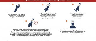
- apply venous tourniquets to the limbs.
In addition, it is necessary to monitor blood pressure, pulse and respiration rates.
Emergency care for acute pulmonary edema after the arrival of an ambulance is intravenous administration of promedol (morphine) and Lasix. For bronchospasm, the use of dexamethasone is justified. Mandatory measures: oxygen therapy and the use of electric suction to prevent foam aspiration.
Wheezing when breathing
Noises of pathological origin may occur in the respiratory tract. Such noises, better known as wheezing, can be detected in any area of the respiratory system: in the lungs, trachea, bronchi, and so on.
Wheezing as a symptom of illness
Wheezing is a characteristic manifestation of most diseases or pathological changes in the respiratory system. Among them:
- bronchial asthma ;
- anaphylaxis (instant allergic reactions);
- COPD;
- broncho- and pulmonary pneumonia. bronchitis. tracheitis. tuberculosis;
- pulmonary infarction, cancer, pulmonary edema. bronchiectasis and other diseases.
Causes of wheezing
The mechanism of wheezing, as well as the location and intensity of its manifestation, differ depending on the reasons for the occurrence of this symptom. Noises in the respiratory organs appear as a result of two main pathological processes:
- narrowing of the lumens in the bronchi as a result of inflammatory changes or spasms in them;
- the lumen of the respiratory tract is clogged with mucopurulent substances of varying degrees of viscosity and density, which means that during inhalation and exhalation these masses will be in constant motion.
Characteristic features of noise
Only a specialist can recognize what type of wheezing this or that noise belongs to.
Wet wheezing
The so-called moist rales are the result of the accumulation of sputum (liquid mucus) in the bronchi. The doctor can determine their type after auscultation: when an air flow passes through the sputum, bubbles form in it, which constantly burst. This “mass explosion” provokes the formation of moist rales. Basically, this manifestation occurs when inhaling, less often it can be recognized when exhaling air from the lungs.
The size of air bubbles may vary. This directly depends on the mass of accumulated mucus, the diameter of the bronchi and the volume of the cavity itself. Accordingly, small-, medium- and large-bubble moist wheezing is distinguished.
Bronchopneumonia. pulmonary infarction and bronchiolitis are characterized by fine-bubble noises, similar to the noise of foaming soda.
Medium vesicles are formed as a result of bronchiectasis or hypersecretory bronchitis. To the ear, such wheezing resembles the sound of air blowing into a liquid through a straw. The same wheezing may indicate small abscesses in the lungs (bronchi) that accompany the development of pneumonia, or can be heard at the first stage of pulmonary edema. A type of medium-bubble noise is the so-called “crackling” noise, they appear due to the opening of the walls of the bronchioles and acini, closed by the surrounding tissue during exhalation. This symptom allows one to diagnose, for example, pneumosclerosis or pulmonary fibrosis.
Large-bubbling or “bubbling” wheezing occurs when mucus accumulates in large bronchi, trachea, or in large cavities of pathological origin. This noise is heard on auscultation when air passes through the organs during inspiration. Note that you can hear bubbling wheezing even without the help of a phonendoscope; they can be heard even at some distance. Such symptoms are characteristic of the late stages of pulmonary edema. Also, similar accumulations in the trachea or bronchi can form in patients who have a weakened (or absent) cough reflex.
Dry wheezing
The second type of wheezing is dry. Among them are “whistling” and “buzzing”.
A whistling noise is a sign of an asthma attack. Such noises are produced by the bronchi as a result of uneven narrowing of the lumens during bronchospasm.
“Buzzing” when breathing is observed in those patients in whom thread-like mucous bridges form in the lumens of the bronchi due to inflammation.
Treatment of wheezing when breathing
To relieve a patient of wheezing, it is necessary first of all to properly treat the disease that causes it. Treatment methods differ significantly for different diseases and types of murmurs. The most commonly prescribed types of drug therapy are:
- Mucolytics are used to thin sputum and simplify its discharge;
- eliminating spasms and relaxing the walls of the bronchi is a task that inhaled beta-adrenergic agonists can do;
- An inflammatory process in the respiratory system caused by a bacterial infection is an indication for the prescription of antibiotics.
Hospital treatment
Treatment of the pathology is carried out in a hospital setting and includes the following measures: oxygen therapy, the prescription of diuretics and cardiac glycosides, intravenous administration of albumin, heparin, atropine, aminophylline. In some cases, hormones are prescribed. All prescriptions are strictly individual and are adjusted taking into account the patient’s condition.
The prognosis for treatment of cardiogenic pulmonary edema depends on the type of pathology, severity, presence of concomitant diseases, as well as the quality of medical care provided.
Pathophysiology
In normally functioning lungs, there is a small influx of fluid from the alveolar capillaries into the interstitial space of the lungs, facilitated by hydrostatic forces and the presence of microscopic gaps between capillary endothelial cells.
The rate of fluid entry is limited by the osmotic pressure gradient of the protein, which promotes the movement of fluid from the interstitial space back into the circulating plasma. As a result, the relatively small physiological movement of fluid from the vasculature into the lungs is usually compensated by the outflow of fluid from the interstitial space of the lung through the pulmonary lymphatic system, which ultimately returns to the systemic venous circulation. As fluid passes through the interstitial space of the lungs, it is delimited from the alveolar space by tight occlusive junctions between alveolar epithelial cells. In the absence of acute lung injury (eg, capillary injury), changes in the rate of fluid flow through the lungs are determined primarily by changes in hydrostatic pressure. Pulmonary capillary wedge pressure (PCWP), recorded from the catheter balloon at the moment of its wedging in the pulmonary artery segment, is a reflection of the filling pressure of the left atrium and is considered to most fully reflect the hydrostatic pressure of the microcirculation of the lung. Starling's equation for filtration mathematically demonstrates the fluid balance between the pulmonary vessels and the interstitial space, which "depends on the net difference in hydrostatic pressure and protein osmotic pressure, and the permeability of the capillary membrane."
Q = K*((Pmv - Ppmv) - (πmv - πpmv))
Where:
- Q = Net filtration of fluid through the vascular wall towards the interstitial space (flow of fluid filtered through the capillary wall into the interstitial space)
- K = Filtration coefficient
- Ppmv = Hydrostatic pressure in the perivascular interstitial space
- Pmv = Hydrostatic pressure inside the capillaries (HPLP)
- πmv = Osmotic pressure of protein in the vascular bed
- πpmv = Osmotic pressure of protein in the perivascular interstitial space
Although the Starling equation is useful for understanding the mechanisms that favor pulmonary edema, it is clinically impractical to accurately measure most of these parameters. However, a basic understanding of this equation is useful for clinicians working with patients with pulmonary edema.
Pathogenesis of cardiogenic pulmonary edema (increased hydrostatic pressure)
In cardiogenic pulmonary edema, hydrostatic pressure in the pulmonary capillaries leads to increased filtration of fluid through the vascular wall and is most often caused by volume overload or impaired left ventricular function leading to increased pulmonary vascular pressure. Moderate increase in pressure in the left atrium, expressed as PCWP 18-25 mm Hg. Art., causes the formation of edema in the perivascular and peribronchial interstitial spaces. With a further increase in left atrial pressure (PAPL > 25 mm Hg), the capacity of the lymphatic vessels and interstitial space (approximately 500 ml of fluid) is exceeded, and the fluid overcomes the pulmonary epithelial barrier, filling the alveoli with protein-containing fluid. The clinical picture of hypoxemia is caused by the accumulation of alveolar fluid, destabilization of the alveolar acini (impaired surfactant function) and, consequently, a violation of the ventilation-perfusion ratio (V/Q).
Pathogenesis of non-cardiogenic pulmonary edema (increased vascular permeability)
Non-cardiogenic pulmonary edema refers to any condition that promotes an abnormal increase in pulmonary vascular permeability, thereby promoting increased fluid and protein flow into the interstitial spaces and air spaces of the lungs. With regard to the Starling equation, pulmonary vascular damage equates to an increase in filtration coefficient and an increase in osmotic pressure in the interstitial space of the lung, which contributes to the formation of pulmonary edema. Another factor contributing to impaired gas exchange in noncardiogenic pulmonary edema is associated with disruption of the alveolar epithelial barrier, for example when the pressure in the interstitial space of the lungs is high enough to disrupt tight junctions, or when there is direct inflammatory or toxic damage to the alveolar epithelial lining. Damaged alveolar epithelium has a reduced ability to actively transfer fluid from the alveolar space to the interstitial space of the lung and causes impaired surfactant production (reduced active surface area), favoring alveolar collapse during normal breathing. An example is direct damage to the alveolar epithelium due to aspiration of gastric contents or pneumonia. Conditions that contribute to acute pulmonary capillary endothelial injury include systemic infections (sepsis), severe burns, trauma, and other systemic inflammatory conditions. Damage to the pulmonary capillary endothelium and/or alveolar epithelium is a hallmark of acute lung injury (ALI) and acute respiratory distress syndrome (ARDS), which are progressive non-cardiogenic lung injuries associated with impaired gas exchange (shunting, V/Q imbalance) and decreased pulmonary compliance (increased work of breathing).
Consequences of pulmonary edema in the elderly, prevention
Considering the severity of the disease, the prognosis in most cases is disappointing. And even if the swelling was managed in a timely manner, there is a high probability of ischemic damage to internal organs and the development of congestive pneumonia. In the absence of an established cause, there is a high risk of re-development of edema.
If we talk about the consequences of pulmonary edema in older people, then we are talking about the development of pneumosclerosis. Quite often such patients fall into a coma.
Prevention of pathology consists of early detection of diseases, the complication of which is edema, and their effective treatment.
If you have any questions, please contact our medical center. Specialists will answer all questions, including “why the lungs swell in a bedridden patient,” help determine the diagnosis and prescribe an effective course of treatment after receiving the examination results.
Related services: Ambulance call 5288
How to fight the disease?
Recovery activities include:
- medications and herbal remedies that promote the active removal of excess fluid;
- systematic inhalation of pure oxygen;
- painkillers;
- agents that dilate vascular channels;
- internal administration of protein-containing solutions;
- antibacterial drugs.
The main condition for getting rid of the anomaly is high-quality treatment of the disease that led to the development of edematous syndrome. If you let the disease take its course, complications such as dysfunction of internal organs and systems, chronic pneumonia, relapse of swelling, disruption of the cardiovascular system and brain function can occur. Half of the complicated cases are fatal. It is necessary to undergo responsible, high-quality treatment.
