There are acute and chronic cor pulmonale.
Acute cor pulmonale is diagnosed when pulmonary embolism occurs and as a consequence of diseases of the respiratory and cardiovascular systems, due to a long sedentary position, for example, prolonged bed rest in severe illnesses. The condition of acute cor pulmonale can be caused by atherosclerotic changes in the pulmonary artery, as complications after operations on the vessels of the lower extremities and pelvis, etc.
Chronic cor pulmonale develops gradually in the absence of signs of heart failure. A number of diseases provoke the process of formation of this condition. Such diseases include chronic bronchitis, bronchial asthma, pneumosclerosis, vasculitis, atherosclerotic changes in the pulmonary artery, etc.
Publications in the media
Acute cor pulmonale (ACP) is a clinical syndrome of acute right ventricular failure caused by sudden pulmonary hypertension due to pulmonary vascular obstruction. A classic example is TELA. Acute cor pulmonale develops over minutes, hours or days.
Etiology • PE • Embolism of fat, gas, tumor • Pulmonary vein thrombosis • Valvular pneumothorax, pneumomediastinum • Pulmonary infarction • Lobar or total pneumonia • Severe attack of bronchial asthma, status asthmaticus • Cancerous lymphangitis of the lungs • Hypoventilation of central and peripheral origin (botulism, poliomyelitis, myasthenia gravis) • Arteritis of the pulmonary artery • Resection of the lung • Massive atelectasis of the lung • Multiple fractures of the ribs, fracture of the sternum (floating chest) • Rapid accumulation of fluid in the pleural cavity (hemothorax, exudative pleurisy, massive infusion of fluid through a subclavian catheter mistakenly inserted into the pleural cavity ).
Risk factors • Thrombophlebitis of the deep veins of the lower extremities • Postoperative or postpartum period • Bronchopulmonary pathology.
Pathogenesis • Acute development of pulmonary hypertension (with massive pulmonary embolism, the right ventricle loses completely or reduces the ability to pump blood into the pulmonary circulation, and therefore acute right ventricular failure develops) • Severe bronchoconstriction • Development of pulmonary-cardiac, pulmonary-vascular and pulmonary-pulmonary coronary reflexes - a sharp decrease in blood pressure, deterioration of coronary blood flow • Acute respiratory failure • See also Secondary pulmonary hypertension.
Clinical manifestations are a sudden deterioration in the patient’s condition within a few minutes or hours (less often days) against the background of complete well-being or a stable course of the underlying disease. Sometimes it develops at lightning speed.
• Severe shortness of breath, feeling of suffocation, fear of death, severe cyanosis, acrocyanosis.
• Pain syndrome: chest pain, with pulmonary embolism - pain in the side associated with breathing (often in combination with hemoptysis). Sharp pain in the right hypochondrium may appear due to an enlarged liver with the rapid development of right ventricular failure.
• Swelling of the neck veins also due to the development of acute right ventricular failure.
• Decrease in blood pressure down to a collaptoid state and tachycardia 100–160 per minute due to decreased cardiac output.
• Auscultation of the lungs - signs of the pathological process that caused ALS: weakening, absence of respiratory sounds or bronchial breathing, dry and/or moist rales, pleural friction noise.
• Auscultation of the heart - accent of the second tone over the pulmonary artery, increased cardiac impulse, often arrhythmia (atrial and ventricular extrasystole, atrial fibrillation), sometimes systolic murmur of tricuspid valve insufficiency, gallop rhythm.
• Sometimes there is a discrepancy between the severity of the patient’s condition and the normal results of percussion and auscultation of the lungs.
Laboratory data • Hypoxia (decreased paO2) • Hyperventilation (determined by a drop in paCO2) • Moderate acute respiratory alkalosis (low paCO2 and elevated pH values).
Special studies
• X-ray examination of the chest cavity •• Signs of pneumothorax, the presence of fluid in the pleural cavity, total pneumonia, atelectasis •• Even with massive embolism, X-ray changes in the lungs may be absent •• Angiography of the pulmonary vessels - determining the localization of a blood clot if emergency embolectomy is necessary.
• ECG (especially informative in dynamics) •• Signs of ALS can be mistaken for MI of the posteroinferior wall of the left ventricle ••• Wide and deep Q wave and negative T wave in standard leads II, III, aVF, V1–V2, increased amplitude of the R wave in leads V1–3, ST segment depression in standard and chest leads ••• Signs of overload or hypertrophy of the right heart: deviation of the EOS to the right, deep S wave in standard lead I, V5–V6, high R in aVR, shift of the transition zone to the left , P pulmonale, partial or complete blockade of the right branch of the His bundle •• Rhythm disturbances (extrasystoles, atrial fibrillation).
Differential diagnosis is acute right ventricular failure in right ventricular myocardial infarction.
TREATMENT etiological; symptomatic is aimed at correcting hypoxia and acidosis, controlling hypervolemia and correcting right ventricular failure.
• Oxygen therapy. The initial stages of treatment for ALS should include the use of oxygen and improving the ventilation ability of the patient's lungs by correcting the underlying pulmonary disease. Since many patients are sensitive to oxygen, it is necessary to avoid the use of high concentrations and maintain saturation at 90%.
• Diuresis. Fluid retention is common and may impair pulmonary gas exchange and increase pulmonary vascular resistance. Improving oxygenation and limiting salt may be sufficient, but diuretics are often necessary.
• Phlebotomy provides a short-term effect and may be useful when Ht levels are above 55–60%.
• Cardiac glycosides do not have a good effect in the absence of left ventricular failure.
• Vasodilators are widely used, especially in cases mediated by occlusive vascular lesions or pulmonary fibrosis. However, the effectiveness of the drugs is questioned.
Abbreviation • ALC—acute cor pulmonale.
ICD-10 • I26.0 Pulmonary embolism with mention of acute cor pulmonale
Treatment
Treatment of pulmonary heart disease by our specialists is based, first of all, on diagnosing the disease and correct assessment of the severity of the situation. In acute forms, emergency resuscitation measures may be required. An individual approach to each case of cor pulmonale and to each patient, a comprehensive system of treatment and rehabilitation, experienced doctors and modern techniques - all this is aimed at the underlying disease, which results in cor pulmonale syndrome, and at restoring normal cardiac activity.
What happens in the body
In patients of groups 1 and 2, all changes develop due to spasm of small arterioles in the lung tissue as a reaction to insufficient oxygen supply. In group 3, in addition to spasm, there is a narrowing or blockage of the vascular bed. The pathogenesis of the disease is associated with the following mechanisms.
- Alveolar hypoxia (lack of oxygen in the alveoli) - scientists associate vascular spasm in response to hypoxia with disturbances in sympathoadrenal regulation. There is a contraction of vascular muscles, an increase in angiotensin-converting enzyme (ACE), calcium in the blood, and a decrease in relaxation factors of the pulmonary vessels.
- Hypercapnia - an increase in the concentration of carbon dioxide in the blood does not directly affect the vascular wall, but through acidification of the environment and a decrease in the sensitivity of the respiratory center of the brain. This mechanism enhances the production of aldosterone (adrenal hormone), which retains water and sodium ions.
- Changes in the vascular bed of the lungs - compression and emptying of capillaries due to developing fibrous tissue is important. Thickening of the muscular walls of the pulmonary vessels contributes to the narrowing of the lumen and the development of local thrombosis.
- An important role is played by the development of anastomoses (connections) between the bronchial arteries, belonging to the systemic circulation, and the pulmonary vessels.
- The pressure in the systemic circle is higher than in the lungs, so the redistribution goes towards the pulmonary vascular bed, which further increases the pressure in it.
- In response to hypoxia, blood cells that carry hemoglobin and erythrocytes change. Their number increases simultaneously with platelets. Favorable conditions for thrombus formation are created and blood viscosity increases.
All taken together leads to increased load on the right ventricle, hypertrophy, and then failure of the right heart. An enlarged right atrioventricular orifice contributes to insufficient closure of the tricuspid valve.
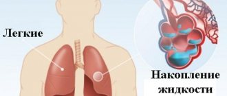
First aid for pulmonary edema
Unlike the chronic form, the acute form of the pathology requires immediate assistance during an attack. A person is not able to help himself, so he needs help from the outside. First of all, you should call an emergency team. While waiting for her, the person should be seated on a chair and given a Nitroglycerin tablet. If the attack occurs indoors, be sure to open all windows to provide fresh air flow.
If a person gets worse, cardiopulmonary resuscitation methods are required. To do this, rhythmically press on the chest area or do 30 presses and mouth-to-mouth breathing (alternating these actions). The main goals of these measures are to normalize blood circulation and restore respiratory function. If you lack the skills, you can simply massage the sternum.
Causes of cardiopulmonary failure
Cardiopulmonary failure never occurs without a reason. As a rule, several reasons lead to its development. There are three main groups of causes that can provoke pathology:
1. Bronchopulmonary diseases. According to statistics, they act as a provoking factor in more than 70% of all cases. Among the most common diseases are bronchial asthma, pulmonary tuberculosis, chronic bronchitis and other severe disorders of the respiratory system (for example, cystic fibrosis).
2. Thoradiaphragmatic diseases. They are directly related to pronounced deformation of the chest and limited mobility of the diaphragm. The appearance is caused by severe curvatures of the spinal column (in particular, kyphoscoliosis), ankylosing spondylitis, and restrictions on the expansion of the lungs (pleuritis).
3. Vascular diseases. This group includes diseases that affect lesions of the pulmonary bed. These include sickle cell anemia, pulmonary vasculitis, compression of the pulmonary veins and arteries by neoplasms.
4. Vascular diseases. Often, the development of cardiopulmonary failure is provoked by aneurysms, atherosclerosis and blood clots in the arteries of the lungs.
Cor pulmonale, folk remedies for treating cor pulmonale with herbs
Cough
in
hypertension
as pulmonary edema of cardiac origin.
We know and are accustomed to the fact that cough is one of the most common signs of respiratory disease, that it occurs when the mucous membrane of the larynx, bronchi, and windpipe becomes inflamed. We often encounter the fact that a cough may be of allergic origin. Sometimes a cough occurs during anxiety or emotional stress. It is extremely rare, but this happens - long-term hypertension can cause disease of the heart, left ventricle, ultimately developing heart failure, ischemia, which leads to insufficient blood supply to the left lung and can cause edema of the left lung. Doctors call this disease cor pulmonale
. therefore, it is necessary to treat the heart and lungs. Folk remedies recommend doing this using various herbs.
Treat hypertension
You can use calendula tincture - 8 tablespoons of inflorescences are infused for a week in 100 ml of vodka and taken 20 - 30 drops 3 times a day for a long time.
Garlic tincture for the treatment of hypertension can be prepared on a vodka basis (crush 2 large heads of garlic and leave for 2 weeks in a glass of vodka), drink 20 drops 5 times a day 15 minutes before meals for 3 weeks. For those who cannot drink vodka infusions, you can grind 3 large heads of garlic and 3 lemons, steep in 5 cups of boiling water in a tightly sealed container in a warm place for 24 hours. Take a tablespoon 2 - 3 times a day.
Pharmacy remedies for the treatment of the disease are adenesitis and Zelenin drops.
An effective remedy can be eating red rowan fruits or in the form of an infusion. A tablespoon of rowan berries is brewed with a glass of boiling water, infused for 1 hour, drink 1/2 glass 2 - 3 times a day.
Since the development of the disease is associated with edematous phenomena in the lungs, one should get rid of this phenomenon. Take the herb Adonis (adonis) spring in the form of an infusion - a teaspoon of the herb is infused for 1 - 2 hours in a glass of boiling water, take 1 - 2 tablespoons of the infusion 3 - 4 times a day before meals or every 2 hours.
For edema of cardiac origin, use corn silk in powder form, mixed with honey 1:2, a teaspoon 3-5 times a day before meals.
Taking an infusion of flax herb. of medicinal herb for 1 hour
in a glass of boiling water and drink 2 - 3 tablespoons 3 - 6 times a day.
It is good to use an infusion of 2 tablespoons of birch leaves: pour 1/2 liter of boiling water and, after steeping for an hour, drink 1/2 cup 4 times a day.
In this case, it is better to eat 3-4 juniper berries per day. 2 tablespoons of them are brewed with a glass of boiling water and after 5 - 6 hours of infusion in a thermos or 10 minutes of boiling, drink the contents per day in 3 - 4 doses.
An infusion of hawthorn fruits is useful to drink as a means of reducing blood pressure and swelling.
In addition, blue cornflower (field cornflower) flowers, flowering heather branches, nettle roots, cloudberry leaves, lingonberries, pine buds, turnip peels, oat straw, flax seeds, and knotweed are used in the form of an infusion or decoction.
Pumpkin juice is good for swelling, you should drink 1/2 glass a day.
Establishment of clinical class and verification of diagnosis
Pulmonary function tests can identify obstructive or restrictive changes for the purpose of differential diagnosis of PH and clarify the severity of lung damage. Patients are characterized by a decrease in the diffusion capacity of the lungs for carbon monoxide (40-80% of normal), a slight or moderate decrease in lung volumes, a normal or slightly decreased PaO2, and usually a decreased PaCO2 due to alveolar hyperventilation.
Ventilation-perfusion lung scintigraphy is a screening method to exclude chronic thromboembolism as a cause of pulmonary hypertension. In patients after thromboembolism, perfusion defects are found in the lobar and segmental zones in the absence of ventilation disturbances. Perfusion scintigraphy has historically become one of the first methods for detecting perfusion defects in the pulmonary parenchyma in pulmonary embolism. The images obtained in acute PE and CTEPH differ significantly. Perfusion defects in acute PE are more clearly defined and contrast sharply with normally functioning tissue. In CTEPH, perfusion defects are not clearly defined and often do not correspond to the blood supply area of the large pulmonary artery.
Computed tomography and CT angiopulmonography
The CT picture of chronic thromboembolism can be represented by occlusions and stenoses of the pulmonary arteries, eccentric filling defects due to the presence of blood clots, including recanalized ones. CT angiopulmonography is performed on spiral computed tomographs during the phase of passage of the contrast agent through the pulmonary arterial bed. Among the methodological features, it should be noted that the study must be carried out using at least an 8-spiral tomograph, with a minimum step (no more than 3 mm) and a slice thickness (no more than 1 mm). A thorough scan should cover both lungs completely, from the apices to the phrenic sinuses. Contrast enhancement of the right chambers of the heart and pulmonary arteries should match or exceed the degree of contrast enhancement of the left chambers of the heart and aorta. The second, arterial phase of scanning is recommended for all patients over 40 years of age, especially if there is evidence of arterial thrombosis and coronary artery disease in the anamnesis. Modern software allows you to reconstruct images of the pulmonary arteries in any plane, construct maximum intensity projections and three-dimensional images. In most cases, to clarify the nature of the lesion, it is enough to analyze cross sections using an image viewer, which makes it possible to determine the presence of changes not only in the lobar and segmental branches, but also in a number of subsegmental arteries. Pathological changes, in addition to the presence of “old” thrombotic material, may include local thickening of the vessel wall, narrowing at the mouth of the vessels and along them, occlusions, intravascular structures in the form of membranes and bridges. If changes are detected in several branches of the pulmonary arteries, we can conclude that there is a high probability of thromboembolic nature of PH. It is important to note that the resolution of modern CT scanners is limited and does not allow the determination of very thin membranous and stringy structures in the lumen of the LA, especially if the size of the object does not exceed 2-3 mm. In some cases, calcification of the “old” thrombotic material develops, and CT can be invaluable in determining the location of the calcification. CT makes it possible to detect not only stenotic changes in the vessels of the lungs, but also disturbances in the perfusion of lung tissue by the nature of the contrast of the parenchyma. In some cases, parenchymal enhancement is so uneven that mosaic enhancement is detected on scans. A clearly defined mosaic pattern of segments usually indicates a good prognosis for surgical treatment. Contrasting exclusively in the root zones is not a true mosaic and is often observed in microvascular forms of PH. By providing detailed images of the lung parenchyma, CT allows the diagnosis of other lung diseases. In addition to the state of the arterial bed, CT can provide comprehensive information about all intrathoracic structures, which is important for confirming the diagnosis and developing a surgical treatment plan. Before performing the operation, the condition of the pulmonary parenchyma, bronchial tree, and pulmonary veins should be taken into account.
Angiographic diagnostics
The main objectives of angiographic diagnosis are to determine the severity of pulmonary hypertension, clarify the nature of the lesion in the pulmonary bed through angiopulmonography, and identify/exclude coronary disease. Carrying out catheterization in isolation without high-quality angiopulmonography in a patient with clear signs of CTEPH is inappropriate. This study should provide clear information to doctors to resolve the issue of the patient’s operability and the severity of his condition. Hemodynamic criteria for post-embolic pulmonary hypertension, identified during catheterization of the right heart, are: mean pulmonary artery pressure (PAP) above 25 mm Hg. Art., pulmonary artery wedge pressure (PAWP) ≤ 15 mm Hg. Art., PVR > 2 units. according to Wood (160 dyn. sec cm-5) in the presence of multiple stenotic and/or occlusive lesions of the branches of the pulmonary artery of various calibers.
Clinical course
Soviet pulmonologists B. Votchal and N. Palev proposed a clinical description of the stages of development of the pulmonary heart:
- in the initial (preclinical) stage - there are no symptoms of hypertension in the pulmonary circulation, hypertension is possible temporarily with an exacerbation of pulmonary disease;
- in the second stage - there is right ventricular hypertrophy, but all signs are compensated; instrumental examination reveals stable pulmonary hypertension;
- third stage - accompanied by decompensation (pulmonary heart failure), there are symptoms of right ventricular overload.


![Rice. 1. Functioning of the glymphatic system of the brain (according to [11]) 1. Glymphatic system functioning (according to [11])](https://expert35.ru/wp-content/uploads/ris-1-funkcionirovanie-glimfaticheskoj-sistemy-mozga-po-11-fig-1-330x140.jpg)