Diagnosis of IHD
Late diagnosis of coronary heart disease is one of the common causes that claim the lives of 700 thousand Russians every year. Only a correct diagnosis, observation by a qualified cardiologist, and regular examinations guarantee that your heart will not let you down. With IHD, mortality (especially in men 55-65 years old) is 70%. And the main reasons are delayed access to a doctor, inappropriate treatment. Don’t risk your life - undergo professional and objective diagnostics at the CBCP Center for Circulatory Pathology!
Treatment of coronary artery disease
Treatment for coronary heart disease includes lifestyle changes, medications and, in some cases, surgery.
All patients are advised to give up bad habits, spend more time in the fresh air, and reduce excess body weight. In your diet, you must avoid foods high in fat, very salty and sweet foods. Smoking and unauthorized discontinuation of prescribed medications are strictly prohibited. All this can lead to a sharp deterioration in the patient's condition. To stop an attack of angina, you need to immediately stop physical activity, provide access to fresh air and take nitroglycerin under the tongue or use nitrate in the form of a spray.
Basic drug therapy includes the following drugs:
- antiplatelet agents – drugs that thin the blood;
- beta blockers;
- ACE inhibitors or sartans;
- statins.
Long-acting nitrates can be used to prevent attacks.
In the presence of concomitant diseases, especially diabetes mellitus and hypertension, their treatment and achievement of target blood pressure and glucose levels are required.
To restore cardiac blood flow, surgical intervention is necessary in some cases:
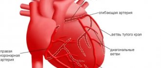
- Coronary artery bypass surgery is the creation of a bypass for blood at the site of narrowing of the coronary arteries using vascular prostheses.
- Coronary angioplasty and stenting - restoration of the diameter of the vessel, and, accordingly, the blood flow in it, by installing a special dilator.
What does the primary diagnosis consist of?
At your first meeting with you, the CBCP cardiologist will:
- will listen to your complaints;
- will ask what medications you are taking;
- will study data from previous studies (if available);
- will conduct an examination to identify cyanosis or swelling;
- listen to noises.
For the most accurate diagnosis of coronary heart disease, your detailed and comprehensive answers are very important, so it is worth preparing for this meeting. Remember the unpleasant sensations in the left side of the chest, their changes over time. Indicate whether you have increased heart rate, irregular heartbeat, shortness of breath, nausea, excessive sweating, dizziness, or loss of consciousness.
Methods for diagnosing transient myocardial ischemia in patients with coronary artery disease
N.V. Shestakova
Department of Cardiology, Russian Medical Academy of Postgraduate Education, Moscow
V.A.Shestakov
Department of Propaedeutics of Internal Diseases, Moscow Medical Dental Institute
Early and timely diagnosis of coronary heart disease (CHD) is an important clinical, social and economic problem. According to American statistics, direct and indirect costs for this group of patients amount to $14 billion per year. Early detection of ischemia, as well as its detection in patients with asymptomatic, latent ischemic heart disease, can bring tangible benefits to the patient [1]. It is well known that in a fairly significant number of patients suffering from coronary artery disease, even with an in-depth survey, its specific subjective signs (angina attacks) cannot be identified or they are of an atypical nature. Detection of transient ischemia is also important in patients with documented ischemic heart disease.
To diagnose myocardial ischemia, various methods can be used: ECG recording at rest, Holter ECG monitoring, various stress tests (treadmill test and bicycle ergometry - VEM), pharmacological tests, stress echocardiography, radioisotope methods.
The choice of method for detecting myocardial ischemia should be determined primarily by the clinical picture and specific tasks facing the doctor [2]. In addition, it is necessary to take into account the features of each specific method: indications and contraindications for its use, advantages and disadvantages, limitations in use, depending both on the characteristics of the method itself and on the patient’s condition, the method’s capabilities in assessing the nature and severity of coronary and myocardial damage , its prognostic capabilities [1,2].
It is very important to consider the cost of the study, as well as whether, in each particular case, a more expensive study is more adequate and informative [2]. The objective value of each test is determined by its sensitivity and specificity.
Sensitivity is the percentage of true positives in the presence of CAD; specificity is the percentage of true negative results in the absence of CAD. The sensitivity and specificity of any test can vary significantly.
The resting ECG is normal in about half of patients with stable angina. Nonspecific changes in the terminal part of the ventricular complex (ST segment and T wave), according to the Framingham study, are quite common in the general population (8.5% of men and 7.7% of women).
The causes of false-positive results may be hypertrophy and dilatation of the left ventricle, electrolyte disturbances, cardiac arrhythmias and conduction disturbances, taking antiarrhythmic drugs, hyperventilation, etc. Transient ischemic changes in ST - T during an attack of angina in patients with documented ischemic heart disease are of greater importance. However, the sensitivity and specificity of this method in diagnosing transient myocardial ischemia is low.
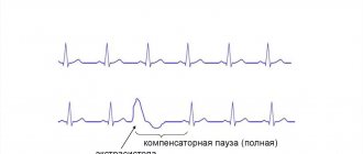
Holter ECG monitoring is used along with stress tests to detect transient myocardial ischemia. The value of this technique lies in the ability to detect transient myocardial ischemia in everyday life. The criterion for myocardial ischemia during Holter ECG monitoring is ischemic-type ST segment depression of 1 mm or more with a duration of ST segment depression of at least 1 minute and time between individual episodes of at least 1 minute. This is the so-called “1x1x1” rule [1]. The method is particularly useful for identifying episodes of vasospastic or spontaneous ischemia, as well as asymptomatic myocardial ischemia. Asymptomatic myocardial ischemia is often a poor prognostic sign. Considering that Holter ECG monitoring often gives false-positive results in patients without angina, it is recommended to use this method as a screening method in patients with a large number of risk factors for coronary heart disease or with a family predisposition to coronary artery disease [1], as well as for assessing individual prognosis.
Due to the fact that the ability to detect transient myocardial ischemia by recording an ECG at rest is very limited, stress tests become much more important.
Load tests
Stress tests provoke myocardial ischemia by increasing myocardial oxygen demand (treadmill test, VEM, dobutamine test) or reducing oxygen delivery to the myocardium (tests with dipyridamole and adenosine). Load testing in the form of a treadmill test or VEM is still the most common research method. This is a relatively simple and inexpensive method for detecting transient myocardial ischemia in patients with suspected or established coronary heart disease [1,3].
The treadmill test has both advantages and disadvantages compared to VEM. The advantage lies primarily in the fact that the load is more physiological and is perceived by patients as more familiar. In addition, when using the standard Bruce protocol, it is possible to perform a greater load than with VEM and more quickly achieve the desired result [4]. The treadmill test is often used in the USA and relatively rarely in Europe and Russia. Possible reasons for this are the higher cost of the treadmill, which is 2–4 times more expensive than a bicycle ergometer, and its large dimensions.
Load on a bicycle ergometer. In case of positive results of the VEM test, the likelihood of a diagnosis of IHD increases. The higher the initial probability of coronary heart disease, the higher the value of this stress test for the patient’s ability to perform adequate physical activity. The most reliable sign of transient myocardial ischemia with a VEM test is horizontal or oblique ST segment depression of 1 mm or more. The probability of diagnosing coronary artery disease is close to 90% if, during exercise, ST segment depression of the ischemic type reaches 2 mm or more and is accompanied by a typical attack of angina pectoris [1]. In patients with an initially high probability of coronary artery disease, detection of myocardial ischemia is more important for assessing the severity of coronary lesions and prognosis.
A positive result of the VEM test in such patients is combined with a significantly higher risk of coronary complications and death (the latter by 3.5-6 times) [5]. When the ST segment rises to 1 mm at the third stage of load according to the Bruce protocol, mortality in the group of such patients is less than 1% per year, and when the ST segment rises by more than 1 mm at the first stage of load it exceeds 5% per year.
The criteria for a high risk of coronary complications when performing a VEM test include:
•depression of the ST segment by 2 mm or more;
•early appearance (at the first stage of load) of ST segment depression of 1 mm or more;
•ST segment depression in several leads;
•reduced tolerance to physical activity;
•ST segment elevation in leads where there is no pathological Q wave;
•long-term persistence of depression or elevation of the ST segment after cessation of exercise (more than 8 minutes);
• low maximum heart rate (HR) during exercise, no more than 120 per minute;
•the appearance of life-threatening ventricular arrhythmias;
•decrease in blood pressure or absence of its increase during exercise.
Due to the relatively low sensitivity of the VEM test for ischemic heart disease, its negative result does not exclude this diagnosis. The rate of false positive results reaches 15%. Numerous studies have reported lower sensitivity of VEM and higher false-positive rates in women compared to men [6]. However, when men and women are stratified according to the prevalence of CAD, the study results are similar [7]. The sensitivity and specificity of the treadmill test and the VEM test are approximately the same. Exercising on a bicycle ergometer poses obvious difficulties for patients who do not have cycling experience. The advantages of VEM include the ability to perform a load both sitting and lying down, which is sometimes necessary according to the research protocol when solving some specific problems. Exercise on a bicycle ergometer and treadmill is an accessible and common test, but from 20 to 40% of patients cannot perform them if necessary due to orthopedic and neurological disorders or vascular diseases of the extremities [3].
Stress echocardiography is a new method widely used in the diagnosis of coronary artery disease. A treadmill or bicycle ergometer, as well as pharmacological drugs, are used as a load. Stress echocardiography using a treadmill or bicycle ergometer competes with radioisotope methods in accuracy and equivalence. This technique should be used if the ECG is initially altered (signs of left ventricular myocardial hypertrophy, intraventricular conduction disturbances, electrolyte disturbances, drug effects, etc.). In these cases, local contractility disorders that occur during the development of myocardial ischemia can be detected using echocardiography [4]. The normal response of the left ventricle to exercise is an increase in the rate of contraction and systolic thickening of the left ventricular myocardium. When ischemia occurs, these indicators can change to varying degrees.
The criteria for a strongly positive test are:
•reduction of ejection fraction to 35% or less;
• increase in ejection fraction at load by less than 5%;
•the appearance of disturbances in local contractility of the left ventricle at a low level of load or at a heart rate of less than 120 per minute;
•the appearance of contractility disorders in several segments of the left ventricle.
The advantages of this test over radioisotope methods: low cost and safety, availability of portable equipment, lack of radiation, quick results, the ability to identify other causes of patient complaints (pain and other sensations in the chest, shortness of breath, weakness). Limitations of the method: the need for a good ultrasound window, difficulties in assessing the basal segments with maximum dyskinesia under heavy load. A significant drawback is that echocardiography is performed immediately after the end, and not at the height of the load; if dyskinesia is detected after the load, it is unclear at what stage of the load it began. In addition, a well-trained ultrasound specialist is required to conduct the study. The sensitivity and specificity of this test have been evaluated in a large number of studies. According to one multicenter study (1150 patients), sensitivity ranged from 74 to 97%, and specificity from 64 to 100% [4]. In a study by R. Cohn et al. (1987, 390 patients) sensitivity averaged 80% and specificity 91%.
Pharmacological stress echocardiography is performed to provoke and identify myocardial ischemia, as well as to determine the functional state of the myocardium and prognosis in a patient with coronary artery disease.
Indications for stress echocardiography are [8]:
• inability to perform a treadmill test or exercise on a bicycle ergometer;
•lack of ability to perform physical activity to the required capacity;
•false-positive results of the exercise test in patients without symptoms of coronary artery disease.
The most commonly used drugs in this test are dobutamine, dipyridamole, adenosine and arbutamine.
Dobutamine is a synthetic catecholamine that predominantly stimulates b-adrenergic receptors, giving a strongly pronounced positive inotropic and to a small extent chronotropic effect. Blood pressure does not change significantly. An increase in myocardial oxygen demand provokes ischemia with significant narrowing of the coronary arteries. The resulting disturbance of local myocardial contractility is detected using echocardiography.
Administration of dobutamine is contraindicated in valvular stenosis, idiopathic hypertrophic obstructive cardiomyopathy, ventricular tachyarrhythmias, and severe arterial hypertension (AH) [3]. The number of side effects is relatively small, but in 10–36% of patients, arrhythmias appear during the test [11].
Dipyridamole, a coronary vasodilator, dilates unchanged coronary arteries to a greater extent, resulting in the development of the phenomenon of intercoronary steal, i.e. the resulting myocardial ischemia is the result of decreased oxygen delivery to the myocardium. The administration of dipyridamole is safe, side effects are rare and are observed in 1 - 2% of cases [3]. Contraindications for the administration of dipyridamole are obstructive pulmonary diseases, severe intraventricular conduction disorders, and severe hypertension.
Adenosine, like dipyridamole, dilates non-stenotic coronary arteries and increases the perfusion of the segments they supply. This leads to pronounced heterogeneity of the blood supply to the myocardium and provokes ischemia. The vasodilating effect appears quickly and disappears quickly. Despite the fact that the administration of adenosine is safe, various adverse reactions often occur (hot flashes, shortness of breath, headache, dizziness, parasthesia), which are subjectively difficult to tolerate by patients.
Arbutamine is a new non-selective β-adrenergic receptor agonist with moderate α1-sympathomimetic activity, developed specifically for stress testing and is well tolerated [3]. Like other beta-agonists, which increase myocardial oxygen demand, it increases heart rate and myocardial contractility. To administer arbutamine, a special system (GenESA) is required that automatically adjusts the dose of the drug based on heart rate.
The system provides constant monitoring of heart rate, blood pressure, heart rhythm, ECG and ECG signs of myocardial ischemia, and conducts their computer analysis.
Dobutamine stress echocardiography. Numerous studies [2] have reported high sensitivity and specificity of this test. In comparison with the results of coronary angiography when dobutamine was administered at a dose of 40 mcg/kg per minute, the sensitivity of the test ranged from 72 to 86%, specificity from 77 to 95%, and accuracy from 76 to 89%. It is noted that reducing the dose of dobutamine was accompanied by a decrease in the accuracy of the study. Many studies indicate that the sensitivity of dobutamine stress echocardiography is higher for multivessel disease than for single-vessel disease, and for stenosis of 70% or more than for vessel stenosis of 50% or less. However, there are other data [7]. A meta-analysis of 7 published studies using high-dose dobutamine in 703 patients shows that dobutamine stress echocardiography has a higher sensitivity (75% versus 63%) in single-vessel disease. In patients with angiographically unchanged coronary arteries, dipyridamole stress echocardiography is characterized by higher sensitivity in combination with high specificity (91 and 83%, respectively). The sensitivity of these two tests for multivessel disease is approximately the same, being 79% with dipyridamole and 81% with dobutamine.
Adding atropine to dobutamine increases the sensitivity of this test. Sensitivity in detecting transient ischemia in patients who have had myocardial infarction is lower. Dobutamine stress echocardiography is used in patients after coronary artery bypass grafting to identify the functional state of the shunt; its sensitivity is 80% and specificity is 90%. A direct comparison of dobutamine stress echocardiography and stress echocardiography with vasodilators in the same patients showed that the test is more sensitive when using dobutamine, dipyridamole (78 and 67%) and adenosine (82 and 52%).
Dipyridamole stress echocardiography is the easiest to interpret among other pharmacological tests. Its accuracy is comparable to that of dobutamine stress echocardiography. Safety and prognostic value have been demonstrated in large multicenter studies [8]. S. Severi et al. (1994) note that the sensitivity of dipyridamole stress echocardiography is higher in multivessel lesions, in patients with well-developed collaterals and in patients with reduced regional myocardial perfusion. The sensitivity of this test, according to R. Mazeika et al. (1992), varies widely: from 0% with single-vessel disease to 100% when using the transesophageal technique, even with single-vessel disease. According to S. Beckmann et al. [4], it averages 57 - 75%. Specificity ranges from 80 to 100%. For multivessel disease, sensitivity reaches 79%.
Adenosine stress echocardiography. The sensitivity of this test averages 40 - 85% and is quite variable in patients who have not had myocardial infarction, and low in patients with single-vessel disease. The specificity of adenosine stress echocardiography ranges from 87 to 100%. The relatively low sensitivity of this test and the frequent occurrence of adverse reactions that are poorly tolerated by patients, although not dangerous, prevent the widespread use of this test.
Arbutamine stress echocardiography is a fairly accurate and safe method for detecting myocardial ischemia, diagnosing coronary heart disease and determining the prognosis in these patients. The results of a multicenter study that assessed the clinical suitability and safety of arbutamine showed that the sensitivity of the test in detecting myocardial ischemia reaches 71%, and in diagnosing coronary artery disease - 82%; the specificity is 100 and 50%, respectively. In a study by Cohen et al., performed in 143 patients, the sensitivity of this test in provoking myocardial ischemia is 76%, in diagnosing coronary artery disease - 84%, and its specificity reaches 96% [9].
In addition, some studies have demonstrated that arbutamine is potentially better in its pharmacological properties, effect on hemodynamics and tolerability than dobutamine, dipyridamole and adenosine [7]. However, the cost of the system used to administer it and evaluate the results is significantly higher than that of other pharmacological stress tests. However, in some European countries this new method is highly valued and is practically the only officially accepted stress echocardiogram test.
Radionuclide stress tests. Myocardial perfusion scintigraphy with thallium-201 or technetium-99m makes it possible to detect defects in their accumulation in the myocardium. The capabilities of the method increase significantly when perfusion scintigraphy is combined with physical or pharmacological stress. The need for this study arises if the patient cannot perform an exercise test, the test has not been brought to diagnostic criteria or its results are questionable, stress echocardiography cannot be performed or its conduct does not give the desired result (for example, poor visualization of the lateral wall of the left ventricle during EchoCG). When comparing perfusion scintigraphy with thallium-201 or technetium-99m with stress echocardiography, one should first of all note the high cost of the study, primarily due to the need for a special laboratory and expensive equipment [3]. Poldermans et al. (1993) provide data on the comparative cost of dipyridamole-thallium-201 perfusion scintigraphy and dobutamine stress echocardiography: 530 and 185 US dollars, respectively. In addition, the study involves radiation and a lot of time. False-positive results reduce the diagnostic value of the method. Their most common causes are: obesity, leading to poor image quality; large mammary glands and a high diaphragm, which contribute to the appearance of overlay artifacts. In addition, perfusion scintigraphy visualizes the posterior wall of the left ventricle worse. But the method also has undoubted advantages: automation of the process, the ability to quantify the results obtained and determine the location of the lesion. When combining myocardial perfusion scintigraphy with thallium-201 and exercise on a bicycle ergometer or treadmill, the appearance of an accumulation defect during exercise indicates the presence of transient myocardial ischemia. The combination of myocardial perfusion scintigraphy with a pharmacological load (dipyridamole or adenosine) leads to pronounced inhomogeneity of thallium absorption and its subsequent redistribution, which is also a sign of transient myocardial ischemia.
In both cases, the criteria for a sharply positive test associated with a high risk of complications are:
•the appearance of accumulation defects against the background of low load or at a heart rate of less than 120 per minute;
•multiple accumulation defects;
•increased accumulation of thallium in the myocardium.
Comparative data on the diagnostic value of stress echocardiography and myocardial perfusion stress scintigraphy with thallium-201 or technetium-99m, based on the results of seven independent studies (390 patients), in which a treadmill test or VEM test was used to provoke myocardial ischemia, show that that the sensitivity of stress tests averaged 80 and 84%, respectively. At the same time, the sensitivity of stress perfusion scintigraphy was higher in single-vessel lesions (80 versus 69%), but the specificity was higher when performing stress echocardiography (91 and 83%, respectively) [2].
Dobutamine stress echocardiography and dobutamine perfusion scintigraphy were compared in four studies in 318 patients. The sensitivity of these methods in detecting myocardial ischemia was comparable (76 and 81%). At the same time, a higher sensitivity of both methods was revealed for single-vessel lesions (83 and 85%) than for multi-vessel lesions (68 and 76%). The specificity of dobutamine echocardiography (85%) was higher than that of stress perfusion scintigraphy (71%) [2].
Stress echocardiography and myocardial perfusion scintigraphy with vasodilators (dipyridamole or adenosine) were compared in five studies in 222 patients. Myocardial perfusion scintigraphy has been shown to be superior in results to stress echocardiography. Its sensitivity is higher than the sensitivity of stress echocardiography and is 90 and 66%, respectively, and specificity is 79 and 91%. The results of the comparative assessment are shown in the table. T. Marwick et al. (1993) reported that the sensitivity of these tests depends on the severity of coronary lesions. According to their data, the sensitivity of perfusion scintigraphy with vasodilators in single-vessel lesions is higher (81%) than the sensitivity of echocardiography with vasodilators (52%). Sensitivity for multi-vessel lesions increases and is 93 and 64%, respectively.
Only two studies (T. Marwick et al., 1993; M. Takeuchi et al., 1993) compare data from dobutamine stress echocardiography and myocardial perfusion scintigraphy with vasodilators. It was shown that both tests have approximately the same sensitivity (85 - 86 and 85 - 89%). However, the specificity of dobutamine stress echocardiography is higher (93 - 85 and 82 - 71%). Based on these results, the authors suggest that the use of dobutamine stress echocardiography will increase in the future.
Assessment of the severity of CAD using dobutamine stress echocardiography and dobutamine perfusion scintigraphy was carried out in two centers. Senior et al. (1997) revealed signs of multivessel disease in 70% of cases with stress echocardiography and in 77% with perfusion scintigraphy. The specificity of the results obtained was 90 and 94%, respectively. T. Marwick et al. (1993) examined patients who had suffered acute myocardial infarction. Signs of multivessel disease were detected using stress echocardiography in 18% of patients, and using myocardial perfusion stress scintigraphy - in 34% of patients. The capabilities of each test were compared with the results of coronary angiography. The results of stress echocardiography and perfusion scintigraphy correlated with angiography data (correlation coefficients 0.45 and 0.35, respectively). These data show that stress echocardiography and perfusion scintigraphy are quite accurate in recognizing multivessel disease. Based on the results obtained, the authors suggest performing dobutamine stress echocardiography in patients with suspected CAD or with documented CAD in the following cases:
• if it is impossible to perform the load;
•if there are contraindications to the use of vasodilators;
•if other studies pose an increased danger to the patient;
•if the degree of coronary stenosis is unclear;
• in the presence of concomitant left ventricular hypertrophy or left bundle branch block;
•to assess the effectiveness of antianginal therapy.
In all these cases, stress echocardiography is more specific than perfusion scintigraphy.
Thus, methods for diagnosing transient myocardial ischemia are numerous. When determining the objective value of a particular method, it is necessary to focus on its sensitivity and specificity. They differ for different methods. Equally important is taking into account the cost of the study. However, the low cost of the method should not be the determining argument when choosing it. Taking into account the specific features of the methods, including their diagnostic value, as well as based on practical expediency, the examination should begin with simpler methods (VEM, daily ECG monitoring). If they cannot be performed or do not solve the assigned tasks, stress echocardiography is necessary. And only if this study does not solve the problem, it is advisable to use stress perfusion scintigraphy of the myocardium with thallium-201 or technetium-99 m.
Literature:
1. Gottlieb SO. Diagnostic procedures for myocardial ischemia. Eur Heart J 1996;17(suppl G):53-8.
2. Geleijnse ML, Salustri A, Marwickt TH, et al. Should the diagnosis of coronary artery disease be based on the evalution of myocardial function or perfusion? Eur Heart J 1997;18(suppl D):68-7.
3. Elhendy A, Geleijnse ML, Roelandt JRTC, et al. Dobutamine without transient wall motion abnormalities: Less severe ischemia or less severe stress? J AM Coll Cardial 1996;27:323-9.
4. Beckmanns, Scharti M, Boksch W, et al. Diagnosis of coronary artery disease and viable myocardium by stress echocardiography. Diagnostic accuracy of different stress modalities. Eur Heart J 1995; 16 (suppl J): 10-8.
5. Scharte M, Beckmann S, Bocksch W, et al. Stress echocardiography in special groups: in women, in left bundle branch block, in hypertension and after heart transplantation. Eur Heart J 1997;18(suppl D):63-7.
6. Proceedings of an WHLBJ Conference: Exercise ECG testing with and without radionuclide studies. Jn Cardiovascular Health and Disease in women. Greenwich, CT, Le Jacg Communications, inc 1993: 74.
7. Picano E, Ostojic M, Sicari R et al. Dipyridamole stress echocardiography: state of the art 1996. Eur Heart J 1997;18 (suppl D):16-23.
8. Afridi J, Kleiman NS, Rainzer AE, et al. Dobutamine echocardiography in myocardial hibernation. Circulation 1995;91:663-70.
9. Ketteler T, Krahwinkel W, Wolfertz J, et al, Arbutamine stress echocardiography. Eur Heart J 1997;18(suppl D):24-30.
Published with permission from the administration of the Russian Medical Journal.
Laboratory diagnostic tests
Coronary heart disease is characterized by many symptoms. Patients feel many of them quite subjectively. Therefore, instrumental and laboratory examinations are important to make an objective diagnosis.
- The CBCP laboratory will determine the level of sugar, cholesterol in the blood, specific enzymes characteristic of IHD (troponins, myoglobin, etc.)
- Among instrumental studies, various types of electrocardiography, as well as ultrasound studies (echocardiography, stress echocardiography), have the greatest accuracy.
Electrocardiography (ECG) at CBCP
Chronic coronary heart disease is traditionally diagnosed using an ECG . Almost every person undergoes this simple and painless procedure several times during their life. You will sit on a comfortable couch, the doctor will attach electrodes to your body and within 10-15 minutes the electrocardiograph will read the performance of the heart muscle. In our clinic you will undergo an ECG using a General Electric MAC 800 electrocardiograph (USA), which determines hidden changes in myocardial metabolism. The transcript is issued immediately.
An expanded option for diagnosing coronary heart disease is Holter ECG monitoring. It is the same ECG, but carried out over 24, 48 or 72 hours. A portable heart rate monitor (fits comfortably under your shoulder or on your belt) with secure electrodes attached to your body, continuously reads and records your readings. The method identifies irregular heart rhythm disturbances, examines the functioning of the heart muscle during sleep, during emotional and physical stress. The procedure allows the doctor to understand the causes of ischemic heart disease.
Causes of the disease
There is still ongoing debate about the causes of “silent” myocardial ischemia. Our American colleagues have been trying to refute the existence of this pathology for the last few years, passing off ECG changes as post-infarction aneurysms, etc.
It is believed that this form of ischemic heart disease occurs against the background of damage to the nerve fibers responsible for the perception of pain impulses. Factors leading to polyneuropathy are:
- long-term alcohol abuse,
- long history of smoking,
- obesity,
- hereditary hyperlipidemia (increased concentrations of cholesterol and other fats in the blood),
- diabetes,
- previous myocardial infarction.
I draw your attention to the fact that silent myocardial ischemia is not necessarily accompanied by a violation of the nerve structure.
No less rare factors leading to pathology are:
- age over 60 years,
- constitutionally determined high pain threshold (in other words, a person does not feel weak impulses),
- hypertonic disease,
- long-term stress,
- sedentary lifestyle,
- chronic overwork and lack of sleep.
Echocardiography (ultrasound of the heart)
Echo-CG, also called cardiac ultrasound, can significantly clarify the diagnosis. Coronary heart disease is accompanied by changes in its size, condition of valves, cavities, pericardium, and large vessels. In addition to visualizing cardiac structures, this safe ultrasound procedure evaluates myocardial contractility and the presence of acoustic noise.
At the CBCP clinic you will undergo a cardiac ultrasound using a modern expert-class ultrasound system MyLab 50 (Italy). You will need to undress to the waist and lie on your left side. The doctor will lubricate the chest with a special gel and will move an ultrasonic sensor over the skin, which will read the information. You receive a conclusion 10 minutes after the end of the procedure.
Stress echocardiography
Stress echocardiography is another ultrasound method that allows you to effectively diagnose the disease. In this case, coronary heart disease is studied under physical or medicinal stress. Disturbances in heart contractions and blood flow are detected that are not noticeable at rest, which is why IHD is asymptomatic.
Under the supervision of a diagnostician who has undergone special training in stress echocardiography, you will perform exercises on an exercise bike. If exercise is contraindicated, you will be given a safe stimulant drug. At this time, the equipment records various indicators. The doctor will decipher it within 15 minutes and give you a conclusion.
Treatment of the disease
During my practice (and this is no less than 14 years), I have become convinced that convincing a patient to treat a disease that does not bother him is extremely difficult. Although it is silent myocardial ischemia, which is considered prognostically unfavorable, that requires timely and adequate therapy. Indeed, in 40-46% of cases it becomes the cause of sudden coronary death.
Only a qualified specialist should prescribe medications; I categorically do not recommend stopping or adding any medications on your own.
Non-drug correction
Non-drug therapy for the disease consists of correcting modifiable risk factors for its development. Namely, losing body weight, reducing the proportion of fatty foods in the diet, quitting smoking and drinking alcohol. Moderate physical activity also has a positive effect on microcirculation in the heart muscle.
Conservative therapy
Drug treatment of the “silent” form of ischemia is based on the basics of therapy for coronary artery disease and is selected strictly individually, based on the patient’s age, premorbid background and examination results.
To improve blood supply to the myocardium, the following groups of drugs are used:
- β-blockers (“Atenolol”, “Bisoprolol”, “Concor”) – reduce the heart rate, thereby reducing the load on the heart;
- calcium antagonists (Nifedipine, Amlodipine, Verapamil) - dilate small vessels;
- antiplatelet agents (“Aspirin-cardio”, “Cardiomagnyl”, “Aspecard”) - reduce blood viscosity;
- hypolipidemic (“Crestor”, “Rosuvastatin”, “Atorvastatin”) - affect the level of harmful low- and very low-density lipoproteins, triglycerides and cholesterol;
- antiari, "Cordarone", "Etatsizin") - prescribed for arrhythmias accompanying ischemia;
- ACE inhibitors (Captopril, Lisinopril, Enalapril) – regulate blood pressure;
- diuretics (“Veroshpiron”, “Indapamide”, “Triampur”) - remove fluid from the body, which puts additional stress on the heart;
- Long-acting nitrates (“Isoket”, “Cardix”, “Efoxlong”, “Sidnopharm”) – have a vasorelaxing effect on the coronary vessels.
Surgical intervention
If the narrowing of the vessels supplying the myocardium is too pronounced and conservative therapy is ineffective, then it is necessary to resort to surgical methods of treating silent ischemia: endovascular stenting or coronary artery bypass grafting.
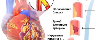
The first operation is minimally invasive and involves inserting a balloon into the affected vessel, inflated under X-ray control. Then a metal stent is installed on its surface - a hollow cylinder, which will maintain the original lumen of the coronary artery. As a result of this intervention, normal blood flow in the myocardium is restored.
In case of critical blockage of the vessel, coronary artery bypass grafting is performed, which consists in creating a “roundabout route” of blood supply. That is, the vascular autograft constructs a “bridge” between the aorta and the normally functioning part of the coronary artery.