What is cardiac extrasystole?
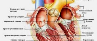
During normal heart function, the main driver of the heart rhythm is the sinus node, and the heart rate is 60-80 beats per minute. With extrasystole, extraordinary electrical impulses occur. This leads to the appearance of extrasystoles - premature contractions of the heart or its individual chambers that do not fit into the overall heart rhythm.
During an extraordinary contraction, the myocardium pumps less blood than during normal contraction of the heart. In this regard, frequent extrasystoles negatively affect blood circulation, in particular coronary and cerebral blood flow.
Prevention of heart rhythm disturbances
The basic rules of prevention are:
- Therapy of concomitant pathologies of the cardiovascular, circulatory, endocrine and nervous systems.
- Do not self-medicate and do not take strong tranquilizers, hormones, or sedatives.
- Conduct control diagnostics in a timely manner.
The disease of extrasystole is characterized by relapses, therefore, after completing the course of therapy, you should regularly undergo control diagnostics of the heart rhythm.
This article is posted for educational purposes only and does not constitute scientific material or professional medical advice.
What are the types of extrasystoles?
Depending on the place of occurrence of extraordinary contractions, extrasystoles are:
- ventricular;
- atrial;
- atrioventricular (nodal).
In rare cases, extraordinary impulses are generated by the sinus node.
Parasystoles are also distinguished, which are characterized by parallel work of an additional cardiac pacemaker (for example, atria, ventricles) and the sinus node. In such cases, there are 2 rhythms simultaneously: sinus and extrasystolic.
Depending on the time of occurrence of extraordinary contractions of the heart, extrasystoles are divided into:
- bigeminy - every normal, physiological contraction of the myocardium is accompanied by a pathological contraction;
- trigeminy - two normal myocardial contractions are followed by one extraordinary contraction;
- quadrigeminy - a pathological contraction follows every three physiological contractions of the myocardium.
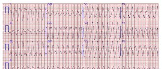
A phenomenon in which three or more pathological contractions occur in a row is called “unstable paroxysmal tachycardia.” Such arrhythmia is quite dangerous and indicates severe electrical instability of the heart muscle. The pathology is characterized by a heart rate from 140 to 220 beats per minute.
Classification of ventricular extrasystoles
Under certain circumstances, ventricular extrasystole causes a severe form of arrhythmia - ventricular tachycardia, which turns into fibrillation. And this condition is the most common cause of sudden coronary death.
Lown classification
The classification of PVCs has changed several times following diagnostic and prognostic needs. Extrasystoles in them were distributed according to quantitative values, location and frequency of occurrence. For about 15 years in cardiology they used the classification of ventricular extrasystoles according to Lown and Wolf (B. Lown and M. Wolf). They proposed it for the gradation of gastric extrasystoles in post-infarction patients. A few years later, it was adapted for patients without a history of heart attack.
This classification reflects the quantitative and morphological signs of PVCs (based on the results of a 24-hour ECG):
| Class | Characteristics of ventricular extrasystole |
| Extraordinary layoffs are not recorded | |
| 1 | Less than 30 single extrasystoles in any 60 minutes during the day |
| 2 | More than 30 single extrasystoles during any hour of monitoring |
| 3 | There are (polymorphic) extraordinary contractions occurring in different foci of excitation |
| 4A | There are two extrasystoles in a row emanating from one focus of excitation (monomorphic) |
| 4B | There are paired polymorphic extrasystoles |
| 5 | There are polymorphic PVCs passing in one gulp of 3–5, registration of paroxysmal ventricular tachycardia is possible |
According to Lown's classification, class 1 is a benign condition, rather considered as a functional disorder without circulatory impairment and clinical signs. Starting from class 2, ventricular extrasystole has a poor prognosis, being associated with an increased risk of developing fibrillation and cardiac arrest.
Gradation of extrasystoles by location, time and frequency of occurrence
There is a classification of ventricular extrasystole according to the place of origin of impulses and the number of foci of excitation:
- extrasystoles emanating from one focus are called monotopic;
- PVCs that originate in several foci are polytopic;
- right ventricular extrasystole;
- left ventricular extrasystole.
According to the time of occurrence, ventricular extrasystoles are divided into early (recorded at the beginning of diastole), interpolated, appearing in the middle between two beats, and late, occurring at the very end of diastole. Early extraordinary contractions according to the ECG sign are called “R on T” (layering of two teeth on top of each other). They are caused by organic changes in the myocardium. According to Lown, this type of ventricular extrasystole is classified as class 5.
In cardiology, there is a concept of a statistical daily “norm” of VES if they are well tolerated. There is no such norm for early extrasystole - it should not exist at all. Average or interpolated is the majority of the VES (up to 80%). Its average normal indicator is considered to be up to 200 extraordinary contractions per day. Late extrasystoles almost overlap the subsequent normal contraction. Their permissible daily quantity is up to 700. Today, the medical community uses the RJ Myerburg classification, which reflects the shape and frequency of ventricular extrasystoles:
| By frequency | According to morphology | ||
| 1 | less than 1 in 60 minutes - rare; | A | Single from one source |
| 2 | 1–9 per hour - infrequent; | B | Single from different foci |
| 3 | 10–30 per hour – moderately frequent; | C | Doubles |
| 4 | 31–60 per hour - frequent; | D | Unsustained ventricular tachycardia |
| 5 | more than 60 per hour - very frequent. | E | Sustained ventricular tachycardia |
Causes of extrasystole
The danger of extraordinary heart contractions directly depends on the reason for which they occurred. Today, extrasystoles are distinguished:
- functional origin, when the heart is not affected by pathologies;
- organic origin, which are associated with structural disorders of the heart. It is organic extrasystoles that are pathological and pose a threat to health.
Functional extrasystoles can be a consequence of:
- cervical osteochondrosis;
- neuroses, stress;
- neurocirculatory dystonia - a functional pathology that is associated with an imbalance in the functioning of the autonomic nervous system;
- drinking a lot of coffee;
- bad habits - smoking, alcohol abuse;
- taking certain medications - bronchodilators, psychotropic drugs, glucocorticoids, diuretics.
Also, extrasystoles can occur for no apparent reason. The heart makes more than 100 thousand contractions per day, so it is difficult to imagine that all of them are rhythmic. In this regard, extraordinary cardiac contractions may well be a variant of the norm.
The reasons for the appearance of organic extrasystoles include:
- cardiosclerosis - proliferation of connective scar tissue in the myocardium;
- coronary heart disease - a discrepancy between the myocardial need for oxygen and its delivery to the heart muscle;
- previous myocardial infarction;
- cardiomyopathies - mechanical or electrical dysfunction of the heart muscle;
- thickening of the myocardial walls;
- congenital and acquired heart defects;
- inflammatory processes of the myocardium;
- cor pulmonale - enlargement and expansion of the right parts of the myocardium as a result of increased blood pressure in the pulmonary circulation. The pathology is a complication of lung disease or chest deformation;
- some systemic diseases that affect the heart;
- surgical operations on the myocardium;
- arterial hypertension.
Symptoms
Ventricular extrasystole can be asymptomatic or have a pronounced clinical picture:
- interruptions in the functioning of the heart - first rapid heartbeat, then freezing;
- dizziness;
- weakness;
- unpleasant, painful sensations in the heart;
- pulsation of the neck veins;
- fast fatiguability;
- reduced performance;
- lack of air;
- shortness of breath.
If you notice similar symptoms or feel unwell, contact a specialist immediately.
Diagnostic methods
A cardiologist may suspect the presence of extrasystole in a patient during a physical examination and history. When collecting an anamnesis, the clinical manifestations of arrhythmia are clarified, at what time and under what circumstances unpleasant symptoms occur (at rest, during physical activity).
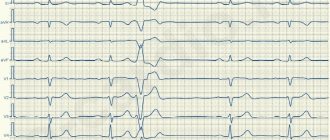
The main method for diagnosing extrasystole is ECG , which allows not only to identify the deviation itself, but also to determine the type and number of extraordinary heart contractions.
In addition, the patient may be prescribed other types of examination:
- daily ECG monitoring, which will give a more complete picture of the disease and will allow you to determine the percentage of extrasystoles in relation to normal heart contractions;
- electrocardiography with exercise - bicycle ergometry, treadmill test;
- ultrasound examination of the heart;
- MRI of the heart;
- biochemical blood test for electrolytes.
Signs on ECG
Supraventricular extrasystole is very easy to recognize on a cardiogram. Main features:
- extraordinary (extrasystolic) appearance of a pathological deformed P wave and the following unchanged QRST complex;
- the presence of a compensatory pause, i.e. a straight line on the film.
If the P wave has a different shape in different leads, this phenomenon is called polytopic atrial extrasystole. Its detection is highly likely to indicate a heart or lung disease and requires a more thorough diagnosis.
It happens that after an extraordinary P wave there is no QRST complex. This happens when atrial extrasystole is blocked. ES from the atrioventricular junction differs in that the P wave is negative or not recorded at all due to overlap with the T wave.
When taking an ECG at rest, extrasystoles may not be detected. Therefore, in order to “catch” them and find out how often they occur, I prescribe Holter monitoring to my patients. In case of concomitant diseases, a person undergoes a cardiac ultrasound (EchoCG).
After supraventricular ES, the pause lasts less than with ventricular ES.
Treatment of extrasystole
Treatment of pathology is carried out in the following cases:
- if extrasystoles account for more than 20% of the total number of heartbeats;
- if the patient has severe symptoms.
Episodic functional extrasystoles that are asymptomatic do not require treatment.
Today there are two methods of treating extrasystole:
- Drug treatment. The patient may be prescribed antiarrhythmic drugs, beta-blockers and other medications.
- Radiofrequency ablation is a surgical treatment method in which the site of extraordinary heart contractions is destroyed using a special electrode. Accordingly, after the operation the person completely gets rid of extrasystole.
Examples of diagnosis formulation:
WPW syndrome, frequent paroxysms of supraventricular tachycardia (up to 2-3 times a month).
DCM, complete block of the left bundle branch, frequent polytopic ventricular extrasystoles, CHF II B, FC III. Cardiac asthma.
- Single polymorphic ventricular extrasystole
3. IHD: exertional angina, FC III. Post-infarction cardiosclerosis (with Q wave, infarction, date, localization). SSSU corrected with a pacemaker (date). CHF II A, cardiac asthma.
Background: Stage III hypertension, stage 2 hypertension, risk 4.
4. IHD: progressive angina. PIX (date, location), aneurysm of the anterior wall of the left ventricle. Incomplete AV block II degree (Mobitz II), CHF III, FC IV.
Prognosis and possible complications
As a rule, with functional extrasystoles the prognosis is positive. With organic extrasystoles, the prognosis depends on the timeliness of diagnosis and treatment. If treatment is started at the wrong time, organic extrasystole can lead to serious complications:
- atrial fibrillation - chaotic contraction of the atrial myocardium. In turn, atrial fibrillation often ends in heart failure;
- ventricular fibrillation, in which there is low cardiac output, arterial hypotension and loss of consciousness. Pathology causes sudden cardiac death in 70-80% of cases.