What is the T3 hormone responsible for?
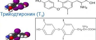
T3 hormone is a thyroid hormone and is the more active of the two main hormones. You can find another name for it – triiodothyronine. The presence of the number three in the definition of a hormone is explained by the fact that each of its molecules contains exactly that amount of iodine.
T3 is formed as a result of the breakdown of another hormone - T4, when one iodine atom is split off from it. The process that occurs after the splitting of an atom can be compared to the process of removing the pin from a grenade. The transformed, previously sedentary T4, having transformed into triiodoitrine, becomes very active.
Its purpose is to control the energy metabolic processes occurring in the human body. The hormone influences the breakdown of energy and sends it to where it is needed. Once in the bloodstream into the cells of the child’s brain, the hormone promotes its rapid development. Thanks to the work of triiodothyronine, nerve conduction is enhanced in an adult.
Triiodothyronine is important for the cardiac system and bone tissue, as it helps activate metabolism in them. General nervous excitability increases under the influence of triiodothyronine.
- Signs of elevated T3 levels
- Difficulties in conducting analysis
Effect of triiodothyronine on the body
Regulation of mitochondrial function is one of the most important functions of T3
The task of this biologically active substance is to control the level of metabolism and energy.
Its action is multifaceted:
- absorption of carbohydrates;
- lipid breakdown;
- regulation of mitochondria;
- participation in the production of vitamin A;
- adjustment of mineral metabolism;
- stimulation of children's intellectual development;
- maintaining normal human body temperature;
- improving memory and performance in adults;
- increasing the passage of signals along nerve fibers;
- prevention of the development of atherosclerotic changes;
- synthesis of proteins, ribonucleic and deoxyribonucleic acids.
In addition, T3 regulates energy conversion and the transport of energy-rich compounds to those cells that need them.
T3 hormone is free and total - what is the difference?
A certain amount of triiodothyronine can be produced by gland cells in an already “ready” state, that is, with 3 iodine atoms. Once in the bloodstream, it communicates with transporter protein molecules. The hormone is transported through the vessels to the tissues that need it. But a small amount of triiodothyronine remains in the blood, unbound to protein molecules. This triiodothyronine is called “free T3 hormone.”
The hormone that remains free in combination with the one that is bound to proteins is defined as the total hormone T3. It is its quantity that is often indicative of questionable results of tests for free hormone, which are carried out to determine thyroid dysfunction in humans.
[Video] Doctor MD Mikhail Yurievich Bolgov, a surgeon of the highest category, will explain the difference between the total and free hormone T3. The features of its action and use in diseases of the thyroid gland are highlighted:
Which one to take - general or free?
The doctor decides which test is best. You can get a referral for it from a therapist, pediatrician or endocrinologist. The form will necessarily indicate the type of hormone.
Donating blood to check the level of free T3 is necessary for diseases of the thyroid gland, or to determine the effectiveness of hormonal therapy. An assessment of total T3 levels is required when diagnosing hypothyroidism or thyrotoxicosis. However, the best option is to take both tests.
Decreased free T3
The lack of free T3 in the body may be due to the following factors:
- hypofunction (insufficiency) of the thyroid gland;
- hypothyroidism (critically low production of endocrine hormones);
- thyroiditis (subacute, acute form);
- adrenal insufficiency;
- severe systemic diseases, recent surgeries or injuries;
- serious eating disorders (fasting, low-calorie diets or low protein content in the daily diet);
- sudden weight loss, anorexia, etc.
The test results are interpreted by an endocrinologist, general practitioner or therapist.
T3 hormone test
When determining pathological conditions of the thyroid gland, the endocrinologist necessarily sends the patient to take tests for three hormones - T4, TSH, T3. Testing for the latter type of hormone is extremely important, as it allows minimizing diagnostic error.
For example, with nodular toxic goiter, very often independently working nodes are engaged in the reproduction of the T3 hormone. Its amount also increases in diffuse toxic goiter, Graves' disease and Graves' disease. If the analysis produces a result that shows a significant increase in triiodothyronine, then doctors talk about T3 toxicosis. This condition is difficult to correct with drugs and manifests itself with more vivid symptoms than those found with an increase in the amount of the T4 hormone.
Possible reasons for deviation from the norm
If the results of a thyroid hormone test deviate from the norm for children and adolescents, this may indicate problems with the thyroid gland itself, as well as with the regulation of its functioning.
Among the most common causes of increased T4 (hyperthyroidism) are:
- diffuse toxic goiter (DTZ);
- thyroid adenoma;
- excess body weight;
- chronic liver and kidney diseases, etc.
An analysis in which T4 is reduced relative to the child’s age norm may indicate the presence of one of the pathologies:
- autoimmune thyroiditis;
- endemic goiter;
- lack of iodine in food and water;
- taking narcotic drugs;
- inflammation of the hypothalamus, pituitary gland or surrounding tissues;
- intoxication of various origins;
- some types of pituitary tumors, etc.
An increase in T3 levels can occur with iodine deficiency, thyroid cancer, exposure to radiation, the development of Pendred's syndrome, thyroiditis and endemic goiter. The hormone often decreases in acute or subacute thyroiditis of various origins, as well as after operations and injuries.
The TSH level in children is also a marker of a number of pathological processes. An increased concentration may indicate the presence of one of the following diseases:
- hypothyroidism;
- Hashimoto's thyroiditis;
- tumor of the pituitary gland or lungs;
- adrenal insufficiency;
- lead poisoning;
- mental disorders, etc.
TSH in children and adolescents may be lower than normal due to the development of:
- diffuse toxic goiter;
- thyrotoxic adenoma;
- autoimmune thyroiditis;
- severe exhaustion (cachexia), etc.
It is important to remember that an isolated deviation from the norm of one or another hormone is not yet a sign of pathological processes. The result of the analysis can be influenced by lifestyle on the eve of blood donation, nutrition, taking a number of medications and other factors. Interpretation is carried out only by the attending physician, who takes into account not only laboratory diagnostic data, but also other data.
Norm of T3 hormone
The norm of total hormone T3:
| Age | Norm in Nmol/l | Norm in ng/dl | Norm in ng/ml |
| 4 months | 1,23 — 4,22 | 80 — 275 | 0,80 — 2,75 |
| 4 months - 1 year | 1,32 — 4,07 | 86 — 265 | 0,86 — 2,65 |
| 17 years | 1,42 — 3,80 | 92 — 247 | 0,92 — 2,47 |
| 7 – 12 years | 1,43 — 3,55 | 93 — 231 | 0,93 — 2,31 |
| 12 – 20 years | 1,40 -3,34 | 91 — 217 | 0,91 — 2,17 |
| 20-50 years | 1,2 — 3,1 | 78 — 201 | 0,78 — 2,01 |
| 50 years | 0.62 to 2.79 | 40 — 181 | 0,40 — 1,81 |
To convert total T3 from nmol/L to ng/mL, multiply the number by 0.651: (nmol/L) x 0.651 = ng/mL.
To convert ng/ml to nmol/l, multiply the number by 1.536: (ng/ml) x 1.536 = nmol/l.
The norm of free hormone T3:
| Age | Floor | Norm in pmol/l | Norm in pg/ml |
| In the first year | Both | 3,5 – 7, 35 | 2,28 – 7,78 |
| 1 – 12 years | 4,1 – 6,5 | 2,67 – 4,23 | |
| 12 – 16 years old | Man | 4,3 – 6,8 | 2,8 – 4,43 |
| Woman | 3,7 – 6,3 | 2,4 – 4,1 | |
| 16 – 18 years old | Man | 3,4 – 6,2 | 2,21 – 4,04 |
| Woman | 3,7 – 5,7 | 2,4 – 3,71 | |
| 18 years | Both | 2,5 – 5,7 | 1,63 – 3,71 |
If the values on your form are indicated in pg/ml, then simply multiply them by 1.536: (pg/ml) x1.536 = pmol/l. To convert the numbers the other way around: (pmol/l) x0.651 = pg/ml.
It must be said that these standards may differ from those that you receive in the laboratory. The fact is that, depending on what equipment the hormone test is performed on, the normal values will vary. Each specific laboratory makes a choice in favor of one or another apparatus and set of reagents. Therefore, the quantity will be considered normal if the results obtained fall within the reference limits indicated on a specific form from a particular laboratory.
Functions of triiodothyronine
- Provides cellular “respiration” of tissues and organs;
- Participates in general metabolism (metabolism);
- Responsible for rhythm and heart rate;
- Activates regeneration processes (cell renewal);
- Regulates nervous excitability;
- Stimulates the synthesis of vitamin A;
- Reduces the concentration of “bad” cholesterol in the blood serum.
TK is responsible for energy metabolism, i.e. promotes the extraction of energy from food and its further rational use.
Also, this hormone takes an active part in the “correct” formation of internal organs and systems in the physical development of the embryo. Therefore, T3 levels are very important to monitor in women planning to conceive and pregnant women.
But basically, a general analysis of T3 (bound by transporter proteins) makes it possible to diagnose disorders in the endocrine system and pathology of the thyroid gland itself.
What does elevated T3 hormone mean?
Free T3 increases if a person has problems with the thyroid gland. The most common of them:
- Multinodular or diffuse toxic goiter develops with isolated T3 toxicosis. Without treatment, the levels of all thyroid hormones will increase, but in the early stages of these diseases, only T3 increases, and T4 remains normal.
- Inflammation of the thyroid gland caused by autoimmune diseases, infection or childbirth.
- Thyroid cancer. Tumors are capable of producing substances similar in structure to hormones, so T3 levels will be increased.
- Endemic goiter caused by iodine deficiency. The proliferation of thyroid tissue provokes an increase in T3 levels.
- Pendred's disease. This is a congenital disease associated with genetic disorders. It gives several symptoms, one of which is an enlarged thyroid gland and hyperthyroidism.
- Resistance to thyroid hormones.
An increase in T3 levels can be caused by nephrotic syndrome, choriocarcinoma, myeloma, and hemodialysis.
In older people, the level of the T3 hormone may be reduced due to existing diseases of the internal organs. In this case, the values of the T4 hormone will remain within normal limits. In this case, the changes detected in the analysis do not indicate damage to the thyroid gland.
Signs of elevated T3 levels
Due to the fact that T3 is an extremely active hormone, its increase in the blood causes a number of very pronounced symptoms:
- The patient becomes overly irritable, nervous, quickly becomes enraged and excited. Against this background, the patient is constantly haunted by fatigue. Doctors sometimes refer to this set of symptoms as irritable weakness;
- Tremor of the fingers on the upper extremities is another common sign of increased triiodothyronine;
- The patient's pulse quickens, symptoms of tachycardia are observed, and disturbances in the heart rhythm occur. Extrasystole is a symptom of increased hormone. This condition is characterized by an increase in the number of heart contractions with a long period of rest. A person feels these disturbances and often complains to the doctor about “interruptions” in the functioning of the heart;
- Loss of body weight is often noted.
Signs of decreased and increased thyroid hormone levels
When the production of thyroid hormones is low, hypothyroidism develops. The disease can occur for a long time without significant symptoms.
The main manifestations of low levels of thyroid hormones:
- decreased performance;
- the appearance of daytime drowsiness and some inhibition of reactions;
- frequent illnesses due to weakened immune defenses;
- swelling in the legs and arms;
- disruption of the female menstrual cycle;
- instability to low temperatures and increased sensitivity to cold;
- deterioration of skin and hair condition.
When the concentration of thyroid hormones increases, the following signs appear:
- weight loss;
- cardiopalmus;
- unstable mental state;
- trembling of fingers;
- increased sweating;
- formation of a goiter in the neck area;
- physical weakness and high fatigue.
Diseases leading to a decrease in the T3 hormone
A decrease in triiodothyronine levels is observed when the production of all hormones produced by the thyroid gland is disrupted. This condition occurs in serious diseases:
- There is such a disease - Hashimoto's thyroiditis, when a person's own immunity begins to destroy some of the cells of the thyroid gland. These cells cannot be restored and in most cases cease to function and produce hormones forever.
- Hypothyroidism. This condition often develops while taking certain drugs used to treat diffuse and nodular toxic goiter. Potentially dangerous drugs include thyreostatics such as: Propicil, Tyrosol, Mercazolil.
- Triiodothyronine levels may be reduced when surgery has been performed to remove either the entire thyroid gland or a specific part of it.
- The level of T3 decreases while a person undergoes treatment with radioactive iodine. Such therapy is carried out when it is necessary to rid the patient of diffuse toxic goiter.
- A drop in hormone production is observed when taking medications containing significant amounts of iodine. Among these are cordarone, amiodarone and others.
It is worth knowing that hormones do not decrease in a chaotic manner. The level of the T4 hormone always drops first and only after that the normal value of triiodothyronine decreases. This condition is caused by the activity of the body. When the T3 hormone drops, it tries to hedge its bets and, as it were, transfers “cash into freely convertible currency,” because triiodothyronine is almost 10 times more active than T4. Doctors call this activity of the body an increase in the peripheral conversion of T4 to triiodothyronine. Thanks to this process, the effects of hypothyroidism are not as severe as they could be. Knowing this, you can independently suspect a laboratory error. If the analysis shows that the level of triiodothyronine is reduced (and it does not matter what hormone it is - total or free), but TSH and T4 remain within normal limits, then you should double-check the data obtained and donate blood for hormones again.
After all, thyroid hormone deficiency is a serious pathology. A disease in which the function of the thyroid gland decreases is fraught with the development of processes such as drowsiness, weight gain, deterioration of thought processes and speech, and disruptions in the menstrual cycle in women. If the disease is severe, cretinism is often observed in childhood, and adults suffer from myxedema. However, peripheral conversion of hormones allows you to avoid these manifestations if treatment was started in a timely manner.
DIFFICULTIES AND ERRORS IN ASSESSING THYROID STATUS
Often, a doctor is faced with a problem when the results of hormonal studies of the thyroid gland in a patient do not correspond to the clinical picture. Of course, the simplest thing in such cases is to blame the laboratory for the error of the result (errors are possible in the work of anyone, even the most experienced specialist). However, first of all, it is necessary to exclude other causes that lead to distortion of laboratory parameters, namely, a violation of the peripheral metabolism of thyroid hormones: euthyroid weakness syndrome; the influence of medications that the patient has recently taken or is taking; contact with environmental pollutants; occupational chemical hazards; resistance to thyroid hormones.
Thyroid status primarily depends on the secretion of T3 and T4.
It is estimated that the thyroid gland normally secretes about 110 nmol of thyroxine (T4) and about 10 nmol of triiodothyronine (T3) per day. T4 has 10 times less affinity for nuclear receptors than T3 and requires deiodination to become biologically active T3 (which is why T4 is now considered a prohormone).
Thyroid status depends on the deiodination process. Deiodination of T4 occurs using the iodothyronine-selenium-deiodinase system, which consists of 3 types of enzymes:
- type 1 functions in the liver and kidneys, is responsible for the conversion of T4 to T3, and is also involved in the inactivation of thyroid hormones, converting T4 into reverse T3 (rT3), T3 and rT3 into inactive T2;
- type 2 is found in the brain and skeletal muscle, where it converts T4 to T3;
- type 3 - the main inactivating enzyme - is found in the liver, central nervous system, and skin. It is no coincidence that insolation often causes thyroid diseases!), converts T4 into rT3, T3 into T2, rT3 into rT2.
About 30-40% of extrathyroidal T3 production is formed by type 1 deiodinase, 60-70% by type 2 deiodinase. A particularly active process of deiodination occurs in the hypothalamus and pituitary gland. It is important to emphasize that deiodinases are iodine-containing and selenium-containing enzymes: with iodine or selenium deficiency, a deficiency of these enzymes occurs and T4 metabolism suffers, and in addition to iodine deficiency goiter, selenium deficiency goiter is now known. Deficiency of these enzymes can develop with liver or kidney pathology. However, thyroid status depends not only on the quantity, but also on the activity of enzymes: free fatty acids (FFA), tumor necrosis factor (FNOα), and many medications (glucocorticoids, amiodarone, beta-blockers, radiocontrast agents, etc.) inhibit their activity. ). Deiodinase activity may vary between tissues, resulting in different T3 concentrations in different tissues.
Thyroid status depends on the connection of T3, T4 with plasma proteins: thyroxine-binding globulin (TBG), thyroxine-binding prealbumin, albumin. In plasma, 99% of thyroid hormones are bound to protein. Bound to T3 and T4 proteins, they do not have biological activity. Normally, the content of free T3 and T4 in the body is maintained at a constant level. The level of TSH and other proteins that bind T3, T4 depends on many factors: human nutrition, liver condition, intercurrent diseases, medication, pregnancy, etc.
Thyroid status depends on the distribution of thyroid receptors in the cells and nuclei of different organs, and on the delivery of T3 to nuclear receptors. There are several isoforms of thyroid receptors. Thus, the TRa isoform is localized in the heart and blood vessels, and the TRb isoform is localized in the liver.
Thyroid status depends on liver function. It plays a central role in the deiodination of thyroid hormones with the formation of their more active and inactivated forms. The liver is able to take up both free T4 and protein-bound T4. It synthesizes plasma proteins that bind lipophilic thyroid hormones, including thyroxine-binding globulin, thyroxine-binding prealbumin, and albumin. With liver diseases, deviations in the metabolism of thyroid hormones inevitably occur.
Thyroid status depends on the medications the patient has recently been treated with or is currently receiving.
Thyroid status depends on the person's age. With age, the concentration of T4 in the blood serum does not decrease, although its secretion by the thyroid gland decreases. In parallel with the decrease in T4 secretion, its metabolism and clearance slow down, and the peripheral conversion of T4 to T3 slows down. TSH concentration does not change with age.
Thus, the blood levels of TSH, T3, T4 total, T3, T4 free depend not only on their secretion, but also on all of the listed factors.
Euthyroid sick syndrome is a change in the level of thyroid hormones in the blood in people without thyroid disease; In most chronic diseases, there is a violation of peripheral metabolism and transport of thyroid hormones. Other designations for this syndrome are also used: “nonthyroidal illness syndrome”; "euthyroid pathological syndrome". Based on the level of thyroid hormones in the blood serum, several variants of this syndrome are distinguished: low level of T3; low levels of T3, T4; high T4 level; other violations.
Decreased concentration of total T3 in blood serum
is the most common variant of euthyroid weakness syndrome. This was initially discovered in obese patients and became known as T3-low syndrome. Then a decrease in T3 levels was detected in elderly people and this was regarded as an adaptation mechanism. Later, in case of heart failure, low T3 levels in the blood were also established. The origin of this syndrome is explained by a decrease in the activity of hepatic deiodinase, as well as a decrease in the entry of T4 into cells. Since in obesity there is always an excess of saturated fatty acids, and in severe somatic pathology there is an excess of tumor necrosis factor (TNFα), this in many cases can explain the inhibition of deiodinase activity. It is now known that 70% of hospitalized patients with non-thyroid diseases have a reduced concentration of T3 in the blood serum, and free T3 decreases to a lesser extent than total T3, while the level of T4 and TSH, the TSH response to thyrotropin-releasing hormone (TRH) remain normal, although Recently, reports have appeared in the literature about a possible slight increase in the level of TSH in the blood, so that there is a need for laboratory differential diagnosis with primary hypothyroidism. However, this does not cause much difficulty. Hormonal diagnosis of T3-low syndrome and hypothyroidism is presented in Table 1.
Table 1. Hormonal diagnosis of T3-low syndrome and primary hypothyroidism
| T3-low-syndrome | Primary hypothyroidism |
| T3 is reduced to a greater extent than T4 | T4 is reduced more significantly than T3 |
| TSH is normal or slightly elevated | TSH increased significantly |
| Free T4 is normal | Free T4 is reduced |
| rT3 increased | rT3 is reduced |
Erased clinical signs of hypothyroidism are possible in T3-low syndrome, but nevertheless it is believed that the detection of an isolated decrease in T3 levels in a patient cannot serve as a basis for a diagnosis of hypothyroidism. The treatment of this pathology remains controversial to this day.
Euthyroid weakness syndrome with low levels of T3, T4. This variant occurs in patients with severe pathology, especially often in intensive care unit patients; this condition can develop acutely. A low T4 level is an unfavorable prognostic sign, indicating a threat of death. The development of this variant of euthyroid weakness syndrome is explained by a violation of the binding of thyroid hormones to plasma proteins. Inhibitors of T3 and T4 binding appear in the blood. Obviously, such inhibitors are cytokines, in particular TNFα and interleukin-2. Polypharmacy also plays a role in the development of this variant: patients are often prescribed glucocorticoids, beta-blockers, heparin, furosemide, dopaminomimetics and other drugs that disrupt the metabolism of thyroid hormones. TSH levels may be normal or (more often) decreased; the TSH response to TRH may also decrease. A simultaneous decrease in the levels of TSH, T3, T4 necessitates differential diagnosis with secondary hypothyroidism. However, with secondary hypothyroidism, the blood level of rT3 also falls, in contrast to euthyroid weakness syndrome, in which the level of rT3 increases due to a slowdown in its metabolism. The phenomenon of inappropriately low TSH secretion with reduced levels of T3 and T4 in such patients is not entirely clear. In case of successful treatment of the underlying disease that caused the development of low T3, T4 syndrome, TSH and thyroid hormone levels spontaneously normalize.
High levels of T4 in euthyroid weakness syndrome develop mainly in liver diseases, when large doses of iodine are administered to patients, for example, radiocontrast agents or amiodarone. They inhibit the uptake of T4 by the liver, thereby its conversion in the liver to T3 slows down, and the level of total and free T4 in the blood serum becomes higher than normal. The TSH level remains normal, but in some cases decreases, and then differential diagnosis with thyrotoxicosis has to be carried out.
TSH abnormalities as a variant of euthyroid weakness syndrome. This variant is most common in patients with diabetes due to irreversible glycation of TSH, which leads to a decrease in its activity. The production of T3 and T4 decreases, and clinical signs of hypothyroidism may appear. To recognize this syndrome, anamnestic data (diabetes) play an important role. You also need to take into account the high level of glycated hemoglobin.
Medicines and indicators of thyroid status. Physicians should consider possible changes in thyroid status under the influence of medications and distinguish transient changes in hormonal status caused by medications from true disorders of thyroid function. The mechanisms of action of drugs on the thyroid system are varied (see Table 2).
table 2
Mechanisms of action of drugs on the thyroid system
| Mechanism of action | Drugs | TSH, T3, T4 indicators |
| Decreased production of TSH by the pituitary gland | Dopamine, dobutamine, dobutrex, glucocorticoids, octreotide, bromocriptine, vitamin B6, phentolamine, haloperidol | Reduced |
| Impaired synthesis and release of thyroid hormones | Glucocorticoids, lithium, glutethimide | T3, T4 total and free decreased |
| Suppression of T4-T3 5'-deiodination | Amiodarone, beta-blockers, glucocorticoids, radiocontrast agents | T3 is normal or decreased, T4 is increased, TSH is normal or slightly decreased |
| Increased levels of thyroid binding globulin | Estrogens, hormonal contraceptives, heroin, tamoxifen, 5-fluorouracil | Total T3, T4 are increased, free T3, T4 are normal; TSH is normal, may increase or decrease slightly |
| Displacement of T3 and T4 from binding to proteins | NSAIDs, furosemide, heparin, phenytoin, carbamazepine, sulfonylurea derivatives | T4, T3 general decrease |
| Release of T4 from tissue depots (primarily from the liver) | Cyclophosphamide, cyclophosphamide, cholecystographic drugs | T4 increases |
| Increased metabolism of thyroid hormones | Rifampicin, phenytoin, carbamazepine, barbiturates, co-trimoxazole | T3, T4 total decrease, T3, T4 free are normal |
| Increased formation of transport proteins | Estrogens, hormonal contraceptives | Total and free T4 are normal or elevated |
| Increased conversion of T4 to T3 | Heroin, somatotropin | T4 decreased, T3 increased |
Many drugs have multiple mechanisms of action on the thyroid system. An example is glucocorticoids, which suppress the secretion of TSH and inhibit the release of thyroid hormones. Simultaneous stimulating and suppressive effects are possible. Thus, iodine-containing drugs (amiodarone, radiopaque agents, topical iodide preparations) stimulate the synthesis of thyroid hormones and suppress their release. It is no coincidence that long-term treatment with amiodarone can cause hypothyroidism in some patients and thyrotoxicosis in others.
Changes in thyroid status depend on the patient’s initial state of the hypothalamic-pituitary-thyroid system. A convincing clinical example is patient K-va, 42 years old, who after 6 months. After undergoing thyroid resection for toxic goiter, a persistent increase in the TSH level (from 13.3 to 56.2 mIU/ml) was recorded for a year, with a simultaneous increase in the content of free and total T4 and a persistently normal level of total and free T3. The patient's health was satisfactory; signs of hypothyroidism were clinically noted (dry skin, hyperkeratosis of the elbows, drowsiness, lethargy, tendency to constipation, chilliness). The patient was examined to exclude thyrotropinoma: the diagnosis was not confirmed. It turned out that the patient had been taking anaprilin for hypertension for 1.5 years. She was diagnosed with decompensated primary postthyroid resection hypothyroidism and a drug reaction to anaprilin (deceleration of the conversion of T4 to T3). Indicators of thyroid status returned to normal after discontinuation of beta-blockers, replacing them with other antihypertensive drugs and replacement therapy with levothyroxine in adequate doses.
Drugs from the same pharmacological group can disrupt thyroid status to varying degrees. Thus, propranolol in therapeutic doses or with a long course of treatment inhibits the conversion of T4 to T3, causing a pronounced decrease in the level of T3 in the blood and an increase in T4. Selective beta-blockers (atenolol, metoprolol) do not cause this effect.
Inconsistency of hormonal results with the clinical picture of the disease when treated with thyroid hormones. Every endocrinologist is familiar with this situation, especially often with subclinical or transient hypothyroidism. This may be due primarily to intercurrent diseases and/or their treatment. An increase in the dose of levothyroxine (even if the patient receives it at the maximum dose) will be required if:
- malabsorption syndrome, increased intestinal motility, protein deficiency: for example, with exacerbation of chronic pancreatitis, nonspecific ulcerative colitis, Crohn's disease, etc.;
- increasing the clearance of thyroxine during treatment with antiepileptic drugs (phenobarbital, phenytoin, carbamazepine), the antibiotic rifampicin, etc.;
- impaired intestinal absorption (use of dietary fiber, almagel, ferrous sulfate, cholestyramine, colestide);
- increase in the level of thyroxine-binding globulin: pregnancy; hormonal contraceptives; estrogen-containing drugs;
- the use of drugs with antithyroid action, for example, treatment with beta-blockers of coronary artery disease or hypertension in a patient with hypothyroidism.
With postpartum or subacute thyroiditis, a change in phases of the functional state of the thyroid gland is possible: destructive thyrotoxicosis can quickly give way to symptoms of hypothyroidism, but the dynamics of TSH levels “do not keep up” with the changes in clinical phases.
The influence of environmental pollutants and occupational chemical hazards on thyroid status and the results of hormonal examinations. The effects of various chemical agents are diverse: they can cause primary (synthetic detergents, bromides, uranium, etc.) or secondary (lead, mercury, other heavy metal salts) hypothyroidism, various variants of euthyroid weakness syndrome. They are able to suppress enzymes involved in the synthesis or conversion of thyroid hormones (phosphorus, nitrates, fluorine, vanadium, carbon monoxide, thiourea, cyanides, etc.), block SH groups and reduce the activity of the transition of T4 to T3 (pesticides, fluorine) , worsen the functions of the liver, kidneys and impede the excretion of thyroid hormones (carbon disulfide, phosphorus, heavy metal salts) from the body. Therefore, the results of hormonal studies of the patient’s thyroid gland may not correspond to the clinical picture.
Resistance to thyroid hormones. Conflicting results of hormonal studies are possible due to resistance to thyroid hormones: despite elevated levels of free T4, and sometimes T3, TSH levels in patients are also elevated, and clinical symptoms of hypothyroidism may be detected. The pathogenesis is based on a decrease in tissue sensitivity to thyroid hormones, caused by genetic disorders - a mutation in the thyroid hormone receptor gene. Resistance can be generalized or partial. The disease was thought to be rare. However, along with the increase in genetic pathology in general, an increase in the prevalence and resistance to thyroid hormones is predicted.
Medical tactics in case of discrepancy between the results of hormonal examination and the clinical picture of the disease. First of all, you need to recheck the result (in the same or another laboratory). If indicators of thyroid status contradict the clinic, then the influence of medications should be excluded (it is rare when goiter is the only pathology; as a rule, it is combined with other diseases).
It is necessary to find out from the patient what medications she used to treat intercurrent diseases and which ones she is currently treating, be sure to clarify whether dietary supplements were used, assess the condition of the patient’s liver, check for the presence of euthyroid weakness syndrome, and analyze the likelihood of other causes of discrepancy.
Of course, treatment with thyroid hormones, correction of their doses, and prescription of thyreostatics should be based not only on laboratory hormonal data, but primarily on the clinical picture of the disease. In cases of discrepancy, the doctor must have a clear concept of what causes the discrepancy.
Irina TERESHCHENKO, professor.
Perm State Medical Academy.
How to prepare for the analysis?
Blood is taken to determine hormone levels in the morning, on an empty stomach. However, the patient is allowed to drink sweetened water.
If the patient has undergone surgery on the thyroid gland, is taking medications, or has recently completed a course of radiotherapy, then blood for the T3 hormone is taken no earlier than 14 days later.
To obtain an accurate result, you need to stop taking medications for 2 days. In particular, oral contraceptives can distort the data.
Difficulties in conducting analysis
Carrying out an analysis to detect the level of triiodothyronine is considered a rather complex procedure. Errors often occur in laboratories. You can think about these on your own if an additional test was carried out to identify the level of other thyroid hormones - TSH and T4. If the results obtained show that TSH is normal and the T3 hormone is elevated, then most likely an error has occurred. The unreliability of the analysis can also be judged on the basis of an increase in T3 and TSH, but normal T4 values. If such results were obtained, then it makes sense to double-check the data. This is due to the fact that when T3 increases, the TSH level decreases and T4 increases.
If the tests were carried out qualitatively and the results clearly indicate an increase in triiodothyronine, it is necessary to consult an endocrinologist.
Standards for the content of various forms of T3
The highest T3 content is observed in children from the 4th day to the 12th month of life
Depending on the gender and age of a healthy person, his blood contains free T3 and total T3 in concentrations from 1.30 to 7.50 nmol/liter. The concentrations of these substances, taking into account age, are given in the following tables.
Triiodothyronine (total):
| Floor | Age (years) | Concentration (nmol/liter) |
| AND | From 4 days to 1 year | 1.300 – 6.300 |
| M | From 4 days to 1 year | 1.300 – 6.300 |
| AND | 1 — 12 | 1.740 – 2.910 |
| M | 1 — 12 | 1.740 – 2.910 |
| AND | 12 — 15 | 1.500 – 2.710 |
| M | 12 — 15 | 1.500 – 2.710 |
| AND | 15 — 17 | 1.440 – 2.150 |
| M | 15 — 17 | 1.450 – 2.390 |
| AND | 17 — 19 | 1.590 – 2.080 |
| M | 17 — 19 | 1.590 – 2.080 |
| AND | Over 19 | 0.890 – 2.440 |
| M | Over 19 | 0.890 – 2.440 |
Important! Only a specialist can interpret the results of the study. Self-diagnosis in the case of hormonal levels is strictly prohibited.
On average, women's results are 5 to 10 percent lower than men's. Pregnancy leads to an even greater decrease in the content of total triiodothyronine, and its restoration occurs no earlier than 7 days after the birth of the child. A further decrease in hormone levels is observed in both sexes upon reaching the age of sixty-five.
Starting at age 65, T3 levels begin to decline noticeably
Triiodothyronine (free):
| Floor | Age (years) | Concentration (nmol/liter) |
| AND | From 4 days to 1 year | 3.600 – 7.500 |
| M | From 4 days to 1 year | 3.600 – 7.500 |
| AND | 1 – 12 | 4.300 – 6.800 |
| M | 1 – 12 | 4.300 – 6.800 |
| AND | 12 – 15 | 3.800 – 6.100 |
| M | 12 – 15 | 4.400 – 6.700 |
| AND | 15 – 19 | 3.600 – 5.700 |
| M | 15 – 19 | 3.500 – 5.900 |
| AND | Over 19 | 2.600 – 5.700 |
| M | Over 19 | 2.600 – 5.700 |
In order to obtain correct results, the influence of a number of factors must be excluded.
After all, indicators can change significantly under the influence of:
- stress;
- physical activity;
- sports training;
- consumption of alcoholic beverages and drugs;
- changes in the seasons of the year (maximum values in autumn and winter, and minimum values in summer);
- errors in the diet that must be followed on the eve of collecting biological material;
- taking medications such as hormones (including contraceptives), salicylates, amiodarone, coumarin (and derivatives), drugs containing iodine and lithium.
Different laboratories may use different systems for determining T3 levels
Important! When taking an analysis for T3, it should be taken into account that each laboratory may have its own internal instructions for carrying out the procedure, since the study can be carried out using several different algorithms and the results, accordingly, may differ and be indicated in different units of measurement.
As a rule, on the forms of each laboratory there is a table of normal, reference values of the indicator for ease of comparison and interpretation of the result by the specialists who ordered the study.
What factors affect triiodothyronine levels?
The T3 hormone, the norm in women is lower than in men, can change its concentration under the following conditions:
- Age. In children, the amount of biologically active substances in the thyroid gland reaches the level of adults by the age of 11-15 years. With age (after 65 years), its concentration gradually decreases.
- During pregnancy, the physiological norm is a slight decrease in the amount of this hormone, which is restored after childbirth.
- Seasonal changes. After taking a test for triiodothyronine in winter, the norm in women and men may be increased. Conversely, during the summer months there is an increase in thyroid hormone levels.
