Read in this article:
- Why does blood pressure change in pregnant women?
- Change in pressure by trimester
- Normal blood pressure in pregnant women
- Increased blood pressure in pregnant women
- Reducing blood pressure in pregnant women
- How to measure blood pressure during pregnancy
- Blood pressure control is the key to the health of mother and child
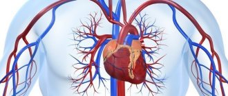
Blood pressure is one of the key indicators of the functioning of the human circulatory system. During pregnancy, the load on the body increases, the heart and blood vessels also react to these changes. Regularly measuring blood pressure in pregnant women allows you to monitor the functioning of the cardiovascular system, and sometimes prevent the development of dangerous complications.
Why does blood pressure change in pregnant women?
The body has to adapt to a special condition - pregnancy. A woman’s body weight gradually increases, and additional blood circulation is activated between the uterus and placenta. The functioning of the heart and blood vessels at this time is affected by:
- Changes in hormonal levels;
- An increasing volume of blood circulating in the body;
- Increased tone of the central nervous system;
- Changing the location of the diaphragm, which rises upward;
- Increased pressure inside the abdominal cavity;
- Displacement of the heart in the chest;
- Increased heart rate.
The body directs all its efforts to improve blood flow in the placenta and in the uterus, because this is necessary to provide the developing fetus with oxygen and nutrients. Under these circumstances, blood pressure changes.
Baby
The 17th week of pregnancy is marked for the baby by an active increase in the mass of his muscles, their gradual strengthening, which allows him to move in the tummy more intensely and variedly. Quite frequent movements of the limbs help the child train muscles and also develop the joints of the knees and elbows. At this stage, the hands are still clenched into fists, rarely unclenched, and only for the purpose of grasping the umbilical cord or sucking a finger. Until 17 weeks, the fetal head was practically motionless, dropped quite low, and the chin was tightly attached to the chest. Strengthening the muscles of the back and cervical region at this stage allows the child to raise his head to an almost vertical position.
Internal organs continue to develop and improve. Their structure only becomes more complex, which contributes to the expansion of functionality. Their preparation will allow you to begin normal, full functioning after the baby is born.
It is important to note the changes that occur in the vascular system and heart of the fetus. The nerve plexuses that surround the heart give rise to the formation of the conduction system; Its uniqueness lies in the fact that the heart does not need signals from the outside to work. All human internal organs obey the central nervous system. The brain sends certain instructions according to which each of the organs works. The heart, in turn, functions with the help of command signals that arise within itself and cause it to contract. The exclusively autonomous mode of operation of the heart allows it to be independent of the level of central nervous system activity throughout a person’s life.
The 17th week of pregnancy is also characterized by the formation of the respiratory system, namely, the strengthening of the lung muscles. Active respiratory contractions resemble inhalation and exhalation and thereby strengthen the muscles of the chest. The formation of the alveolar apparatus of the lungs also occurs. Alveoli are presented in the human body in the form of small bubbles, which are located on the surface of the respiratory apparatus and are responsible for gas exchange. Each breath helps saturate the blood with oxygen. At the 17th week, the alveoli are in a compressed state, but immediately after birth, with the first breath, they will straighten out and be ready for normal functioning.
The weight of the fetus at this stage is about 150 g, and the body length is 12-13 cm.
Change in pressure by trimester
Pressure indicators and its changes are determined by the duration of pregnancy. The entire period of pregnancy is divided into three trimesters, and in each trimester certain changes in blood pressure occur:
- In the early stages, the heart and blood vessels work as usual. But already from the first weeks of pregnancy, the female body actively produces the hormones estrogen and progesterone, which contribute to the dilation of blood vessels. Because of this, the pressure decreases slightly;
- In the second trimester, blood pressure levels decrease even more. This is due to the formation of a new blood circulation between the placenta and the uterus. A decrease in indicators also provokes a decrease in vascular resistance;
- In the third trimester, the volume of blood circulating in the body becomes maximum. Blood pressure gradually increases. Closer to childbirth, blood volume stops increasing, the fundus of the uterus and the diaphragm descend. As a result, the pressure in pregnant women in the later stages returns to the normal level familiar to the woman.
The periods from 4 to 12 weeks are considered especially dangerous, when a drop in pressure combined with toxicosis can significantly worsen the well-being of the expectant mother, as well as from 28 to 32 weeks, when the load on the blood vessels and heart becomes maximum. But regular blood pressure measurements should not be neglected during any period of pregnancy, especially if a woman has pathologies of the cardiovascular system or is predisposed to them.
Arterial hypertension during pregnancy: diagnosis, treatment, prognosis
R.I.STRYUK,
Doctor of Medical Sciences, Professor,
Y.V. BRYTKOVA
, Candidate of Medical Sciences, Moscow State Medical and Dental University named after. A.I. Evdokimov" of the Ministry of Health of the Russian Federation
The article presents the classification of arterial hypertension (AH) in pregnant women, provides an algorithm for diagnostic studies, discusses the issues of pharmacotherapy of hypertension during pregnancy, and emphasizes that the presence of hypertension during pregnancy is a risk factor for the development of cardiovascular pathology in women in subsequent periods of life.
Arterial hypertension (AH)
during pregnancy remains the leading cause of maternal, fetal and neonatal mortality in developing and industrialized countries. In different regions of Russia, its frequency ranges from 7 to 29%; in Western Europe, hypertension occurs in approximately 15% of pregnant women [1, 2]. Women with hypertension have a higher risk of severe complications - both from the mother and from the fetus. Thus, when analyzing 15,945 births, researchers significantly more often noted in women with hypertension (888 women, or 5.5%) premature births, abruption of a normally located placenta, cases of surgical delivery by cesarean section, and a low newborn Apgar score (1-1). 1st and 5th minutes <7 points), newborn weight ≤ 2,500 g, need for intensive care of newborns, immaturity and prematurity of the fetus, birth trauma, neonatal mortality [3]. Risk factors, in addition to hypertension, also include maternal age ≥ 30 years, low level of education, number of previous births ≥ 3, weight gain ≥ 16 kg during pregnancy, dyslipidemia, obesity, family history, antiphospholipid syndrome and impaired glucose tolerance [ 4].
It must be emphasized that the diagnostic criteria for hypertension in pregnant women are the same as for other categories of patients: an increase in systolic blood pressure ≥ 140 mm Hg. or diastolic blood pressure ≥ 90 mm Hg. [5, 6]. The presence of hypertension before pregnancy or the detection of elevated blood pressure in a woman during pregnancy requires a thorough examination of her to decide on the possibility of prolonging pregnancy, selecting adequate antihypertensive therapy and monitoring the condition of the fetus. In this case, it is necessary to take into account a number of physiological features of hemodynamics during gestation, caused by the influence of a complex of neurohumoral factors, an increase in body weight due to the placenta and the increasing weight of the fetus, increased metabolism, the development of physiological hypervolemia, and the formation of uteroplacental blood flow. During pregnancy, physiological myocardial hypertrophy develops - myocardial mass increases by 10–31% by the end of the gestational period and quickly returns to its original level after childbirth. The gestation period is accompanied by an increase in minute (by 15–50%) and stroke (by 13–29%) heart volume, blood flow velocity (by 50–83%), heart rate (by 15–20 beats/min), and a decrease in general peripheral resistance vessels (on average by 12–34%). During normal pregnancy, systolic blood pressure (BPs) changes slightly, diastolic blood pressure (BPd) at a period of 13–20 weeks decreases by 5–15 mm Hg. The lowest blood pressure is usually observed at the 28th week of pregnancy, then it increases, and in the third trimester blood pressure and blood pressure return to pre-pregnancy blood pressure values [7].
The gestational period is characterized by a physiological increase in the activity of the renin-angiotensin-aldosterone system, which contributes to an increase in plasma volume and total water volume in the pregnant woman’s body. An important factor in the adaptation of the cardiovascular system to pregnancy is systemic vasodilation, in the development of which increased secretion of nitric oxide and other vasodilating factors plays a role. A change in the reactivity of the vascular bed towards the predominance of vasodilation reactions is also associated with an increase in the level of estrogen and progesterone, which contribute to an increase in the sensitivity of adrenergic receptors to hormones of the sympathetic-adrenal system [8, 9]. In addition to a thorough history taking and assessment of physical data, pregnant women with hypertension must undergo ECG and EchoCG studies, blood and urine tests, biochemical blood tests to determine glucose levels, levels of liver enzymes, creatinine, and, if necessary, indicators of the hemostatic system. The protein content in daily urine should be determined (if it exceeds 2 g/day, careful monitoring is recommended; if proteinuria exceeds 3 g/day, the feasibility of delivery should be discussed). Uterine artery Doppler imaging in the second trimester reveals uteroplacental hypoperfusion, which is associated with a higher risk of preeclampsia and intrauterine growth restriction in both high- and low-risk women [208].
Arterial hypertension in pregnant women is not a homogeneous condition; it includes the following forms [5, 6]: - arterial hypertension existing before pregnancy (hypertension or symptomatic hypertension - chronic hypertension (CAH)); - gestational hypertension; - arterial hypertension existing before pregnancy and combined with gestational hypertension and proteinuria; - unclassified hypertension.
The criterion for diagnosing CAH is an increase in blood pressure ≥ 140/90 mmHg. before pregnancy or in the first 20 weeks of pregnancy. Arterial hypertension usually persists for more than 42 days after delivery and may be accompanied by proteinuria. In women with undiagnosed hypertension, blood pressure may be normal at the beginning of pregnancy due to its physiological decrease in the first trimester.
Gestational hypertension, with or without proteinuria, develops after 20 weeks of gestation and in most cases resolves within 42 days after birth. Gestational hypertension complicates pregnancy in 6-7% of cases; it leads to deterioration of organ perfusion.
Preeclampsia (in Russian literature - gestosis)
– this is gestational hypertension, which is accompanied by proteinuria (≥ 0.3 g/day in daily urine). Preeclampsia is considered a systemic disease that causes changes in the body of both mother and fetus. Overall, preeclampsia complicates pregnancy in 5–7% of cases, but its incidence increases to 25% in women with pre-pregnancy hypertension [10]. Preeclampsia is more common in first pregnancies, multiple pregnancies, hydatidiform moles, and diabetes mellitus. It is combined with placental insufficiency, which often leads to poor fetal growth and is one of the most common causes of prematurity. The share of preeclampsia in the structure of causes of birth of children with very low body weight (less than 1,500 g) is 25% [11]. Symptoms of severe preeclampsia include: - pain in the right hypochondrium or epigastric region due to liver edema + liver bleeding; - headache + blurred vision (cerebral edema); - blindness associated with damage to the occipital lobe; — strengthening of reflexes + clonus; - convulsions (cerebral edema); - HELLP syndrome: hemolysis, increased liver enzymes, decreased platelet count.
Edema today is no longer considered a diagnostic criterion for preeclampsia, because their frequency during normal pregnancy reaches 60%. Arterial hypertension existing before pregnancy, in combination with gestational hypertension and proteinuria, is characterized by a further increase in blood pressure and the appearance of proteinuria ≥ 3 g/day after 20 weeks of gestation.
Hypertension is classified as unclassifiable if blood pressure is first measured after 20 weeks of pregnancy and hypertension is detected (with or without systemic manifestations). In such cases, it is necessary to continue monitoring blood pressure for 42 days after birth and beyond.
The tactics for managing pregnant women with hypertension consists of measures aimed at ensuring a protective regime, which includes the woman’s employment with the exception of shift work, physical and psycho-emotional overload, sufficient sleep, and rest during the daytime. Nutrition should be complete in composition and not excessive in calorie content, especially in obese women. About half of the food composition should be animal proteins. It is recommended to consume foods containing predominantly complex carbohydrates - wholemeal bread, cereals, vegetables, fruits, berries, and easily digestible carbohydrates (sugar, confectionery) should be limited. Food must contain a sufficient amount of vitamins, because... the need for them during pregnancy increases significantly. It is advisable to have a varied menu, including low-fat dairy products, unsalted cheeses, meat, fish, liver, kidneys, eggs, legumes, etc. The diet should not include fatty meats and fish, smoked meats, pickles, chocolate, strong tea and coffee. . Weight loss for obese pregnant women is not recommended, because... it can lead to a decrease in the newborn's body weight and a subsequent slowdown in his growth. However, obesity can cause adverse outcomes for both the woman and the fetus, so the suggested ranges for weight gain during pregnancy should be adhered to: for women with a normal body mass index (< 25 kg/m2), the recommended weight gain is 11.2 –15.9 kg, in overweight women (25.0–29.9 kg/m2) – 6.8–11.2 kg, in obese women (≥ 30 kg/m2) – < 6.8 kg [12].
The issues of drug therapy for hypertension during pregnancy are quite complex; the range of antihypertensive drugs is narrow, because any impact, incl. medication may have a negative effect on the fetus, and with a beneficial effect on the fetus, the drug may be suboptimal for the mother. The embryotoxic and teratogenic effects of drugs appear at different stages of pregnancy. Thus, up to 20 days after conception, the “all or nothing” principle applies, based on the fact that at this stage the pluripotent cells of the embryo capable of regeneration have not yet been differentiated. At 3–8 weeks of pregnancy, during the period of active organogenesis, the most sensitive period in relation to the negative effects of drugs, a teratogenic effect appears. After the 8th week before birth, the embryotoxic effect of the drugs may be detected. During this period of growth, structural defects, as a rule, do not occur, but disruption of postnatal functions and various behavioral abnormalities are possible. However, a recent analysis of the effectiveness of antihypertensive therapy in pregnant women with mild and moderate hypertension (46 studies (4,282 women) and 28 studies (3,200 women), respectively) showed that the prescription of one or more antihypertensive drugs led to a 2-fold reduction in the risk of developing severe hypertension (19 studies, 2409 women, relative risk (RR) 0.50, 95% confidence interval (CI) 0.41 to 0.61; risk difference (RR) -0.10 (-0 .12 to -0.07)) [13].
According to the recommendations of the VNOK and the European Association of Cardiology for the treatment of pregnant women with cardiovascular diseases, in patients with a blood pressure level of 140/90 mm Hg. antihypertensive therapy is not carried out, but is prescribed in such cases as: - gestational hypertension ± proteinuria; — CAH combined with gestational hypertension; — Hypertension, accompanied by subclinical target organ damage or clinical symptoms.
Antihypertensive therapy is recommended to begin if blood pressure exceeds 150/95 mmHg.
The general principles of drug treatment of hypertension are: - maximum effectiveness for the mother and safety for the fetus; — starting treatment with minimal doses of one drug; - switching to drugs of another class if the treatment effect is insufficient (after increasing the dose of the first drug) or is poorly tolerated; - if a woman is taking an antihypertensive drug at the stage of pregnancy planning, adjustment of drug therapy (cancellation of angiotensin-converting enzyme inhibitors, angiotensin II receptor blockers, direct renin inhibitors) and dose of the drug (achieving a target blood pressure level < 140/90 mm Hg); - use of long-acting drugs to achieve a 24-hour effect with a single dose.
The use of such drugs provides a milder and longer-lasting antihypertensive effect, more intense protection of target organs, as well as high patient adherence to treatment. It is very important not to sharply reduce blood pressure, because this may impair uteroplacental blood flow. The blood vessels of the placenta function in a maximally dilated state and are not capable of autoregulation, so a drop in blood pressure in the mother’s bloodstream can worsen the condition of the fetus.
In accordance with the classification of foods and drugs adopted by the Food and Drug Administration (FDA) [14], there are 5 categories of drugs according to the level of safety for the fetus:
A. Controlled studies have shown no risk to the fetus. B. Lack of evidence of risk to the fetus: a risk to the fetus has been found in animals, but not in humans, or there is no risk in the experiment, but there are insufficient studies in humans. C. Risk to the fetus cannot be excluded - side effects have been identified in animals, but there are insufficient studies in humans. The expected therapeutic effect of the drug may justify its use, despite the potential risk to the fetus. D. Conclusive evidence of risk—in humans, there is a proven risk to the fetus, but the expected benefit to the expectant mother may outweigh the potential risk to the fetus. X. Use during pregnancy cannot be justified: the negative effects on the fetus outweigh the potential benefits of this drug for the expectant mother.
In accordance with the recommendations of the All-Russian Scientific Society of Cardiology (2010), ESH/ESC experts currently use 4 groups of antihypertensive drugs that meet the criteria for pharmacotherapy during pregnancy for the treatment of hypertension during pregnancy [5, 6]: - centrally acting drugs (methyldopa) ; - calcium antagonists of the dihydropyridine series (long-acting nifedipine); - cardioselective β-blockers (metoprolol, bisoprolol); - α-β-adrenergic blockers (laβlol).
Combination therapy is carried out in case of ineffectiveness of monotherapy at the maximum dose. A rational combination is long-acting nifedipine + β-blocker; if this combination is ineffective, it is possible to add chlorothiazide (hypothiazide) in small doses (12.5–25.0 mg/day).
The main medications recommended for use during pregnancy for the treatment of hypertension are presented in the table.
If we talk in more detail about each group of antihypertensive drugs used in pregnant women, then we should first of all talk about methyldopa, a centrally acting antiadrenergic drug classified as category B according to the FDA classification. A single study conducted about 30 years ago demonstrated the safety of methyldopa based on long-term follow-up of newborns (7.5 years), and since then it has been considered a first-line drug in patients with hypertension [15, 16]. However, it must be remembered that the side effects of methyldopa are drowsiness, the development of depression, fluid retention and edema, orthostatic hypotension; in the 16th–20th week of pregnancy, the use of the drug is not recommended due to its effect on the content of dopamine in the fetal nervous system.
In 2007, data were published evaluating the effectiveness of antihypertensive therapy in women with mild to moderate hypertension during pregnancy: 19 studies (1282 women) compared β-blockers with methyldopa in reducing the risk of developing severe hypertension (10 studies, 539 women , RR 0.75 (95% CI 0.59 to 0.94); RD -0.08 (-0.14 to 0.02); NNT 12 (6,275)). β-blockers were found to be better than methyldopa in reducing the risk of severe hypertension (10 studies, 539 women, RR 0.75 (95% CI 0.59 to 0.94); RD -0.08 (- 0.14 to 0.02); NNT 12 (6,275)). Along with this, there was no clear difference between any of the alternative drugs with respect to the risk of developing proteinuria/preeclampsia in patients with CAH [17].
The second group of drugs used for hypertension in pregnant women are cardioselective β-blockers. They belong to category C (FDA), and are recommended for use from the second trimester of pregnancy. The mechanism of action of drugs in this group is due to the fact that they competitively and selectively inhibit the binding of catecholamines to β-adrenergic receptors. Drugs in this group are divided into cardioselective, predominantly blocking β1-adrenergic receptors, and non-selective, blocking β1- and β2-adrenergic receptors. The principal mechanism of their inhibitory effect on adrenoreactive structures is to weaken or eliminate the effects associated with excitation of β1-adrenergic receptors by catecholamines, which cause increased heart rate, increased automaticity of the atrioventricular node and myocardial excitability, increased impulse conduction speed, increased myocardial contractility, decreased refractory period , activation of lipolysis. Excitation of β2-adrenergic receptors by catecholamines leads to dilation of arterioles, decreased tone of smooth muscles of the bronchi, bladder, uterine tone during pregnancy, tremor of skeletal muscles, inhibition of the release of histamine, leukotrienes in mast cells during allergic reactions of type I, hypokalemia, increased hepatic glycogenolysis. During pregnancy, β-blockers can cause intrauterine growth retardation due to a decrease in uteroplacental and fetoplacental blood flow and induction of premature labor. This is especially true for atenolol, which belongs to FDA category D and is not recommended for use during pregnancy. When using drugs in this group, the fetus may experience various side effects: bradycardia, hypoglycemia, apnea, metabolic disorders. However, the frequency of these adverse events is quite low, and if it is necessary to prescribe β-blockers, the balance of benefit and risk is considered.
In 2003, Magee LA et al. based on an analysis of systematic reviews of the Cochrane Library and data from various registries, they summarized the experience of using β-blockers for the treatment of mild and moderate hypertension in pregnant women [18]. The authors analyzed 29 clinical studies (approximately 2,500 women) that compared the effectiveness and safety of β-blockers with placebo, no therapy, or other antihypertensive drugs. Oral β-blockers were shown to reduce the risk of developing severe hypertension (RR 0.37, CI 95% (0.26–0.53); 11 CIs, N = 1,128 women) and the need for additional antihypertensive therapy (RR 0.44, CI 95% (0.31–0.62); 7 CI, N = 856 women). The authors do not draw conclusions on the effect of β-blockers on mortality rates and premature birth of the fetus due to the lack of sufficient information in the analyzed CTs. However, β-blocker use was associated with an increase in the number of small gestational age neonates (RR 1.36, CI 95% (1.02–1.82); 12 CIs, N = 1,346 women), with an increase in neonatal bradycardia and reduction in neonatal respiratory distress, but these endpoints were analyzed in a small number of CTs. The authors’ conclusion that blood pressure control in a mother with hypertension using β-blockers can be justified only if there is significant benefit for the mother and/or child is absolutely justified, but the analyzed clinical studies did not answer the question.
Von Dadelszen P., Ornstein MP (2000) analyzed data from 45 randomized controlled trials (RCTs) and found an inverse relationship between the reduction in mean blood pressure during treatment with beta-blockers and the incidence of births of newborns whose size is not appropriate for their gestational age. Decrease in average blood pressure for every 10 mm Hg. was accompanied by a decrease in birth weight by 145 g (with a 95% confidence interval from 5 to 285 g). However, no relationship was found between the duration of antihypertensive therapy and mean birth weight [19]. Calcium antagonists (CA), the first representative of which is verapamil, synthesized in 1962 in Germany, are the drugs of choice for the treatment of hypertension; according to the FDA classification, drugs in this group belong to category C. Calcium antagonists are heterogeneous in chemical structure, electrophysiological properties, pharmacological effects and clinical use. To treat hypertension during pregnancy, dihydropyridine calcium antagonists are used, the main mechanism of action of which is associated with peripheral vasodilation due to the blockade of slow voltage-dependent L-type calcium channels and a decrease in the intracellular concentration of Ca2+, as well as stimulation of endothelial synthesis of NO and bradykinin [20, 21]. The body's compensatory response to systemic vasodilation is the activation of the sympathetic-adrenal system. However, a number of studies have shown that not all drugs in this group have a similar effect. As a rule, short-acting dosage forms of nifedipine lead to sympathetic activation with the development of reflex tachycardia, while no increase in heart rate was recorded with the use of a long-acting form of nifedipine [21, 22]. The results of a multicenter randomized study indicate that routine administration of long-acting nifedipine for mild to moderate hypertension in the second trimester of pregnancy does not have a positive effect on pregnancy outcomes, but is not associated with an increased risk of negative effects on the fetus. Nifedipine did not lead to an increase in the incidence of congenital anomalies in 57 children exposed to it in the first trimester of pregnancy [23].
Along with this, it is necessary to emphasize that to relieve a hypertensive crisis in pregnant women, it is advisable to use short-acting nifedipine, excluding its sublingual administration, because a rapid decrease in blood pressure can provoke a deterioration in cerebral hemodynamics in the mother, a sharp decrease in uteroplacental blood flow and lead to fetal distress. In addition, you should pay attention to the categorical contraindication of the simultaneous administration of magnesium sulfate and other magnesium preparations with AK, because this can lead to the development of irreversible hypotension and other severe maternal and fetal complications.
When considering the management of patients with hypertension after childbirth, it should be noted that blood pressure usually increases in the first 5 days after childbirth. In women with hypertension during pregnancy, blood pressure may be normal after delivery, but then rise again during the 1st week. If hypertension occurred during the first pregnancy, the risk of its development in subsequent pregnancies increases [24]. There are observations that show that women with gestational hypertension or preeclampsia have an increased risk of developing hypertension, stroke and coronary artery disease in older age [25–27]. The highest risk is observed in women with early-onset preeclampsia (delivery before 32 weeks of gestation), stillbirth, or intrauterine growth restriction [28]. Due to the fact that hypertension during pregnancy is considered an important risk factor for the development of cardiovascular complications in women in subsequent periods of life, after childbirth, women with hypertension and risk factors are recommended to adjust their lifestyle, regularly monitor blood pressure and metabolic parameters in order to prevent complications during pregnancy. subsequent pregnancies and reduce the risk of cardiovascular outcomes in the future.
Literature
1. Kulakov V.I. Reproductive health of the Russian population. Obstetrics and Gynecology 1998; 5:3–6. 2. James PR, Nelson-Piercy C. Management of hypertension before, during, and after pregnancy. Heart 2004;90:1499–1504. 3. Madi JM, Araújo BF, Zatti H, Rombaldi RL et al. Hypertension Pregnancy. 2010 Dec 21. 4. Madi JM, Araújo BF, Zatti H, Rombaldi RL et al., BJOG. 2010 Aug; 117(9):1080-6. Epub 2010 May 19. 5. Diagnosis and treatment of cardiovascular diseases during pregnancy. Recommendations of the All-Russian Scientific Society of Cardiologists. M., 2010. 6. Guidelines on the management of cardiovascular diseases during pregnancy. European Heart Journaldoi:10.1093/eurheartj/ehr218. 7. Stryuk R.I. Cardiovascular diseases and pregnancy. M.: GEOTAR-Media, 2010. 278 p. 8. Sundlof G., Wallin BG, Stromgren E., Nerhed S. Acute effects of metoprolol on muscle sympathetic activity in hypertensive humans. Hypertension 1983; 5: 749–56. 9. Wallin BG, Sundlof G., Stromgren E., Aberg H. Sympathetic outflow to muscles during treatment of hypertension with metoprolol. Hypertension 1984; 6: 557–62]. 10. Steegers EA, von Dadelszen P, Duvekot JJ, Pijnenborg R. Pre-eclampsia. Lancet 2010;376:631–644. 11. Hiett AK, Brown HL, Britton KA Outcome of infants delivered between 24 and 28 weeks' gestation in women with severe pre-eclampsia. J Matern Fetal Med2001;10:301–304. 12. Leddy MA, Power ML, Schulkin J. The impact of maternal obesity on maternaland fetal health. Rev Obstet Gynecol 2008;1:170–178. 13. Abalos E, Duley L, Steyn DW, Henderson-Smart DJ Cochrane Database Syst Rev. 2007 Jan 24;(1):CD002252. 14. Drugs in Pregnancy and Lactation: A Reference Guide to Fetal and Neonatal Risk by Gerald G. Briggs, Roger K. Freeman, and Summer J. Yaffe. Phyladelphia, LippincottWilliamsandWilkins, 2005: 1858 p. 15. Redman CW. Fetal outcome in trial of antihypertensive treatment in pregnancy. Lancet 1976;2:753–756. 16. Cockburn J, Moar VA, Ounsted M, Redman CW. Final report of study on hypertension during pregnancy: the effects of specific treatment on the growth and development of the children. Lancet 1982;1:647–649. 17. Abalos E, Duley L, Steyn DW, Henderson-Smart DJ. Cochrane Database Syst Rev. 2007 Jan 24;(1):CD002252 18. Magee LA, Duley L. 22. Wenzel RR, Allegranza G, Binggeli C, Shaw S, Weidmann P, Luscher TF, Noll G. Differential activation of the cardiac and peripheral sympathetic nervous system by nifedipine: role of pharmacokinetics. J Am CollCardiol 1997; 29: 1607-14. 23. Magee LA, Schick B, Donnenfeld AE et al. The safety of calcium channel blockers in human pregnancy: a prospective, multicenter cohort study. Am.J.Obst. Gynecol 1996; 174: 823-28. 24. Hargood JL, Brown MA. Pregnancy-induced hypertension: recurrence rate in second pregnancies. Med J Aust 1991;154:376–377. 25. Wilson BJ, Watson MS, Prescott GJ, Sunderland S, Campbell DM, Hannaford P, Smith WC. Hypertensive diseases of pregnancy and risk of hypertension and stroke in later life: results from cohort study. BMJ 2003;326:845. 26. Ray JG, Vermeulen MJ, Schull MJ, Redelmeier DA. Cardiovascular health after maternal placental syndromes (CHAMPS): population-based retrospective cohort study. Lancet 2005;366:1797–1803. 27. McDonald SD, Malinowski A, Zhou Q, Yusuf S, Devereaux PJ. Cardiovascular sequelae of preeclampsia/eclampsia: a systematic review and meta-analyses. Am Heart J 2008;156:918–930 28. Mosca L, Benjamin EJ, Berra K, Bezanson JL, Dolor RJ, Lloyd-Jones DM, Newby LK, Pina IL, Roger VL, Shaw LJ, Zhao D., Beckie TM, Bushnell C., D'Armiento J., Kris-Etherton PM, Fang J., Ganiats TG, Gomes AS, Gracia CR, Haan CK, Jackson EA, Judelson DR, Kelepouris E., Lavie CJ, Moore A., Nussmeier NA, Ofili E., Oparil S., Ouyang P., Pinn VW, Sherif K., Smith SC Jr., Sopko G., Chandra-Strobos N., Urbina E.M., Vaccarino V., Wenger N.K. Effectiveness-based guidelines for the prevention of cardiovascular disease in women 2011 update: a Guideline from the American Heart Association. Circulation 2011; 123:1243–1262.
Appendix table
Normal blood pressure in pregnant women
Blood pressure levels in pregnant women also depend on the trimester. In general, the optimal indicator is 120-130/80-85. The upper permissible limit is 130-139 mmHg. for systolic and 85-89 mm Hg. for diastolic pressure. Blood pressure exceeding 140/90 is considered elevated.
By trimester, the table of normal values looks like this:
| Pressure norm | Index |
| First trimester | 110-120/70-80 |
| Second trimester | Not higher than 130/85 |
| Third trimester | From 140/90 to 100/60 |
It is important to consider that each body is individual, and each woman has her own normal blood pressure. That is why blood pressure readings should be compared not only with norms, but also with the usual pressure at which a woman feels good.
conclusions
The pulse rate in pregnant women in the third trimester differs most greatly from the average values in the population, exceeding them by 15-20 beats per minute. But this is a small price to pay for the coexistence of two organisms in one body. Such changes are physiological and explainable from a scientific point of view.
You should be concerned when your heart rate changes suddenly and for no apparent reason. If an expectant mother in the eighth month of pregnancy climbed the stairs on foot to the fifth floor, and at the same time her pulse jumped to 100 and dropped to 75 after a short rest, this is the norm. And if it’s up to 115, she has a headache, the patient is lying in bed, they go to the doctor.
Increased blood pressure in pregnant women
High blood pressure during pregnancy is dangerous for both mother and baby. In such a situation, the blood vessels narrow and the flow of blood to vital organs, including the placenta, is disrupted. A deficiency of oxygen and essential nutrients creates a risk of complications, slower fetal growth, and placental abruption.
An increase in blood pressure is called arterial hypertension, but such a diagnosis is made only with a constant or regular increase in the indicator. A one-time increase in pressure is a reason to monitor indicators more carefully, but not a reason for such a diagnosis.
The reasons for increased blood pressure during pregnancy are:
- More active blood circulation while maintaining its previous volume - in the later stages, this condition indicates the body is preparing for childbirth;
- Hypertension – women with chronic hypertension have a particularly high risk of blood pressure problems during pregnancy;
- Preeclampsia is a disruption of the functioning of capillaries and internal organs, which manifests itself in the appearance of severe swelling and protein in the urine;
- Concomitant diseases, for example, diabetes or pathologies of the kidneys, thyroid gland, adrenal tumor;
- Hormonal imbalance.
Increased pressure is usually accompanied by dizziness, headache and tinnitus, palpitations, and nosebleeds may occur. This condition is very dangerous; you must immediately consult a doctor, and if your health deteriorates significantly, call an ambulance.
Future mom
The abdomen is actively growing, and the fundus of the uterus occupies a position midway between the navel and the woman’s pubis. The normal weight gain for a given week is 5 kg. Also, the 17th week of pregnancy is characterized by an increase in the total blood volume of the expectant mother, because her fetus needs intensive nutrition. You may also notice a rapid heartbeat due to large amounts of blood passing through the heart. Additional heart contractions or tachycardia may not be felt at all by the pregnant woman or may cause her discomfort. In case of active manifestation of tachycardia, you should seek help and advice from your gynecologist or cardiologist, who can identify the symptoms of heart disease in a pregnant woman, as well as prescribe the correct treatment to maintain normal well-being.
A sharp increase in the volume of blood in the usual circulation can cause the expectant mother to have an increased level of bleeding gums, as well as nosebleeds. Increased load on the capillaries often leads to a malfunction in their operation. The mucous membranes of the nose and mouth are especially vulnerable. Properly selected vitamin complexes will correct this situation and strengthen the walls of blood vessels. In addition, doctors recommend using a soft toothbrush for hygienic purposes, which will reduce the risk of bleeding gums. It is also recommended to rinse the nasal passages with solutions such as AQUAMARIS, PHYSIOMER, SALIN. You can prepare a similar solution at home if you wish. For this you will need 1 teaspoon of salt and a glass of water. All ingredients should be mixed before use. If these simple recommendations are ineffective and bleeding becomes more frequent, you should seek help from a specialist.
Reducing blood pressure in pregnant women
A slight decrease in blood pressure during early pregnancy is normal. However, if the levels decrease significantly, it can be dangerous for both mother and child.
Low blood pressure can be a symptom of the development of serious diseases, from stomach ulcers to adrenal insufficiency. Hypotension is especially dangerous due to impaired blood circulation in the placenta. This impedes the supply of oxygen and nutrients to the fetus, hypoxia, non-developing pregnancy and the risk of premature termination of pregnancy.
Blood pressure during pregnancy may decrease due to:
- Constant stress;
- Dehydration of the body;
- Infectious diseases;
- Hormonal changes;
- Sedentary lifestyle;
- Serious blood loss;
- Malfunctions of blood vessels and heart.
In this case, the woman feels weak and drowsy, gets tired quickly, dizziness, darkening of the eyes and fainting are possible. Self-medication in such cases is unacceptable; you must definitely visit a doctor.
How to identify the problem in time
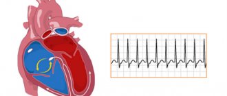
Bradycardia leads to slight dizziness and even loss of consciousness. This type of arrhythmia includes a rate below 60 beats per minute. But if the expectant mother is an athlete and had a low heart rate before pregnancy, then there is nothing to worry about: in this case, this is a variant of the norm. Bradycardia is relatively rare.
Tachycardia is dangerous, even if it does not exceed the critical values described above, but is accompanied by the following symptoms:
- deterioration of health;
- weakness;
- we cloud our consciousness;
- nausea;
- headache;
- shortness of breath not caused by physical exertion.
Sometimes tachycardia is not associated with pregnancy and the accompanying changes in the body. And it occurs as a result of ARVI (acute viral infection), against the background of an increase in temperature (even low-grade fever has a dangerous effect on the child, especially immediately before birth) due to intoxication. In this case, they fight the virus, and not the change in heart rhythm. The pulse level should be understandable even in a pregnant woman. Do not attribute sudden changes in heart rate to hormonal changes in the body.
In order to identify a dangerous pathology in time, the described symptoms are correlated with a possible rhythm disturbance and a parallel is drawn between the clinical picture and the current pulse rate.
To control heart rate, you need to know the places where the arteries pass close to the skin (where they can be easily felt). It is advisable that there are solid bone elements nearby (by pressing the vessels against them, it is easier to catch the vibrations of the walls during the pulse wave).
Places for pulse examination:
| Artery | Place of palpation | Methodology | Peculiarities |
| Radial | Groove between the styloid process of the bone of the same name and the tendon of the brachioradialis muscle | The index and middle fingers are placed on the palmar surface of the wrist of the other hand. Place of application: near the base of the first finger (thumb) | Palpation of the pulse on the radial artery is the standard for clinical examination of the patient |
| Sleepy | At the level of the upper edge of the larynx, at the site of bifurcation (bifurcation) of the common carotid artery | Place two fingers on the border of the upper and lower thirds of the neck, between the sternocleidomastoid muscle and the larynx (at the level of the Adam's apple) | If you strongly press the carotid artery on both sides, the carotid reflex is triggered, which leads to a sharp drop in heart rate and loss of consciousness |
When it is too difficult to detect the pulsation of the arterial walls, heart rate is calculated based directly on the heart beats. To do this, find the left fifth intercostal space and at its intersection with a conventional perpendicular lowered from the middle of the collarbone, two fingers (middle and index) are placed on the same side of the body. At this point, the impulses of the apex of the heart are better felt.
Simplified algorithm: place your right hand under the base of the left breast, leaning slightly forward.
Because breasts are affected by hormones during pregnancy and their size increases, it is difficult to find the apex impulse. Therefore, preference is given to monitoring the pulse on the vessels.
If the peripheral artery was palpable before pregnancy (there are no congenital anomalies of its passage), and in the second half the pulse on it ceased to be palpable - this is an alarming sign. This situation is typical for severe swelling - one of the symptoms of gestosis (preeclampsia). It is necessary to tell the doctor about this and adhere to the recommendations prescribed by him (usually they do not include drug correction and at first are limited to diet changes).
How to measure blood pressure during pregnancy
The blood pressure measurement procedure takes only a few minutes, but can help prevent very serious health problems. Modern automatic blood pressure monitors do almost everything themselves; you need to use them regularly and follow some rules:
- Take measurements at the same time of day so that the indicators do not change under the influence of hormonal fluctuations and biological rhythms of the body;
- Do not measure blood pressure immediately after exercise, including walking - in this state the pressure may temporarily increase, there is nothing to worry about;
- Do not take measurements less than an hour after eating - due to the active activity of the digestive system, the blood volume is redistributed, so the result will be biased;
- Measure the pressure in a sitting position, the hand lies on the table top, the muscles are relaxed;
- Do not move or talk during the measurement.
You should not measure your blood pressure in a state of strong excitement. First you need to calm down, lie down for a few minutes. Repeated measurement is carried out no earlier than after 10 minutes, if necessary.
Rules for measuring pressure
Putting on a tonometer cuff and pressing a button on the device is not enough to accurately measure blood pressure. Doctors adhere to a number of rules that should be used when taking home measurements using an electronic device:
- The pressure should be measured at complete rest, in a sitting position, preferably on both the right and left arms.
- The blood pressure cuff should be approximately at the level of the woman's heart.
- It is advisable to avoid physical activity about an hour before measurements and spend at least five minutes in a calm state.
- While the device is operating, you need to relax, straighten your legs and arms, and lean back in a chair or armchair.
- The first measurements may be too high, so to be sure, measure your blood pressure again.
Regarding blood pressure, doctors advise preventing its elevated levels. For pregnant women, this means proper sleep, rest after lunch, psychological calm, walks outside and exercises feasible for the expectant mother, as well as a moderate diet and control of weight gain.
Deviations from normal blood pressure levels in pregnant women are a reason for the close attention of doctors. A pregnant patient can be hospitalized at any stage. And women who were diagnosed with hypertension before pregnancy are placed “for preservation” at least three times: in the first trimester to analyze the ability to carry a pregnancy to term, at a period of 28-32 weeks, since at this time the maximum increase in blood in the body occurs , and after 38 weeks to determine how the patient will give birth.
Expectant mothers with blood pressure problems often give birth in the department of pathology of pregnant women or specialized maternity hospitals, where they can be assisted by experienced cardiologists.
Blood pressure control is the key to the health of mother and child
Often problems with pressure do not manifest themselves until the situation becomes truly threatening. Sometimes pregnant women attribute the deterioration in their health to their “interesting situation” and hormonal changes and fatigue. But pressure surges during such a period can have quite serious causes and dangerous consequences.
The sooner such prerequisites are identified, the easier it will be to normalize the state of health. This is why pregnant women are advised to regularly monitor their blood pressure, especially since it is very easy to do this with modern home blood pressure monitors.
Baby
The 19th week is characterized for the fetus by intensive growth of the limbs in length, an increase in size, and a slowdown in head growth. Its length is already 25 cm, and its weight is approximately 250-300 g. The child changes greatly in appearance, his body no longer looks disproportionate.
The fat under the skin allows the relief of the body to change slightly and become rounded. This brown fat is formed at 19-20 weeks. After birth, it is he who will perform the function of thermoregulation and protect the child from hypothermia or overheating. With age, this fat disappears and remains only in certain places: in the thickness of a person’s cheeks, kidney area, under the armpits, and shoulder blades.
At this stage, the formation of permanent teeth, which are located in the dental plate under the rudiments of milk teeth, and the maturation of the brain also occur. Special processes allow connections between neurons to be established. Cells intertwine with each other and create a whole system for transmitting impulses, ensuring the synchrony of the brain. The structures responsible for touch, taste, vision, hearing, and smell are actively developing.
Changes also occur in the hematopoietic system. The spleen is connected to the process of formation of blood cells, produces leukocytes, the task of which is to protect the child’s body from external and internal factors. Until 19-20 weeks, the fetal blood consisted only of red blood cells, and at this stage its composition changes sharply and approaches the composition of the blood of a newborn.