directions
An important advantage of ultrasound scanning is the low cost and simplicity of the procedure with good information content.
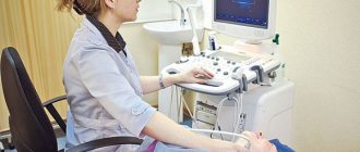
The reliability of the result of any diagnostic study is directly proportional to the professionalism of doctors and the quality of the equipment on which it is performed. The Medicenter employs highly qualified functional diagnostic doctors who have extensive experience in performing ultrasound scanning and have knowledge of all the nuances of this difficult procedure. Having analyzed data from expert-class ultrasound equipment, Medicenter specialists provide simple and understandable explanations of the study results to each patient.
Our clinics in St. Petersburg
Structural subdivision of Polikarpov Alley Polikarpov 6k2 Primorsky district
- Pionerskaya
- Specific
- Commandant's
Structural subdivision of Zhukov Marshal Zhukov Ave. 28k2 Kirovsky district
- Avtovo
- Avenue of Veterans
- Leninsky Prospekt
Structural subdivision Devyatkino Okhtinskaya alley 18 Vsevolozhsk district
- Devyatkino
- Civil Prospect
- Academic
For detailed information and to make an appointment, you can call +7 (812) 640-55-25
Make an appointment
Dopplerography (duplex, ultrasound, Doppler) of the abdominal vessels is usually recommended by a doctor. Examination of the vessels from the inside and the blood flow in them is necessary for a specialist to diagnose the pathology of internal organs:
- Liver (cirrhosis, hepatitis, tumors);
- Spleen (blood diseases);
- Gallbladder (congenital malformations, cholelithiasis, cholecystitis, neoplasms, polyps).
More information about the Doppler method
Doppler of abdominal vessels includes examination of the abdominal aorta and its branches, iliac arteries, celiac trunk, common hepatic and splenic arteries, superior mesenteric artery, inferior vena cava, portal venous system.
The specialist evaluates each of these vessels according to their diameter and patency, the condition of the valves, the thickness of the walls of the arteries and veins, and the compliance of these indicators with the norm.
Important! Duplex of abdominal vessels shows their condition in real time!
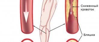
Using ultrasound, the doctor measures the parameters of blood flow and its changes and difficulties: narrowing of the lumens, spasms, the presence of blood clots, atherosclerotic plaques and their sizes.
What information can be obtained using ultrasound of the abdominal aorta?
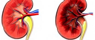
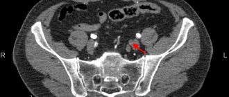
Ultrasound examination of blood vessels makes it possible to examine the vascular wall in detail, identify an aneurysm, signs of atherosclerotic disease and other abnormalities. The doctor examines the aorta from all sides and measures its diameter. Thanks to the wide technical capabilities of ultrasound equipment, our specialists promptly diagnose circulatory disorders, identify atherosclerotic plaques in the vascular cavity, wall thickening, occlusion, tortuosity of the aortic arch and prevent the occurrence of serious complications. Vascular disorders, including atherosclerosis, affect the functioning of the cardiovascular and circulatory systems. Do not delay going to the doctor, especially if you have signs of dangerous diseases or a hereditary predisposition to their development.
Possibilities of abdominal ultrasound
- Assessment of the condition, speed and direction of blood flow in the veins and arteries of the abdominal cavity;
- Detection of early vascular changes (thrombosis, stenosis - that is, narrowing of the arteries);
- Diagnosis of aneurysms;
- Pathologies of the diaphragm;
- Increased pressure in the portal vein;
- Evaluation of the results of therapy and implantation;
- Determination of indications for surgery;
- Detection of blood supply disorders to the abdominal organs.
In addition, vascular duplex is prescribed if it is impossible to determine changes in internal organs using standard ultrasound diagnostics; to confirm the diagnosis (for example, vascular ischemia of the gastrointestinal tract).
Doppler ultrasound is an important diagnostic procedure for assessing the effectiveness of collateral (roundabout) blood flow, which sometimes occurs when the main arteries are blocked.
Preparatory process
To prepare for the scan, you simply need to exclude factors that may blur the clinical picture. This includes pharmacological drugs that are designed to help during the course of treatment:
- hypertension;
- heart block;
- angina pectoris.
But only the doctor should note the medicine, since sometimes it is easier to postpone the procedure by a couple of days than to interrupt a pre-agreed therapeutic program in the middle of treatment.
It is also worth holding off on drinking coffee, which can affect your blood pressure.
The procedure begins with the patient being placed on a couch with a cushion placed under his head. Next, the doctor lubricates the sensor with gel, which is designed to improve the conductivity of incoming ultrasonic waves through the subcutaneous fat layer.
The doctor will have to visually assess the area under study in order to correctly distribute the ascending and descending parts, as well as the arc and extensions in the thoracic region. The calculation is made based on the fact that the arc has three branches:
- brachiocephalic trunk, located on the right;
- common carotid artery;
- left subclavian artery.
Using the anatomical principle of zone separation, the condition of the vascular network is assessed using a transducer sensor. To get a clearer picture, the expert can switch examination modes.
Preparing for diagnosis
To obtain a clear image, it is necessary to exclude from the diet foods that increase gas formation (raw vegetables and fruits, legumes, sauerkraut, brown bread, baked goods, dairy products and carbonated drinks). For diseases that require a strict diet or constant use of medications (diabetes mellitus, hypertension, coronary heart disease), these restrictions are lifted.
The fact is that gases generated from flatulence impede visibility, which can negatively affect the results of the examination. Two to three days before the study, it is recommended to take enterosorbents (espumisan, activated carbon).
Abdominal vascular duplex is performed on an empty stomach. The minimum interval between the study and food intake is at least 6 hours. The optimal time is in the morning on an empty stomach.
Carrying out an ultrasound scan immediately after a colonoscopy or fibrogastroscopy does not make sense, because due to their specificity, air enters the abdominal cavity, limiting the display of ultrasonic waves on the screen.
Scan classification
Dopplerography for the aortic arch provides two main categories that are similar to each other. This is about:
- mapping;
- energy type.
But the second version is considered more suitable for studying vessels smaller than the aorta, so it is used much less often.
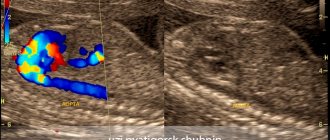
When mapping, technologies are used to color the studied blood flow in two colors. Red is responsible for indicators of the approach of blood elements to the sensitive sensor. The lighter the shade, the higher the speed of movement of the particles under study. And the blue color indicates the direction coming from the sensor.
When using the ultrasound method, you can also use one of two modes to choose from, based on current suspicions:
- B-mode;
- M-mode.
The first variation involves continuous production and reception of waves. Due to such a thorough study of the problem area, it is possible to study the volume on an ongoing basis.
The second approach is non-permanent, which works great if you just want to find the affected area.
How does the procedure work?
There are two options for performing an ultrasound scan of the vessels of the extremities at the Medicenter:
- At home
- In the center
The quality of the equipment, the high qualifications of the doctors and the reliability of the results obtained do not change. The choice depends on the wishes and capabilities of the patient himself.
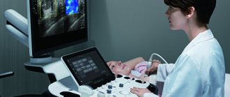
Ultrasound scanning of abdominal vessels is a harmless (without the use of ionizing radiation) and painless non-invasive procedure, taking no more than 30 minutes.
Any doctor’s appointment begins with taking an anamnesis. Ultrasound ultrasound is no exception. Taking into account the data collected during the patient’s interview and the clinical picture as a whole, the doctor draws conclusions about what he sees on the monitor during ultrasound examination.
The patient needs to free the upper body from clothing and jewelry. The patient lies on the couch; the anterior abdominal wall is covered with a transparent gel, ensuring the closest contact between the ultrasound probe and the patient’s skin. During the examination, the patient breathes freely, sometimes at the request of the doctor, inflating the anterior wall of the abdomen. Images (slices) appear on the monitor, the changes of which the doctor carefully examines. Do not be alarmed by unusual sounds that periodically appear during the study. This is how the device measures blood flow in arteries and veins.
The results of the study are recorded on thermal paper and given to the patient with a transcript from the ultrasound examination doctor, but the final word in making a diagnosis remains with the doctor who referred for diagnosis.
Immediately after the examination, the clear gel is removed from the skin and the patient can begin his normal activities.
Principle of operation
Content:
- Principle of operation
- Scan classification
- Indications and contraindications
- Preparatory process
- Decoding the result
- Benefits of Duplex Scanning
Scanning a specified part of a large segment of the cardiovascular system is just one of many methods for confirming or refuting a number of ailments. But it is precisely this that is considered one of the most productive, as it is based on a couple of reliable mechanisms for obtaining the necessary information.
The first option provides a basis from the observations of Mr. Doppler, who was an Austrian physicist. In honor of him, the technique was named Dopplerography. The main principle is based on the difference in the resulting frequency when taking into account the distance from the sensor to the particles. With its help, it is possible to evaluate the elements located inside the vessels: red blood cells, leukocytes, platelets, and the liquid plasma part.
By adjusting the frequency of incoming ultrasonic vibrations, the doctor will be able to check not only the presence of blood flow, but also its speed and direction.
The second component of the technique is the so-called “B-mode”. Typical ultrasonic spectrum diagnostics operate using high-frequency pulses. When reflected from the part being studied, they are captured by the apparatus.
Based on the results obtained, it is possible to record possible pathologies that occur in the vascular structure of potentially affected walls. The technology allows you to monitor changes in real time, provided that a two-dimensional image is transmitted.
Together, this guarantees a high-quality and complete assessment of the anatomy of the cardiovascular system and hemodynamics. No other modern methods can provide such accuracy.
Advantages and disadvantages
The main advantage of this method is its information content and extensive diagnostics. It is worth noting the fact that this procedure does not have any contraindications, which simplifies the research several times. The duration of the procedure can also be called an advantage, because the scanning does not last long. In addition, after the scan, the person is not limited in anything, and can immediately go home.
Types of aortic aneurysms
There are “true” and “false” aortic aneurysms. A true aneurysm develops due to the gradual weakening of all layers of the aortic wall. A false aneurysm is usually the result of trauma. It is formed from the connective tissue surrounding the aorta. The cavity of the false aneurysm is filled with blood through a crack in the aortic wall. The aortic walls themselves do not participate in the formation of an aneurysm.
Depending on the form there are:
- saccular aneurysm - expansion of the aortic cavity on only one side;
- spindle-shaped (fusiform) aneurysm - expansion of the aneurysm cavity on all sides;
- mixed aneurysm - a combination of saccular and fusiform forms.
Criteria for damage to the branches of the aorta
In order to diagnose lesions, visualization is used in conventional black and white mode and in color mapping mode. The combination of two modes allows you to examine the geometry and structure of the walls of the vessel, as well as draw conclusions about the degree of vasoconstriction. An important criterion for vessel stenosis is an increase in peak systolic velocity. In this case, it is important to pay attention to the ratio. This refers to the speed in the vessel to the speed of blood movement in the abdominal aorta. Exceeding this value by 3 times may indicate stenosis.
What influences the success of the procedure?
In addition to the quality of the equipment and the professionalism of the specialist who conducts the study, there are several other important factors that can affect the possibility of high-quality visualization.
First of all, this concerns a person’s body weight. Flatulence and shortness of breath may also affect the results. Therefore, it is extremely important to properly prepare for this study.
More fresh and relevant information about health on our Telegram channel. Subscribe: https://t.me/foodandhealthru
We will be grateful if you use the buttons:
You might be interested
Facelift without surgery? It's real.
Time dictates its own laws. Today, those who look well-groomed and young achieve success. The well-known proverb “You are greeted by your clothes” can be supplemented – and by your face too! But what to do if age takes its toll?