In what situations should you undergo a cardiac ultrasound?
- Changes in ECG indicators.
- Heart murmurs.
- A slight increase in body temperature in the absence of inflammatory processes in the throat, nose, ear or kidneys.
- Arrhythmia.
- An x-ray of the heart showed a change in its shape and size, as well as in the vessels extending from it.
- Hypertension.
- Predisposition to hereditary heart diseases.
- Possible changes in cardiac structures.
- Fainting, swelling, shortness of breath, dizziness.
- Pain in the left half of the chest and behind the sternum.
- Risk of heart tumors.
- After myocardial infarction.
- Effusion pericarditis.
- Angina pectoris.
- Diagnosis of cardiomyopathy and its type.
- To detect true or false cardiac aneurysm.
It is extremely important to carry out this procedure after a myocardial infarction to determine how many muscle cells have died.
ECG and ultrasound of the heart should be performed by people in stressful situations, experiencing severe emotional and physical stress.
EchoCG has no age restrictions. It is prescribed to children if a congenital heart defect or a change in its structure is suspected, which often occurs during the period of active growth of the child.
EchoCG is prescribed for pregnant women, because allows you to identify heart defects in the fetus in the womb. The procedure is painless and does not cause harm.
Heart ultrasound is a mandatory procedure during pregnancy if:
- There was a history of spontaneous abortions.
- Diabetes.
- Hereditary predisposition to heart disease.
- In the 1st and 2nd trimester, the woman was forced to take antiepileptic drugs and antibiotics.
- The analysis revealed high titers of antibodies to rubella, or the woman had it during pregnancy.
Fetal echocardiography is prescribed at 18-22 weeks. Children undergo it when the above symptoms appear.
Why do you need an ultrasound?
Sonography is very often used to diagnose diseases and disorders because it has no side effects, no harmful effects on the body and is performed quickly - usually in less than half an hour. It is used to study the condition:
- any soft tissue: fat, connective and muscle, as well as cartilage, joints, ligaments, nerves and blood vessels;
- abdominal organs - gall bladder, liver, pancreas, bile ducts, stomach, spleen, small and large intestines;
- thyroid gland;
- kidney;
- lymph nodes - tiny “filters” of the lymphatic system The lymphatic system complements the cardiovascular system. The lymph circulating in it - the intercellular fluid - washes all the cells of the body and delivers the necessary substances to them, taking away waste. In the lymph nodes, which act as “filters,” hazardous substances are neutralized and removed from the body, which retain and neutralize substances dangerous to the body;
- hearts;
- eye;
- mammary glands;
- pelvic organs: bladder, uterus and its tubes, ovaries, rectum and prostate;
- and brain in infants.
In addition, ultrasound is used:
- to assess fetal development;
- as a way to distinguish tumors from cysts - fluid-filled blisters;
- to identify various neoplasms that are poorly visible on x-rays;
- with a biopsy - taking a small amount of tissue to study the properties of their cells;
- during surgical operations to control instruments inserted into the patient’s body;
- to assess the speed of blood flow in the vessels and identify blockages and other disorders in them.
Methods for performing echocardiography of the heart
- M – method (one-dimensional). With its help, the dimensions of the heart chambers and the work of the ventricles during their contraction are accurately determined. The indicators are presented in the form of a graph.
- B – method (two-dimensional). Allows you to see tumors, blood clots and aneurysms. With its help, the thickness of the heart walls and valves is measured, and the degree of contractility of the ventricles is determined.
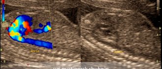
- Electrocardiography with Doppler. The procedure is necessary to diagnose heart defects and other serious disorders.
Ultrasound can be performed through the esophagus or during periods of active physical activity.
Echocardiography through the esophagus is prescribed in situations such as:
- Infectious endocarditis.
- Routine examination before installation of an artificial valve.
- After a stroke and in cases of cerebral circulation disorders and atrial fibrillation.
- Cardio version.
- Defect of the septum between the atria.
- The impossibility of traditional ultrasound of the heart. This occurs in the presence of costal ossification and other disturbances in the structure of the chest.
It is prohibited to perform cardiac echocardiography through the esophagus if there is:
- Varicose veins of the esophagus.
- Strong gag reflex.
- Radiation therapy of the esophagus.
- Osteochondrosis of the cervical spine in acute form.
- Enlarged hernia on the diaphragm.
- Gastric and intestinal bleeding.
- Intestinal perforation.
- Spasms, tumors, intestinal diverticula.
- Pathological mobility of the cervical spine.
Rules for preparing and conducting echocardiography through the esophagus:
- Stop eating and drinking at least 4 hours before the ultrasound
- Pull out dentures and gastric tube
- Before the procedure, the patient's mouth and throat are irrigated with lidocaine.
- The patient lies on his left side, a mouthpiece is inserted into his mouth and an endoscope is carefully inserted into the esophagus, through which ultrasonic waves penetrate
- The procedure takes from 15 to 20 minutes and is recorded on video.
Preparation for the procedure
If urgent diagnosis is necessary, an ultrasound of the heart can be performed without prior preparation. However, in order for the results to be as correct as possible, it is better to prepare. It is important to tell your doctor about the medications you are taking. The doctor will take this point into account and, perhaps, adjust the medication intake for a while. The day before the test, avoid drinking alcohol, tea and coffee. If you have thick hair on your chest, shave it off. Try not to smoke two to three hours before Echo-KG. Don't rush into the office right away. Sit in the hall for 10-15 minutes, relax, calm down after walking along the street.
Ultrasound is the fastest way to confirm or refute the presence of pathology of the heart and large vessels, which has a high safety profile. The result is issued immediately after the examination by a doctor. The doctor has the opportunity to prescribe appropriate treatment, give recommendations on the regimen, physical activity and nutrition on the day of the examination.
← All articles
In what cases is stress echocardiography prescribed?
- To diagnose coronary artery disease.
- To assess the extent to which narrowed coronary arteries in patients with coronary artery disease affect their quality of life and exercise endurance.
- To assess the degree of blood supply to the myocardium.
- To monitor the effectiveness of treatment.
- Uncomplicated heart attack or chronic ischemic heart disease.
- Risk of negative consequences during complex operations.
It is prohibited to perform echocardiography with stress if:
- Acute myocardial infarction (less than a month).
- Aortic aneurysm of dissecting type.
- Respiratory, cardiac, renal and liver failure.
- Blockage of blood vessels by blood clots.
The normal ultrasound of the heart with a load is considered to be:
- Evenly moving walls of the left ventricle (LV) during exercise.
- Decrease in end-systolic volume (ESV).
- Raising the exile faction.
- Absence of disturbance in the functioning of the walls during movement, if a disturbance was recorded at rest.
- Increase in wall thickening in systole.
Negative indicators are an enlarged right ventricle (RV), the appearance of new areas with inactive walls, and a decrease in the ejection fraction to 35%.
Transrectal ultrasound, or TRUS
The examination is carried out using an ultrasound probe inserted into the rectum and provides a much more accurate image of internal tissues than conventional external ultrasound.
It is prescribed to both men and women for:
- frequent, painful or difficult urination;
- suspected presence of tumors, neoplasms, cancer or other diseases of the pelvic organs: bladder, prostate, seminal vesicles, rectum, uterus, its tubes and ovaries;
- routine examinations before surgery;
- discomfort and pain in the lower abdomen, perineum, rectum or scrotum;
- detection of blood in urine or semen;
- identifying abnormalities in the results of blood, urine or sperm tests;
- identifying the causes of infertility and impotence;
- monitoring the patient’s condition after surgery on the pelvic organs, kidneys or intestines;
- preventive examinations carried out during routine medical examination.
With the help of TRUS, specialists discover:
- tumors, including cancer, and other neoplasms, such as cysts - fluid-filled blisters;
- injuries or anomalies - abnormal tissue development;
- inflammatory diseases - prostatitisProstatitis is an inflammation of the prostate gland, cystitisCystitis is an inflammation of the bladder or vesiculitisVesiculitis is an inflammation of the seminal vesicles, which contain the secretion produced by the prostate gland.;
- foreign bodies.
Before performing a transrectal ultrasound, you need to cleanse the intestines using an enema or special medications recommended by your doctor. Before the procedure begins, the patient lies on the couch on his left side, bending his legs and pulling them towards his stomach. Then the doctor conducts a digital examination of the rectum, places a condom on the sensor, inserts it into the anus and examines the condition of the internal tissues for about 10-15 minutes.
What does an ultrasound of the heart show?
The resulting parameters allow you to determine:
- hypertrophy of the LV walls;
- regurgitation of blood through the valves;
- tumors, blood clots, scars, aneurysms;
- blood flow condition;
- valve condition;
- degree of myocardial contractility;
- size of the heart cavities;
- state of the LV pumping function in a dynamic state.
It is considered normal if:
- there is no fluid in the pericardium;
- the size of the final section of the pulmonary artery is within 1.8-2.4 cm; trunk - up to 3 cm;
- ejection fraction is 55 – 60%;
- the volume of the pancreas at the final stage of relaxation of the heart (diastole) ranges from 0.9 to 2.5 cm;
- thickness of the interventricular septum in the final stage of diastole from 0.5 to 1.12 cm
- the final section of the aorta in diameter is 2 – 3.7 cm
- the thickness of the posterior wall of the LV at the final stage of relaxation of the heart (diastole) varies from 0.6 to 1.12 cm
- the change in the movement of the posterior wall of the LV towards contraction (systole) is 0.91 – 1.41 cm
- the size of the LV cavity in the final stage of relaxation (diastole) ranges from 3.51 to 5.7
- cardiac output (MCV) must be no less than 3.5 and no more than 7.5 l/min
- cardiac index ranges from 2 to 4.1 l/m2
- the diameter of the terminal part of the pulmonary artery is from 1.8 to 2.4 cm, its trunk is up to 3 cm
- the blood flow speed through the carotid artery is 22 cm/s with an error of +-5 cm/s
- there are no manifestations of regurgitation and dysfunction of the papillary muscles, growth on all valves.
The degree of discrepancy with normal values is determined by a sonologist, but the final diagnosis is made by a cardiologist taking into account the information received and based on other symptoms. The disease cannot be diagnosed by ultrasound alone.
Interpretation of results and normal indicators
At the end of the procedure, the doctor gives the patient a conclusion with the results of the study. A person who is not related to cardiology will not be able to decipher the presented abbreviations and numbers on his own. Therefore, you should not try to figure it out on your own, but it is better to turn to an experienced cardiologist for interpretation of the echogram, who can correctly diagnose. Interpretation of the data obtained allows you to diagnose the presence of various pathologies. It should be understood that the echocardiograph itself cannot make mistakes, because it simply displays an image of the heart on the monitor . But drawing up a conclusion will require some calculations, so the reliability of the data depends on the experience and qualifications of the doctor who does the research.
Ultrasound Dopplerography of blood vessels
The test uses machines that send ultrasound waves bouncing off oxygen-carrying red blood cells, measure their speed and combine the data with information obtained from a regular ultrasound. This method allows specialists to:
- assess the condition of the tissues of the blood vessels of the neck, head, spine, kidneys, lower and upper extremities, not only in children and adults, but also in the fetus;
- identify diseases of the heart, mammary glands, pelvic organs and abdominal cavity: bladder, uterus and its tubes, ovaries, rectum, prostate and pancreas, liver, gall bladder, adrenal glands and spleen;
- detect blood flow disturbances;
- find out the diameter of blood vessels;
- identify the presence of a blockage and determine its size.
Doppler ultrasound does not require any diet, special diet or preparation. During this procedure, the doctor applies a special gel to the area being examined, then places a sensor on it at different angles and examines the image displayed on the screen.
The procedure is painless, safe and is also prescribed for pregnant women.
The only temporary limitation for its implementation may be the patient’s serious condition and his inability to maintain a supine position throughout the study, which lasts from 10 to 30 minutes. Ultrasound in oncology. Diagnostic secrets.