Vacuum systems: equipment and purpose
A standard vacuum blood collection system consists of three components:
- Double-edged needle for venipuncture.
- Holder (adapter) for fixing the needle.
- Vacuum tube with reagent.
All components of the system are intended for one-time use only.
The principle of operation of the system: under the influence of a vacuum, blood is drawn into a test tube with a reagent and immediately mixed with it. A certain amount of reagent and the volume of blood required for the study provide the exact ratio of components for the study.
Analysis for tumor markers
Today, comprehensive cancer screening is not possible without laboratory tests, in particular, blood tests for tumor markers. Regular testing allows you to detect a serious disease at an early stage.
Indications for passing
The test is prescribed by a doctor both during a course of treatment to assess its effectiveness and to prevent relapses.
If you conduct an examination yourself, without a doctor’s prescription, its results should be provided to specialists for correct interpretation!
Analysis results
It is important to understand that diagnosing cancer cannot be based solely on the results of laboratory blood tests, since benign neoplasms and inflammatory processes can also provoke an increase in the level of tumor markers.
Only a comprehensive study under the supervision of experienced doctors will help you reliably identify a malignant tumor at an early stage.
Our clinic specialists will provide you with a sufficient set of examinations to monitor your health.
Needles
The wall of the blood collection needle is ultra-thin, which increases its internal lumen. The outer and inner surface of the needle wall is coated with silicone to reduce trauma to the patient and improve free blood flow. The V-shaped laser sharpening of the cut facilitates painless, smooth entry of the needle into the vein through the skin.
On the tip side for piercing the test tube, the needle is equipped with a thread and a protective rubber membrane. The needles are sealed in two plastic cases (caps) and sealed with a label to prevent their reuse. The color coding of the caps helps in choosing the needle diameter.
Hormonal blood test
This type of study helps determine the general functioning of the organs of the human endocrine system, as well as identify diseases of the pituitary gland, thyroid gland, adrenal glands and other human endocrine organs. Blood is drawn on certain days depending on the production of the hormone that will be tested. This can be an analysis of thyroid hormones (T4, T3), pituitary gland (TSH, LH, FSH), sex glands (testosterone, estriol), adrenal glands (cortisol), as well as pregnancy hormone (hCG). The results will depend entirely on the age and gender of the person, as well as whether there are any diseases in his endocrine system.
Preparing for a hormone test
The amount of hormones in the blood depends on the time of day, since there is a daily rhythm of secretion (release of hormones). Blood for hormonal analysis should be taken in the morning, on an empty stomach.
In women, hormonal levels also depend on the stage of the menstrual cycle. The most favorable days for analysis are days 5-7 of the cycle, starting from the first day of menstruation.
On the eve of the test, you should not drink alcohol, and you should also avoid increased physical activity and stressful situations. It is advisable not to smoke for an hour before the test.
A week before the test, you must stop taking hormonal medications. If you are prescribed medication, discuss this with your doctor; the test may have to be postponed.
How to take a hormone test correctly?
The human hormonal system is connected to all organs and systems of the body. Therefore, the level of hormones in the blood may depend on various external influences and changes in the human body. General recommendations from doctors are as follows: you need to take a blood test for hormones in the morning on an empty stomach. Before donating blood for hormone testing, stop smoking, drinking alcohol and strenuous physical activity. Women need to have many hormones analyzed on certain days of the menstrual cycle. To ensure that the results of your hormone tests are as reliable as possible, consult your doctor and follow his recommendations.
When is a blood test for hormones prescribed?
Hormonal analysis is usually carried out if there is a suspicion of dysfunction of the endocrine glands or if an increase in the size of the glands is detected.
Indications for testing for female sex hormones (estrogens) are:
- menstrual irregularities;
- infertility;
- miscarriage;
- acne;
- overweight;
- fibrocystic mastopathy (breast disease).
Indications for testing for male sex hormones (androgens) are:
- suspicion of the development of tumor processes;
- ovarian dysfunction;
- kidney dysfunction;
- overweight (obesity);
- infertility;
- acne;
- in women – excessive growth of body hair.
Hormonal analysis is prescribed during pregnancy if pathological development of the fetus is suspected. An analysis for the hormone hCG (human chorionic gonadotropin), produced by the cells of the membrane of the embryo, can detect pregnancy already on the 6-10th day after fertilization.
Interpretation of blood test results for hormones
Blood from a vein is used to test hormones.
Depending on the clinical signs indicating a specific pathology, an analysis with tests for specific hormones is usually prescribed.
The most complete picture of your health can be obtained by taking a test for the following hormones.
Thyroid hormones:
- T3 (triiodothyronine) free – stimulates oxygen metabolism in tissues. Normal values: 2.6 - 5.7 pmol/l.
- T4 (thyroxine) – stimulates protein synthesis. Normal values: 0.7-1.48 ng/dl.
- Antibodies to thyroglobulin (AT-TG) are an important parameter for identifying a number of autoimmune diseases. Normal values: 0-4.11 U/ml.
- Some others.
Pituitary hormones:
- TSH (thyroid-stimulating hormone) – stimulates the production of thyroid hormones (T3 and T4). Normal values: 0.4-4.0 mU/l. An elevated TSH value usually indicates decreased thyroid function.
- FSH (follicle stimulating hormone) . Normal values: in women - depends on the phase of the menstrual cycle. Phase I – 3.35-21.63 mU/ml; Phase II – 1.11-13.99 mU/ml; postmenopause – 2.58-150.53 mU/ml; girls under 9 years old 0.2-4.2 mU/ml. In men – 1.37-13.58 mU/ml.
- LH (luteinizing hormone) . Normal values: in women - depends on the phase of the menstrual cycle. Phase I – 2.57-26.53 mU/ml; Phase II – 0.67-23.57 mU/ml; postmenopause - 11.3-40 mU/ml; girls under 9 years old - 0.03-3.9 mU/ml. In men – 1.26-10.05 mU/ml.
- Prolactin . The main function is to stimulate the development of mammary glands and lactation. Normal values: in women (from the first menstruation to menopause) - 1.2-29.93 ng/ml; in men - 2.58-18.12 ng/ml. Increased concentrations of prolactin are called hyperprolactinemia . There are physiological and pathological hyperprolactinemia. Physiological hyperprolactinemia can be caused by breastfeeding, pregnancy, severe physical activity, and stress. An increased concentration of prolactin in women leads to menstrual irregularities and may cause infertility. In men, hyperprolactinemia leads to decreased libido and impotence.
- ACTH (adrenocorticotropic hormone) – stimulates the synthesis and secretion of hormones from the adrenal cortex. Normal values: 9-52 pg/ml.
- Some others.
Sex hormones:
- Testosterone (male sex hormone) is produced by the adrenal glands and in the gonads (in men - in the testicles, in women - in the ovaries). Affects the development of the genital organs, the formation of secondary sexual characteristics, the growth of bones and muscles. Normal values: for men - 4.94-32.01 nmol/l, for women - 0.38-1.97 nmol/l.
- Estrogens (female sex hormones). The main estrogens - progesterone and estradiol - are produced by the adrenal glands and ovaries. Normal values of progesterone in women depend on the phase of the menstrual cycle: Phase I - 1.0-2.2 nM/l; Phase II - 23.0-30.0 nM/l; for postmenopause - 1.0-1.8 nM/l. Similarly for estradiol: phase I - 198-284 pM/l: phase II - 439-570 pM/l; for postmenopause - 51-133 pM/l. Elevated estrogen levels may indicate tumors of the ovaries and adrenal cortex, as well as cirrhosis of the liver. Reduced - for insufficient development and sclerosis of the ovaries.
Adrenal hormones:
- DEA-s (dehydroepiandrosterone sulfate) is necessary for the synthesis of testosterone and estrogens. The range of normal values for the concentration of this hormone: 3591 – 11907 nmol/l; in women - 810 - 8991 nmol/l. However, this is the general picture; when processing analysis data, the patient’s age must also be taken into account.
- Cortisol is involved in many metabolic processes and is actively produced as a result of the body’s response to hunger or stress. Normal values: for children under 16 years of age - 3-21 mcg/dl, for an adult - 3.7-19.4 mcg/dl.
- Aldosterone is responsible for regulating water-salt balance in the body. Normal values: 35 - 350 pg/ml.
Preparation for the procedure
- Check that all necessary equipment is available.
- Check the expiration date and integrity of the vacuum system packaging.
- Check the expiration date and tightness of antiseptic wipes.
- Take the needle by the long colored cap in one hand, and with the other hand remove the short colored cap from the side of the rubber membrane.
- Insert the freed end of the needle with the rubber membrane into the holder and screw it until it stops.
- Place the needle and holder on the tray.
- Prepare the necessary test tubes in sufficient quantity.
- Wear sterile gloves.
table 2
| Index | x 109/l | % |
| Band neutrophils | 0,04-0,3 | 1-6 |
| Segmented neutrophils | 2-5,5 | 45-72 |
| Basophils | up to 0.065 | up to 1 |
| Eosinophils | 0,02-0,3 | 0,5-5 |
| Lymphocytes | 1,2-3 | 19-37 |
| Monocytes | 0,09-0,6 | 3-11 |
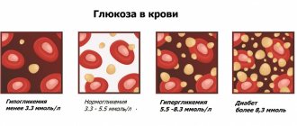
The results of a general blood test (Table 1) show many indicators. Let's consider the main ones:
- RBC – total number of red blood cells (erythrocytes). The pathological increase in these cells is associated with impaired hematopoiesis. A decrease in red blood cells is usually a consequence of anemia, hemolysis and blood loss.
- HGB stands for hemoglobin, which is a protein containing iron. It transports oxygen to tissues, and carbon dioxide from them, and also maintains acid-base balance. A decrease in hemoglobin most often occurs due to anemia.
- HCT – hematocrit. It is defined as the ratio between the red blood cells that have settled to the bottom after taking the test and the total blood volume. An increase in this indicator indicates polyuria, erythrocytosis or erythremia. A decrease in hematocrit level occurs with anemia and an increase in circulating blood volume.
- PLT – platelets. These cells are responsible for blood clotting. If their number decreases, then the cause may be viral diseases, bone marrow lesions, bacterial infections and other pathologies. An increase in the number of platelets is caused by a wide variety of ailments: from joint diseases to cancer.
- CPU – color indicator. It determines the saturation of red blood cells with hemoglobin. If it is insufficient, this may indicate iron deficiency anemia, anemia or lead poisoning. When CP rises above normal, the cause is oncology, gastric polyposis and deficiency of vitamins B9 and B12.
- Erythrocyte indices: MCV - average volume of erythrocytes, used to determine water-salt balance and type of anemia;
- RDW is the degree of red blood cell diversity, which determines how different cells differ from each other in volume;
- MCH – average hemoglobin content in erythrocyte; this criterion is considered analogous to the color indicator;
- MCHC – average concentration and content of hemoglobin in red blood cells; this indicator is calculated taking into account the level of hematocrit and hemoglobin.
Now let's move on to the leukocyte formula (Table 2). It determines the percentage of different types of white blood cells in the blood, that is, the relative content of each type of white cell. What is this formula for? It is very important because with any changes in the body, the percentage of certain types of white cells in the blood decreases or increases. This is associated with a decrease or increase in other types. Based on the information obtained from the leukocyte formula, one can judge the course of a particular pathology, the occurrence of complications, and also more accurately predict the outcome of the disease.
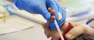
Carrying out manipulation
- Ask the patient to make room for the upcoming venipuncture (most often the elbow, possibly the forearm, the back of the hand).
- Place an oilcloth pillow under the venipuncture site.
- Apply a tourniquet 7-10 cm above the venipuncture site on top of clothing or a diaper. The tourniquet is applied so that the pulsation of the nearby artery is maintained.
- Ask the patient to make a fist.
- Inspect and palpate the vein for venipuncture.
- Treat the venipuncture site with antiseptic wipes.
- Hold the vacuum system in one hand and remove the protective cap from the needle with the other hand.
- With your free hand, stretch the skin below the venipuncture site at a distance of 4-5 cm to secure the vein.
- Puncture the vein at an angle of 10-15º, insert the needle along the vein no more than 1/2 of the length.
- Take the test tube and insert it into the holder until it stops.
- Once the blood begins to flow into the tube, remove or loosen the tourniquet.
- Ask the patient to unclench his fist.
- Draw the required amount of blood into a test tube.
- Remove the tube from the holder when blood stops flowing into it.
- Carefully invert the tube several times to mix the blood with the filler. Do not shake the test tube!
- Place the test tube in a rack.
- If necessary, connect the next test tube to the system and repeat all steps.
- Draw the required amount of blood into all tubes.
- Apply a sterile napkin with an antiseptic to the venipuncture site and remove the needle.
- Ask the patient to press down on the tissue with his free hand.
- Place the needle in a sharps container - Class B waste.
- Place the holder in another container for collecting Class B waste.
- Apply a pressure bandage or adhesive bandage over the napkin.
- Inquire about the patient’s well-being and make sure everything is okay.
- Label the tubes containing the collected blood samples with the patient's last name and initials.
- Place used consumables in Class A and B waste containers.
- Make the necessary notes in medical documentation on paper and electronic media.
- Send the collected material with accompanying documents to the laboratory.
Possible errors when using vacuum tubes
| Problem | Possible reasons | Solution |
| Blood does not flow into the tube after connecting to the holder | The needle did not enter the vein | In all of these cases, it is necessary to carefully adjust the position of the needle. There is no need to disconnect the tube from the holder if there is no need to remove the needle and under the skin. |
| The tip of the needle rests against the venous wall | ||
| The vein is pierced through | ||
| The blood in the test tube was received in less quantity than required for the analysis. | The venous vessel collapsed due to low pressure | It is necessary to disconnect the tube from the holder and wait a while until the vein is filled again |
| The system needs to be replaced and the procedure repeated | Air got into the test tube |
You can order high-quality laboratory consumables. When collecting blood using a vacuum system, follow the algorithm. This will ensure the safety of the procedure and the reliability of the research results.
Factor No. 7. I worked hard and got nervous
Physical activity, especially intense or unusual for the patient, can increase the CBC indicators, namely the number of red blood cells, white blood cells and hematocrit. Psycho-emotional stress will also affect the number of leukocytes. In this case, chronic severe stress is considered as a basic pathological condition, and acute stress is taken into account, for example, with sensitivity to service in municipal institutions.
How it affects: For biological variation, the most sensitive indicator is leukocytes. Physical overexertion in combination with deviations in the time of sampling can change the number of leukocytes by ± 4 × 10⁹/l.
Factor No. 6. “Easy”
It is recommended that blood collection be done early in the morning after a 12-hour fast and no changes in diet for 24 hours. The patient should go to bed at the usual time the day before and get up no later than an hour before blood collection (baseline).
How it affects: Important for hemoglobin levels. Sample turbidity, even slight, distorts photometric data. Hemoglobin is likely to be elevated, which will affect the control of anemic patients. The MCHC indicator can serve as a control over the correctness of hemoglobin measurement.
Types of venous blood studies
Blood from a vein is suitable for various types of research. Each of them is prescribed depending on the clinical picture of the disease, medical history and other factors. It is worth understanding that blood tests are only one component of a comprehensive diagnostic scheme. Despite the fact that this fluid contains information about the condition of all organ systems, it is impossible to make a final diagnosis based only on these studies.
Types of tests that can be performed with venous blood:
- clinical (general) - based on counting blood cells, as well as identifying pathological components in the bloodstream, carried out by microscopy;
- biochemical - determination of the concentration of various biologically active substances, enzymes, lipids and other fractions that cannot be detected by simple microscopy (carried out on a special analyzer);
- immunological - necessary if various autoimmune diseases are suspected, as well as for differentiating potential allergens;
- hormonal - prescribed to monitor the functioning of organs that produce hormones; it is regularly performed for patients with diabetes mellitus, thyroid pathologies and other problems.
REFERENCE! Suitable for some tests, including blood from a finger. However, if the patient has to undergo several studies at the same time, it will be much easier to take it from a vein.
How to donate blood correctly?
It is recommended to donate venous blood in the morning on an empty stomach. You should not eat food for 8 hours, and abstain from drinking alcohol and medications for several days. If there is a need to take medications on a regular basis, you must notify your doctor. On the eve of the analysis, you should not undergo active physical activity, it is advisable to ensure yourself peace. The timing of the analysis depends on the laboratory. If necessary, results can be ready within a few hours. In most cases, the patient can pick them up the next day, and the analysis is deciphered by the attending physician.
Taking venous blood is indicated for suspicion of many diseases, as well as during routine examinations. It is not recommended to take it without prior preparation - this may affect the test results.
Factor No. 8. “Casual” smoking
A mandatory point that must be taken into account by the clinician is smoking. The condition where the patient is a chronic smoker is regarded as the basic condition. If a person was forced to quit smoking and this fact is unknown to the attending physician, then the number of erythrocytes, platelets and hematocrit are subject to changes in the CBC, and the changes can be of a different nature. Particular attention should be paid to adolescents - a group of patients who smoke rarely and irregularly.
How it affects: “Sudden” smoking before taking a blood test can overestimate the number of red blood cells by 10%.