Express diagnosis of syphilis, abbreviated EDS, refers to serological non-treponemal methods of blood testing. This test was proposed by the German immunologist A. Wasserman and was named after him - the Wasserman reaction (RW). Its main advantages are simplicity, low cost, and quick results. Blood for EDS (or RW) is donated during mass examinations (screenings) to identify diseases in seemingly healthy people without any symptoms. Today this test is considered obsolete. It has been replaced by others, although until now, when talking about non-treponemal diagnosis of syphilis, they use the expression “donate blood for RW”.
What is the non-treponemal method?
As you know, syphilis is caused by Treponema pallidum, which is a spirochete (spiral-shaped bacteria). After its introduction, the body begins to produce antibodies (IgG and IgM immunoglobulins) to lipoproteins and phospholipids, which are released as a result of cell damage by the spirochete, and to the membrane lipids of the pathogen itself. Antibodies to lipids appear in the blood about a month after infection. Using this test for primary syphilis, they are detected in the blood in 90% of cases 6 weeks after infection. With secondary syphilis, a positive result is recorded in almost 100% of cases.
The causative agent of syphilis is the spiral-shaped bacterium Treponema pallidum.
Positive result
For example, a positive EDS blood test result. It occurs when a person’s blood, after mixing it with a special solution, produces antibodies to syphilis.
Accordingly, it can be judged that the patient’s body has encountered a disease. It makes no sense to talk about the norm for this analysis. After all, it shows whether there is syphilis or not.
Typically, laboratories simply provide the public with a printout that tells them whether antibodies to the disease have been found or not. But sometimes clinics offer an extended display of the result.
Disadvantages of the method
The main disadvantage of EDS is its low specificity and the possibility of false-positive results. The fact is that antilipid antibodies can be produced not only during syphilis, but also for other reasons. Among them:
- chronic and acute diseases: tuberculosis, hepatitis, systemic lupus erythematosus, tumors, inflammatory heart diseases, blood diseases, diabetes mellitus, scleroderma, dermatoses and others;
- physiological conditions: menstruation, pregnancy;
- anesthesia, taking certain medications;
- drug addiction, alcoholism;
- consuming fatty foods and alcohol the day before donating blood.
Another disadvantage is the false negative results that can occur early in the disease. That is, the patient is infected, but the analysis showed a negative reaction.
In addition, such testing is useless at the late stage of syphilis due to lack of sensitivity.
Errors
EDS (blood test) - what is it? A modern, although not entirely accurate, method for diagnosing syphilis in humans. It is worth paying attention to the fact that a positive result may be false. Therefore, there is no need to panic when you see that the body is producing antibodies to the disease.
First, it is recommended to retake the test. Or choose a different method for diagnosing syphilis. EDS is a study that has certain errors.
Secondly, as already mentioned, the presence of antibodies to a disease may mean the development of immunity to the disease. For example, due to the characteristics of the organism.
Thirdly, the presence of bad habits, as well as chronic diseases, can lead to a positive test result. In girls, even menstruation affects it.
Accordingly, EDS is a fairly quick rapid test for syphilis, which has certain errors. 100% accuracy is given only to perfectly healthy people. But in practice, such research is in great demand. Now it is clear how to donate blood for EDS, what it is, how the analysis is deciphered, and what are the factors influencing the accuracy of the study.
How is the analysis carried out?
Venous blood is usually used for research. It is taken in the morning on an empty stomach, that is, the last meal should occur no later than 8 hours before the procedure. It is not recommended to drink alcohol or eat fatty foods the day before.
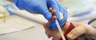
Venous blood is taken to test for syphilis.
Two to three drops of the patient's blood serum are mixed with the cardiolipin antigen in a well of a glass plate and the reaction is observed, which lasts for 30 minutes. The conclusion is made based on the amount of sediment and the size of the flakes formed. The intensity of the reaction is assessed on a scale from one to four (+, ++, +++, ++++).
If the reaction is positive, confirmation of the result using treponemal tests is required.
Indications for EDS
Direct indications for testing blood for syphilis are symptomatic manifestations of the disease:
- Primary syphilis. In the area of penetration of the bacterium (the mucous membrane of the genital organs, anorectal area or oral cavity), a hard chancre appears - a painless erosive compaction, regional lymph nodes enlarge, the temperature periodically rises to subfebrile (37-38℃) values, and neurological symptoms (myalgia, arthralgia) are disturbing.
- Secondary syphilis. It is characterized by syphilitic skin rashes (syphilides) in any area of the body, stable low-grade fever, redness of the tonsils, hair loss, and hoarseness.
Important! It is necessary to donate blood for possible syphilis infection after unprotected intimate contact with a questionable sexual partner.
Routine testing for infection is carried out by:
- catering workers, food industry workers, doctors;
- pregnant women when registering at the antenatal clinic and as part of three perinatal screenings.
- organ donors for transplantation, IVF and blood donors;
- patients who have undergone treatment for syphilis;
- potential hospital patients (in preparation for hospitalization).
A blood test for the Wasserman reaction is carried out in a comprehensive diagnosis of other sexually transmitted infections. It is mandatory to check blood for RW in newborns from mothers diagnosed with syphilis.
Analysis transcript
The EMF result can be either negative or positive. The more advantages, the more serious the defeat:
- + and ++ – weakly positive reaction (with one plus, the result is considered doubtful);
- +++ – positive;
- ++++ – strongly positive.
As a result of the study, antibody titers are indicated. If the analysis is carried out to monitor treatment, then the titers determine whether recovery has occurred. As a rule, after treatment the patient is monitored for a year. During this time, it is tested several times. The effectiveness of treatment is indicated if titers have decreased fourfold or more over the course of a year. If there are no IgM immunoglobulins in the blood, then there is no resumption of infection. IgG immunoglobulins may be present in the blood for a long time after treatment, sometimes even throughout life.
The test result may be negative or positive
Laboratory detection of the causative agent of syphilis
Treponema pallidum (the causative agent of syphilis) belongs to gram-negative bacteria of the spirochete species. In 95% of cases, infection occurs through unprotected intimacy. Contagiousness (infectiousness) is manifested by the concentration of bacteria in the secretory fluids of an infected person (sperm, vaginal secretions) and microscopic damage to the mucous membranes of the partner.
The bacterium enters the intercellular junctions of the endothelium, cells and tissues, and multiplies in the lymph nodes. The high virulence of treponema pallidum ensures the rapid spread of infection throughout the body through the blood and lymph flow.
Treponema pallidum is not detected under a light microscope and does not grow on nutrient media, which excludes conventional microscopic analysis and bacteriological culture (bacteriological culture) from laboratory practice. The bacterium can only be detected in blood serum through serological tests.
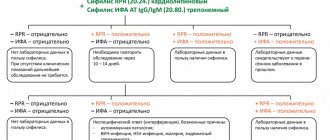
RPR in the list of diagnostic methods for determining Treponema pallidum
Serological blood testing is based on the body’s immune reactions: the interaction of antigens (bacteria or viruses) that have entered the blood and Ig immunoglobulins - specific blood plasma proteins responsible for the differentiation and destruction of antigens.
Reference! Antibodies to foreign pathogens are called immunoglobulins, and serological reactions are called antigen-antibody reactions.
Treponemal and non-treponemal tests are used to diagnose syphilis. Treponemal testing is a complex and expensive method using actual Treponema pallidum antigens. They are necessary to confirm the diagnosis in case of false positive results.
The list includes reactions:
- immunofluorescence (RIF);
- immobilization of Treponema pallidum (RIBT);
- passive hemagglutination (RPHA).
Enzyme-linked immunosorbent assay (ELISA) and immunochemiluminescence (ICL) are similar. In non-treponemal tests, cheap nonspecific antigens are used for the antigen-antibody reaction and immunoglobulins are not separated by titers, but their total amount is determined. Non-treponemal studies include RPR, which is used to quickly diagnose syphilis.
Electropulse therapy in the treatment of arrhythmia
Defibrillation and cardioversion are types of electrical impulse therapy. Despite all their similarities, they have some differences. Defibrillation is the process of stopping ventricular fibrillation by applying an electrical discharge; it is the most important resuscitation measure. Cardioversion is a method of treating tachyarrhythmias, which is based on stopping the circulation of excitation in the myocardium by applying an electrical discharge at a certain phase of the cardiac cycle. Cardioversion requires synchronization - applying an impulse at the moment of registration of the R wave, since otherwise, applying a discharge in another phase of the cardiac cycle can lead to ineffectiveness of the procedure and even to the development of ventricular fibrillation. Cardioversion can be planned, when the rhythm is restored with stable hemodynamic parameters if other treatment methods are ineffective, and emergency - with paroxysms with unstable hemodynamics, with ventricular tachycardia without a pulse (in the latter case, it is carried out without synchronization and is equivalent to defibrillation).
Electrical methods for treating arrhythmias have been known since the beginning of the second half of the 18th century. The first officially documented case of using electrical impulses to help with sudden death dates back to 1774, when Mr. Squires, a resident of London, tried to help a three-year-old girl who had fallen from the first floor, using discharges of electricity from Leyden jars. Over the next few days, the girl experienced stupor, but after about a week she was completely healthy.
Defibrillation was subsequently studied by Luigi Galvani, Charles Kight, John Snow, Jean-Louis Prevost and Frederic Batelli and other scientists. In 1947, American surgeon Claude Beck performed successful defibrillation during heart surgery on a fourteen-year-old boy. Developed by Claude Beck, the defibrillator operated on alternating current and allowed only open defibrillation.
Laying the scientific foundations for understanding EIT, as well as the first serious experiments in this area, were carried out by Paul Zoll. While studying cardiac pacing, he hypothesized that the application of a strong external electrical shock could interrupt ventricular fibrillation, and already in 1956 Zoll and his colleagues made the first clinical demonstration of successful transthoracic defibrillation. In his research, he used a self-designed defibrillator that generated alternating current. In 1960, Bernard Lown developed his first DC defibrillator. This defibrillator was the first in a line of modern devices of this type. Lown also proposed the method of cardioversion - the use of electrical discharges synchronized with the cardiac cycle to treat tachyarrhythmias.
Preparation for planned EIT
- If AF lasts more than 48 hours and there is no adequate anticoagulant therapy during the last 3 weeks, before restoring sinus rhythm with ECV, preliminary transesophageal echocardiography is necessary to exclude intra-atrial thrombosis.
- All patients should refrain from eating for 6-8 hours.
- Cancel cardiac glycosides 3–4 days before the procedure
- Normalization of electrolyte balance (EIT for hypokalemia is less effective and is more often complicated by ventricular fibrillation)
EIT methods
External EIT is the main method. Both electrodes are placed on the chest so that the heart is covered by the electric field of the capacitor discharge. The ERC and AHA guidelines establish recommended energy values for the first shock during defibrillation. They are (for adults): when using a monopolar impulse - 360 J, when using a bipolar impulse - 120-150 J., in children, shocks are used at the rate of 2 J / kg of body weight. When carrying out defibrillation, the anterior or standard arrangement of electrodes is now predominantly used; the electrodes must be lubricated with a special conductive gel, and care must be taken that it does not spread over the surface of the chest between the electrodes. It is permissible to use wipes moistened with saline solution. During the procedure, one electrode marked “Apex,” or red (positive charge), is placed exactly above the apex of the heart or below the left nipple; another electrode labeled “Sternum,” or black (negative charge), is placed just below the right collarbone. An anteroposterior arrangement of electrodes is also used - one electrode plate is located in the right subscapular region, the other in front above the left atrium. There is also a postero-right subscapular location of the electrodes. The choice of electrode location depends on the specific situation; the benefit or harm of any of the described locations has not been proven.
Before administering a shock, make sure that no one touches the patient or the bed on which he is lying. Modern control and diagnostic equipment is protected from defibrillator impulses. At the moment the discharge is applied, the monitor readings change and the patient’s reaction is noted - muscle contraction, shuddering, and sometimes a scream. It is strictly forbidden to touch the patient or objects in contact with him at the time of delivering the shock, as this is dangerous for the personnel. After the discharge is completed, the monitor readings are assessed and, if necessary, the issue of repeated discharge is decided.
If the patient is conscious, then general anesthesia is mandatory. The objectives of general anesthesia during cardioversion are to ensure the loss of consciousness for a short period of time and provide amnesia for the period of the manipulation. As a rule, they are limited to the use of short-acting hypnotics in small doses administered intravenously (thiopental 100-250 mg or propofol 50-100 mg).
Internal EIT – electrodes are applied directly to the heart. In this case, a significantly smaller discharge value is required (for an adult patient, about 500 V or 12.5–25 J).
Transesophageal EIT - one of the electrodes is inserted into the esophagus to the level of the atria, the other is placed in the precordial region. Discharge energy 12–25 J. Transesophageal EIT is indicated for severe supraventricular tachyarrhythmias that are resistant to transthoracic discharges, as well as for suppressing severe ventricular tachyarrhythmias with low-energy discharges.
Transvenous intracardiac EIT using a multipolar electrode, which is installed in the right ventricle, is used in intensive care units for recurrent ventricular tachycardia. The discharge energy during endocardial EIT varies from 2.5 to 40 J. To relieve atrial fibrillation, intracardiac EIT can also be used, which can be of two types: high and low energy. When using high energy (200–400 J), one electrode is placed in the right atrium, the other on the surface of the body. Efficiency up to 100%. When using low energy 2–4.5 J, one electrode is placed in the right atrium, the other in the coronary sinus.
Complications of cardioversion
ECV may be complicated by thromboembolism and arrhythmias, and complications of general anesthesia may also occur. The incidence of thromboembolism after defibrillation is 1-2%. It can be reduced by adequate anticoagulation before elective cardioversion or by ruling out left atrial thrombosis. A common complication is skin burns. Patients with sinus node dysfunction, especially older adults with organic heart disease, may develop prolonged sinus arrest. Dangerous arrhythmias such as ventricular tachycardia and ventricular fibrillation may occur in the presence of hypokalemia, cardiac glycoside toxicity, or inadequate timing. The use of anesthesia may be accompanied by hypoxia or hypoventilation, but arterial hypotension and pulmonary edema are rare.
Electrical cardioversion in patients with implanted cardiac pacemakers and defibrillator
It is clear that the presence of such a device in a patient somewhat changes the technique of the procedure, but is by no means a contraindication to external defibrillation. If the patient has an implanted pacemaker-cardioverter, then the position of the electrodes should be slightly changed. The electrode for external cardioversion must be located at a distance of more than 6-8 cm from the implantation site of the pacemaker or cardioverter-defibrillator. Anterior-posterior placement of electrodes is recommended. It is preferable to use a biphasic defibrillator, since in this case a lower energy discharge is required to stop AF. In pacemaker-dependent patients, it is necessary to take into account a possible increase in the stimulation threshold. Such patients should be closely monitored. After cardioversion, the implanted device should be tested using an external programmer.
Recurrence of arrhythmia after electrical cardioversion
Factors predisposing to recurrent AF include age, duration of AF before cardioversion, number of previous relapses, enlarged left atrium or decreased left atrium function, presence of coronary artery disease, pulmonary disease, or mitral valvular disease. Atrial extrasystoles with changing coupling intervals and the so-called early extrasystoles “P” to “T”, sinus tachycardia, intra-atrial and inter-atrial conduction disorders also increase the risk of relapse of AF. Antiarrhythmics given before cardioversion increase the likelihood of restoration of sinus rhythm and reduce the risk of immediate and early relapses. To prevent late relapses, constant long-term use of antiarrhythmic drugs is necessary. The most effective means of such prevention is amiodarone, which is superior in its effectiveness to all other means of antiarrhythmic therapy. 69% of patients maintain sinus rhythm within a year of amiodarone use. For sotalol and propafenone this figure is 39%. Some patients whose episodes of AF occur with severe clinical symptoms, but do not recur frequently (1-2 times a year), prefer repeated cardioversion to long-term anti-relapse antiarrhythmic therapy or treatment aimed at reducing heart rate in conditions of persistent arrhythmia.
Automatic external defibrillators and the concept of early defibrillation
In this regard, the concept of early defibrillation using a “publicly available defibrillator-monitor” has recently become increasingly popular among specialists. According to this concept, automatic defibrillators should become publicly available, allowing even an unqualified user to provide first aid to a patient with cardiac arrest before the arrival of a medical team. Several case reports of successful defibrillation at airports have already been published. At two Chicago airports, automatic defibrillators are located throughout the terminal and in the baggage area. All airport personnel, including security, are trained to use defibrillators and have the appropriate certificates. As a result of this organization of assistance, 69% of passengers who suffered cardiac arrest as a result of ventricular fibrillation at the airport survived. Thus, only early defibrillation is the only chance in these situations to restore hemodynamically effective cardiac contractions and save the patient.