Low hemoglobin is not an independent disease. It is the result of an underlying disease, therefore, if anemia is detected in a patient, the doctor is obliged to prescribe a comprehensive examination.
Concepts of pseudoanemia and hidden anemia
Pseudoanemia is the entry of tissue fluid into the bloodstream during the resorption of edema.
Hidden anemia is the result of the loss of a significant part of the fluid composition of the blood due to dehydration (can be caused by diarrhea, vomiting, hyperhidrosis). In this situation, the blood begins to thicken, so laboratory analysis shows that the number of red blood cells and hemoglobin is normal, even if this is not the case.
Severity of anemia
Based on severity, anemia is classified into:
- light. Hemoglobin level is more than 100 g/l, erythrocytes - more than 3 T/l;
- average Hemoglobin level from 66 to 100 g/l, erythrocytes - from 2 to 3 T/l;
- heavy. Hemoglobin level is less than 66 g/l.
Classification of anemia
All anemias manifest themselves differently. Taking into account the cause of the pathological condition and its symptoms, there are four main types:
- posthemorrhagic anemia (caused by chronic/acute blood loss);
- hemolytic anemia (develops due to the destruction of red blood cells). This group includes hereditary hemolytic anemias: - with a lack of glucose-6 phosphate dehydrogenase; - thalassemia; - sickle cell; — Minkowski-Shoffar;
- deficiency anemia (caused by a deficiency of microelements/vitamins/iron, that is, any elements that play an important role in the process of hematopoiesis);
- hypoplastic anemia (results from impaired hematopoiesis in the bone marrow, the most dangerous form).
According to the color indicator of blood, anemia can be:
- normochromic (hemoglobin is normal). This group includes hemolytic and posthemorrhagic anemia;
- hyperchromic (hemoglobin increased). These include folate deficiency and B12 deficiency anemia;
- hypochromic (hemoglobin is low). This refers to thalassemia, iron deficiency and chronic posthemorrhagic anemia.
Based on the diameter of red blood cells, anemia can be:
- normocytic (almost all hemolytic and acute posthemorrhagic anemia);
- megaloblastic (B12-deficiency anemia);
- macrocytic (folate deficiency, hemolytic disease of the newborn);
- microcytic (chronic posthemorrhagic anemia).
Based on the iron content in the blood serum, anemia is divided into:
- normosideremic (acute posthemorrhagic anemia);
- hyposideremic (chronic posthemorrhagic and iron deficiency anemia, thalassemia);
- hypersideremic (hemolytic and B12-deficiency anemia).
Causes of anemia
Common causes of low hemoglobin are:
- large blood loss;
- disruption of the formation of new blood cells;
- activation of pathological processes of blood destruction.
Causes of posthemorrhagic anemia
Acute posthemorrhagic anemia is the result of the loss of a large amount of blood in a minimal period of time. The chronic form of this disease is caused by:
- prolonged blood loss;
- polyps, hernias and stomach ulcers;
- kidney diseases;
- malignant tumor neoplasms;
- uterine bleeding;
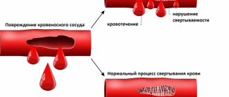
- disorders of the blood clotting system;
- liver failure, cirrhosis.
What causes hemolytic anemia
All hemolytic anemias occur if “old” red blood cells begin to destroy faster than new ones can form.
The sickle cell form is caused by the synthesis of a defective hemoglobin molecule. The emerging defective molecules form peculiar crystals that stretch the red blood cell, causing the latter to take on the shape of a sickle. Sickle erythrocytes are non-plastic. It leads to increased blood viscosity and blockage of small blood vessels. With their sharp ends they can destroy each other.
Thalassemia is a hereditary disease. It occurs due to a decrease in the rate of hemoglobin formation. Hemoglobin that is not fully formed is unstable. It resembles small inclusions that give the red blood cell the appearance of a target cell.
Deficiency anemias and their causes
Iron deficiency anemia develops for reasons such as:
- lack of iron/increased need for iron (this is why degree 1 anemia is often diagnosed in premature babies and pregnant women);
- chronic blood loss;
- problems associated with the absorption of iron from the gastrointestinal tract, its transportation and disposal.
B12 deficiency anemia is caused by a lack of vitamin B12. The disease can be caused by poor nutrition (refusal to eat meat) or disruption of the absorption of cyanocobalamin in the gastrointestinal tract (parasites, diseases of the small intestine, stomach). Also, symptoms of B12 deficiency anemia can appear while taking hormonal drugs and anticonvulsants.
Folate deficiency anemia is a consequence of a lack of vitamin B9. It is usually observed in pregnant and lactating women, cancer patients, teenage children, premature newborns, liver disease, alcohol abuse, etc.
Causes of hypoplastic anemia
Hypoplastic anemia is manifested by a decrease in the content of all cells in the blood. External and internal factors lead to such a pathological and life-threatening condition:
- vibration and radiation effects on the body;
- serious injuries;
- taking certain medications;
- ingestion of poisons into the body;
- herpes, fungi, bacteria;
- genetic mutations;
- rheumatoid arthritis, lack of elements involved in hematopoietic processes;
- endocrine diseases, etc.
Blood test indicator table
Decoding a biochemical blood test is a comparison of the results obtained with normal values. The analysis form contains a complete list of indicators determined by the biochemical laboratory and their reference values.
The norms for biochemical blood test indicators in adults are shown in the table:
| Analysis: | Men: | Women: |
| Total protein | 64-84 g/l. | 64-84 g/l. |
| Hemoglobin | 130-160 g/l | 120-150 g/l. |
| Haptoglobin | 150-2000 mg/l | 150-2000 mg/l |
| Glucose | 3.30-5.50 mmol/l. | 3.30-5.50 mmol/l. |
| Urea | 2.5-8.3 mmol/l. | 2.5-8.3 mmol/l. |
| Creatinine | 62-115 µmol/l | 53-97 µmol/l. |
| Cholesterol | 3.5-6.5 mmol/l. | 3.5-6.5 mmol/l. |
| Bilirubin | 5-20 µmol/l. | 5-20 µmol/l. |
| AlAT (ALT) | up to 45 units/l. | up to 31 units/l. |
| ASAT (AST) | up to 45 units/l. | up to 31 units/l. |
| Lipase | 0-190 units/l. | 0-190 units/l. |
| Alpha amylase | 28-100 units/l. | 28-100 units/l. |
| Pancreatic amylase | 0-50 units/l. | 0-50 units/l. |
Each of the criteria indicated in the table reflects the condition of one or more human organs. And a combination of some of them sometimes makes it possible to make an accurate diagnosis or direct the diagnostic process in the right direction.
Sometimes it is enough to establish a final diagnosis based on deviations from the norm of one or more parameters. But much more often, a full diagnosis requires other results of additional research methods and an assessment of the clinical picture of the disease.
Total protein
Total protein is the total concentration of proteins found in the blood. Proteins take part in all biochemical reactions of the body - they transport various substances, act as catalysts for reactions, and participate in immune defense.
Normal levels of protein in the blood are 64-84 g/l. If the protein is higher than this, the body may be susceptible to infection. In addition, the cause of increased protein may be arthritis, rheumatism, or the onset of cancer. With a low protein content in the blood, the likelihood of liver disease increases many times, as well as problems with the intestines and kidneys. The most difficult diagnosis for low protein is cancer.
Albumen
This protein is produced by the liver and is considered the main protein in the blood plasma. In general, experts distinguish albumins as a separate protein group, called protein fractions.
An increase in the concentration of albumin in the blood (hyperalbuminemia) may be associated with the following pathologies:
- dehydration, or dehydration (loss of fluid from the body through vomiting, diarrhea, profuse sweating);
- extensive burns.
A reduced albumin level is observed in smoking patients and in women during pregnancy and breastfeeding. In other people, a decrease in albumin may indicate various liver pathologies (for example, cirrhosis, hepatitis, or oncology), or intestinal inflammation of an infectious nature (sepsis). In addition, in case of heart failure or cancer, burns or fever, various injuries or drug overdose, the albumin in the blood will be lower than normal.
Glucose (sugar)
The most common indicator of carbohydrate metabolism is blood sugar. Its short-term increase occurs during emotional arousal, stress reactions, pain attacks, and after eating. The norm is 3.5-5.5 mmol/l (glucose tolerance test, sugar load test).
Sugar is elevated - diabetes, endocrine disorders, pancreatitis, pancreatic tumor, cerebral hemorrhage, chronic liver and kidney damage, myocardial infarction, cystic fibrosis.
Sugar is low - damage to the liver and pancreas, hypothyroidism, stomach or adrenal cancer, arsenic poisoning or certain medications, alcohol intoxication.
Uric acid
The main breakdown product of the main component of nucleic acids - purine bases. Since it is not used further in metabolic processes, it is excreted unchanged by the kidneys. The normal level in blood plasma is 0.16-0.44 mmol/l.
An increase in uric acid levels in the blood indicates:
- renal failure;
- leukemia, lymphoma;
- prolonged fasting;
- alcohol abuse;
- overdose of salicylates and diuretics.
A decrease in the level of uric acid in the blood can be observed during treatment with piperazine drugs, allopurinol, prebenecid, ACTH, sometimes with hepatitis, anemia.
Urea
It is a consequence of the breakdown of proteins. The permissible amount of this substance in a person’s blood changes with age. Often, the level of urea goes through the roof in patients who have pathologies in their kidneys: doctors prescribe a similar blood test to diagnose and predict the disease.
A decrease in the level of urea in the blood can be triggered by reasons that are physiological (pregnancy, fasting, excessive exercise), or pathological (celiac disease, cirrhosis of the liver, heavy metal poisoning).
Creatinine
This substance, like urea, is a product of protein metabolism and is also excreted by the kidneys. Creatinine is a product of metabolic processes occurring in skeletal muscles, and to a lesser extent in the brain. Accordingly, its level will depend on the condition of the kidneys and muscles.
Increased creatinine is observed in renal failure, severe injuries with muscle damage, increased thyroid function, and after the use of certain anti-inflammatory and antibacterial agents. Moderately high creatinine is found in athletes.
Alanine aminotransferase (ALT, AlAt)
This indicator, along with AST, is used in medical practice for laboratory diagnosis of liver damage. Alanine aminotransferase is synthesized intracellularly, and normally only a small part of this enzyme enters the blood. When the liver is damaged (hepatitis, cirrhosis) as a result of cytolysis (cell destruction), this enzyme enters the blood, which is detected by laboratory methods.
The level of this transaminase may also increase during myocardial infarction and other conditions. An increase in ALT that exceeds an increase in AST is characteristic of liver damage; if the AST indicator increases more than the ALT increases, then this, as a rule, indicates problems with myocardial (heart muscle) cells.
Aspartate aminotransferase (AST, AST)
A cellular enzyme involved in amino acid metabolism. AST is found in the tissues of the heart, liver, kidneys, nervous tissue, skeletal muscles and other organs. An AST blood test may show an increase in AST in the blood if the body has a disease such as:
- myocardial infarction;
- viral, toxic, alcoholic hepatitis;
- angina pectoris;
- acute pancreatitis;
- liver cancer;
- acute rheumatic carditis;
- heavy physical activity;
- heart failure.
AST is elevated in skeletal muscle injuries, burns, heat stroke, and as a result of cardiac surgery.
Alkaline phosphatase
Many laboratories automatically include this enzyme in their biochemical analysis. From a practical point of view, only an increase in the activity of this enzyme in the blood may be of interest.
This is evidence of either intrahepatic stagnation of bile in the small bile ducts, which occurs with mechanical and parenchymal jaundice, or progressive osteoporosis or destruction of bone tissue (myeloma, aging of the body).
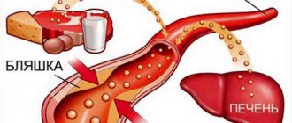
Cholesterol
A component of fat metabolism, it is involved in the construction of cell membranes, the synthesis of sex hormones and vitamin D. There is total cholesterol, low-density lipoprotein (LDL) and high-density lipoprotein (HDL) cholesterol.
Degrees of increased cholesterol in the blood:
- 5.2-6.5 mmol/l – mild degree of increase in the substance, a risk zone for atherosclerosis;
- 6.5-8.0 mmol/l – a moderate increase, which is corrected by diet;
- over 8.0 mmol/l – a high level requiring drug intervention.
An increase in cholesterol levels in the serum or blood plasma is an argument in favor of atherosclerosis, hypothyroidism (low activity of the thyroid gland), chronic hepatitis, decompensated diabetes mellitus, obstructive jaundice.
This indicator decreases when:
- malignant liver tumors;
- cirrhosis of the liver;
- rheumatoid arthritis;
- hyperfunction of the thyroid and parathyroid glands;
- starvation;
- malabsorption of substances;
- chronic obstructive pulmonary diseases.
Bilirubin
Bilirubin is a yellow-red pigment that is formed when hemoglobin breaks down in the spleen, liver and bone marrow. Its normal level in the blood of children and adults is 3.4–20.5 µmol/l.
If the table with the examination results contains an elevated level of bilirubin, then the doctor can diagnose one of the following diseases in adults:
- cholelithiasis;
- pancreatic tumors;
- inflammatory diseases of the biliary tract.
If bilirubin is below normal, then the patient may have one of the following diseases:
- acute viral hepatitis;
- bacterial liver damage (leptospirosis, brucellosis, etc.);
- toxic hepatitis;
- medicinal product;
- neoplasms in the liver and primary biliary cirrhosis;
- hemolytic anemia of various etiologies.
Bilirubin, formed as a result of the breakdown of hemoglobin (indirect), is released into the blood, where it binds to albumin and is transported to the liver. In liver cells, bilirubin combines with glucuronic acid. This bilirubin bound to glucuronic acid is called direct bilirubin.
Amylase
Breaks down carbohydrates from food and ensures their digestion. Contained in the salivary glands and pancreas. There is alpha-amylysis (diastase) and pancreatic amylase.
- alpha-amylase rate: 28-100 units/l.
- pancreatic amylase rate: 0-50 units/l.
A high amylase content in a biochemical blood test indicates: peritonitis, pancreatitis, diabetes mellitus, pancreatic cyst, stone, cholecystitis or renal failure.
Decreased alpha-amylase: thyrotoxicosis; myocardial infarction; complete necrosis of the pancreas; toxicosis of pregnant women.
Potassium
Another important intracellular electrolyte. Its normal content in the body ranges from 3.5 to 5.5 mmol per liter.
Reduced potassium content:
- excess hormones of the adrenal cortex (including taking dosage forms of cortisone);
- chronic fasting (failure to receive potassium from food);
- prolonged vomiting, diarrhea (loss with intestinal juice);
- renal dysfunction;
- cystic fibrosis.
Increased potassium content:
- dehydration;
- acute renal failure (impaired renal excretion); ,
- adrenal insufficiency.
- cell damage (hemolysis - destruction of blood cells, severe starvation, convulsions, severe injuries).
The condition when potassium is elevated is called hyperkalemia, and when it is low, hypokalemia.
Sodium
Sodium does not directly participate in metabolism. It is completely abundant in the extracellular fluid. Its main function is to maintain osmotic pressure and pH. Sodium excretion occurs in the urine and is controlled by the adrenal hormone aldosterone.
Sodium reduction:
- decreased concentration due to increased fluid volume (diabetes mellitus, chronic heart disease)
- failure, liver cirrhosis, nephrotic syndrome, edema).
- loss of an element (abuse of diuretics, kidney pathology, adrenal insufficiency).
Increased sodium content:
- increased function of the adrenal cortex;
- excess salt intake;
- loss of extracellular fluid (profuse sweat, severe vomiting and diarrhea, increased urination in diabetes insipidus);
- violation of the central regulation of water-salt metabolism (pathology of the hypothalamus, coma).
An increase in a microelement is called hypernatremia, and a decrease is called hyponatremia.
Symptoms of anemia
Conventionally, the symptoms of low hemoglobin are classified into:
- specific (manifest only in specific types of anemia);
- nonspecific (the same for all types of disease).
Nonspecific symptoms of anemia are as follows:
- pallor of the skin/mucous membranes;
- migraine;
- increased fatigue;
- noise in ears;
- refusal of food;
- constant drowsiness;
- feeling of lack of air;
- dizziness;
- cardiopalmus;
- decreased libido, sexual impotence in men;
- disruption of the menstrual cycle;
- heart failure.
Specific signs of different types of anemia
Each type of anemia has its own specific symptoms. Thus, the acute posthemorrhagic form manifests itself:
- increased heart rate;
- fainting;
- cold sweat;
- attacks of dizziness;
- pale skin;
- decrease in body temperature.
In chronic posthemorrhagic anemia the following are observed:
- very white skin;
- craving for or intolerance to certain smells;
- swelling of the face;
- change in taste sensations;
- dry skin;
- brittle nails;
- nausea;
- lack of air;
- sweating;
- involuntary urination when sneezing or laughing;
- temperature increase.
Hemolytic anemias are characterized by:
- yellowness of the skin and mucous membranes (when red blood cells are destroyed, bilirubin enters the bloodstream);
- increased size of the spleen/liver;
- increased levels of bilirubin in the blood;
- darkening of urine and feces;
- high body temperature;
- weakness.
Sickle cell anemia - a type of hemolytic - has the following symptoms:
- jaundice;
- shortness of breath;
- formation of inflamed areas on the legs;
- blurred vision;
- the presence of hemoglobin in the urine.
Doctors diagnose thalassemia if the patient:
- the shape of the skull bones changes;
- the skin becomes very pale with a yellowish/greenish tint;
- eyes become narrow and swollen;
- there is a lag in mental/physical development;
- bones are deformed;
- the spleen/liver enlarges.
Signs of iron deficiency anemia include:
- buzzing in the head;
- lack of oxygen;
- migraine;
- constant desire to sleep, rest;
- dry skin;
- peeling of nails;
- hair section;
- desire to eat chalk, smell paint, etc.;
- violation of the act of urination;
- hyperhidrosis.
Also, during laboratory tests, it is discovered that the level of hemoglobin and red blood cells in the blood is significantly lower than the established norm.
B12 deficiency anemia affects the gastrointestinal tract and central nervous system. As a result, the patient experiences:
- swelling of the legs;
- crawling sensation on the arms/legs;
- change in gait;
- memory impairment;
- problems swallowing food;
- enlarged liver/spleen;
- drying of the gastrointestinal mucosa.
In turn, folate deficiency anemia manifests itself:
- inability to eat acidic foods;
- glossitis;
- difficulty chewing and swallowing food;
- enlarged spleen/liver;
- atrophy of the mucous membranes of the gastrointestinal tract.
The symptoms of hypoplastic anemia are:
- the appearance of ulcers in the mouth, on the skin of the face;
- bruising on the skin;
- severe bleeding gums;
- increased fatigue;
- desire to sleep;
- tachycardia;
- decrease in the number of leukocytes, red blood cells and platelets in a blood test.
If you experience similar symptoms, consult your doctor
. It is easier to prevent a disease than to deal with the consequences.
Normal hemoglobin level in blood during pregnancy
During pregnancy, a woman’s amount of enzyme in her blood will differ from the values indicated in the table above. Also, pregnant women should know what medications can and cannot be taken during pregnancy. These deviations are associated with various changes that occur in the body.
Approximate indicators of enzyme content in pregnant women are shown in the table:
| Trimester | Amount of hemoglobin g/l. |
| 1 | 115 — 165 |
| 2 | 110 — 145 |
| 3 | 110 — 140 |
These differences are associated with the end of menstruation and the subsequent growth of the placenta, which requires constant nutrition. Therefore, it is important for the expectant mother to systematically undergo blood tests, since a critical decrease/increase in hemoglobin can become a source of dangerous complications (hypoxia, abnormal fetal development, early birth, underweight of the baby, retardation in mental/physical development, etc.).
Doctor's advice
Often the cause of increased hemoglobin is non-compliance with the drinking regime. The body receives little fluid (less than 1.5-2 liters of clean water per day, tea, coffee and other liquids do not count), this causes the blood to thicken. In this condition, not only hemoglobin, but other indicators also increase - hematocrit, red blood cells, platelets. Hematologists advise that if you have anemia, your diet should focus on meat. Liver and buckwheat are in second place in importance. Next are apples, nuts, cocoa. These same principles apply not only to adults, but also to children.
Victoria Druzhikina Neurologist, Therapist
Diagnosis of anemia
To identify anemia in a patient, the doctor conducts a comprehensive diagnosis, which includes:
- clarification of the circumstances under which health deteriorated for the first time, the symptoms that are most pronounced;
- studying the patient's life history. The doctor clarifies whether there are chronic diseases, a hereditary predisposition to any pathological conditions, whether there are bad habits, whether the patient has recently taken any medications, or whether there has been contact with poisons or toxic compounds;
- examination of the skin, determination of its color. Measurement of pressure, pulse;
- laboratory tests of blood and urine.
In some situations, the patient is prescribed a bone marrow test. It is necessary to understand whether there are any disturbances in the hematopoietic processes. Also, using special equipment, a trephine biopsy can be performed, during which the doctor correlates the bone marrow with the tissues surrounding it.
An ECG is usually performed to examine the heart. If necessary, the patient is referred to a hematologist.
Laboratory diagnosis of low hemoglobin
In blood biochemistry, if anemia is suspected, the levels of:
- glucose;
- cholesterol;
- uric acid;
- creatinine;
- electrolytes.
This is necessary in order to assess the condition of the patient’s internal organs. A urine test is also performed to exclude the presence of concomitant diseases.
Laboratory signs of different types of anemia:
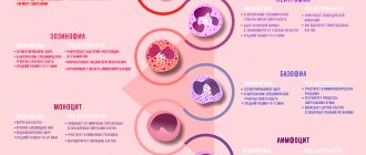
- posthemorrhagic acute anemia. Reticulocytes more than 11%, the presence of immature red blood cells and red blood cells of abnormal shapes. Leukocytes - 12 g/l (increased), shift in the formula - to the left;
- chronic posthemorrhagic anemia. Presence of tiny, poorly colored and oval red blood cells. Unexpressed lymphocytosis. Iron - below 9 µmol/l. A decrease in the amount of calcium and copper against the background of an increase in the concentration of manganese, nickel and zinc;
- sickle cell anemia. Hemoglobin is reduced to a level of 50 to 80 g/l, red blood cells - from 1 to 2 T/l. Reticulocytes are increased by about a third. The presence of red blood cells, the shape of which resembles a sickle;
- Thalassemia. Detection of target-like red blood cells in a blood sample, an increase in the number of reticulocytes. Hemoglobin - 20 g/l, erythrocytes - 1 T/l. Decreased concentration of platelets and leukocytes;
- B12 deficiency anemia. The presence of giant erythrocytes. Their color is bright, there is no highlighted area in the center. Shape: pear-shaped or oval. Life expectancy is shortened. Detection of huge neutrophils, a decrease in the number of eosinophils (may be completely absent). The concentration of basophils and leukocytes also decreases. Bilirubin, on the contrary, is increased.
How to boost a low level
Most often, older people suffer from iron deficiency anemia, which can be determined by the color index of the blood, as well as the ratio of various elements. When wondering how to increase hemoglobin, it is necessary to treat not the consequences of the disease, but to eliminate the factors that caused it.
To increase protein levels, a patient with low hemoglobin in the blood is prescribed iron supplements. It is important that they contain a sufficient amount of divalent iron, as well as components that improve the absorption of its ions. Often, these medications are taken twice a day with meals. At the same time, you should never prescribe medications yourself, since the content of the drug can cause side effects. Old people may experience disturbances in the functioning of the digestive tract, inflammation of the vascular walls of the veins, hypotension and an allergic reaction. Another fairly common side effect is chest pain. Therefore, drugs to increase hemoglobin should be prescribed exclusively by a specialist who will take into account all the individual characteristics of the body.
You can also get rid of anemia by following a proper and balanced diet. Products that increase hemoglobin in the blood of the elderly are:
- sausages and meat products;
- offal;
- black currant;
- fish;
- egg yolks;
- wheat sprouts and wheat germ;
- greens and cabbage;
- whole grain baked goods;
- nuts and seeds;
- beet;
- dried fruits (figs, prunes, dried apricots, dates).
Such nutrition with low hemoglobin can raise its level and help permanently get rid of the manifestations of anemia at an early stage of the disease.
Treatment of anemia in children and adults
To get rid of the symptoms of anemia, you need to eliminate the factor that provoked the decrease in hemoglobin. So, if the pathological condition is associated with the presence of parasites in the body, you need to get rid of them, if with poor nutrition, start following a diet, if with a malignant/benign tumor, it should be removed.
In other words, treatment of low hemoglobin in men, women and children may be conservative or require specialized surgical procedures. Usually, in order to improve the condition of patients and reduce the severity of negative symptoms, doctors adhere to the following therapeutic regimen:
- drugs are prescribed that can compensate for the resulting deficiency - B12 for B12-deficiency anemia, iron for iron deficiency, B9 for folate deficiency, etc.;
- normalize the level of red blood cells. This can be accomplished by transfusion of red blood cells or washing of red blood cells. However, these measures when providing assistance to people with low hemoglobin levels are extreme and are carried out only if the resulting disease is life-threatening.
Treatment of acute and chronic posthemorrhagic anemia
Treatment of the acute form of posthemorrhagic anemia is carried out in a hospital or hematology clinic
. The medications prescribed to the patient help normalize the amount of blood and the level of formed elements, and are also aimed at preventing relapses of the disease. Taking into account the amount of blood lost, the patient may need a transfusion, the introduction of blood substitutes or red blood cells.
As for the chronic form of this type of anemia, it is impossible to get rid of its symptoms without eliminating the cause. After the factor that provoked the pathological condition has been eliminated, the patient will be prescribed a diet that includes eating foods rich in iron. Medicines that can be used are Sorbifer Durules, Ferrum-Lek, vitamins B12 and B9, etc.
Treatment of sickle cell anemia
The main goal of therapeutic measures when it comes to a patient with sickle cell anemia is to prevent the development of hemolytic crises. To do this, the patient must avoid being in places with low oxygen levels. In parallel, blood substitutes and red blood cell infusions can be used.
Elimination of iron deficiency anemia
Treatment of iron deficiency anemia includes eating foods rich in iron and treating existing gastrointestinal diseases. The patient should regularly eat:
- cheeses;
- porridge;
- chicken eggs;
- meat;
- dairy products.
Iron supplements can also help quickly get rid of the symptoms of anemia. The tablets usually used are “Ferrum-Lek”, “Totem”, “Sorbifer Durules”, etc. Injections are prescribed only for severe forms of the disease. It is important that the medicine used does not cause problems with the gastrointestinal tract. If constipation or flatulence occurs, the product needs to be replaced.
Treatment of B12 deficiency anemia
Complex therapy of gastrointestinal diseases and adherence to the principles of proper nutrition help eliminate the manifestations of B12-deficiency anemia. Most often, patients are prescribed vitamin B12 injections. They allow you to quickly restore hematopoietic processes in the bone marrow.
How to get rid of folate deficiency anemia
Folate deficiency anemia is treated by taking vitamin B9 and following a diet. The patient should include foods that contain high amounts of folic acid in their diet. This means citrus fruits, vegetables, herbs, asparagus, nuts, seeds, tomatoes, watermelons, corn, avocados, eggs, animal liver, cod liver, cereals, grain bread.
Treatment of hypoplastic anemia
A hematologist treats hypoplastic anemia. Depending on the age, gender and condition of the patient, he can use different methods - bone marrow transplantation, stimulation of hematopoietic processes, blood transfusion, etc.
Anemia risk group
Low hemoglobin is most often diagnosed in:
- people with gastrointestinal diseases (oncology, gastritis, stomach and duodenal ulcers, inflammatory process in the large/small intestine, etc.);
- persons with poor nutrition, vegetarians;
- people with a genetic predisposition to a certain type of anemia (the disease can be passed from parent to child).
What are the dangers of low hemoglobin?
Anemia can be simultaneously called both a dangerous and harmless disease. So, if we are talking about a mild degree of the disease in a pregnant woman or a small child, then this is more likely a variant of the norm than a deviation. After taking a course of iron supplements, the baby/future mother will become absolutely healthy.
At the same time, moderate and severe degrees of some forms of anemia can lead to such dangerous conditions as:
- anemic coma (the patient loses consciousness and does not respond to any external stimuli, death may occur);
- malfunctions of internal organs (kidneys and liver are especially often affected by anemia);
- decrease in hemoglobin level to less than 70 g/l (death is also possible).
Prevention of anemia
To prevent the development of anemia, it is necessary:
- avoid contact with poisons and toxic substances;
- refuse to visit areas contaminated with radiation;
- do not contact with sources of ionizing radiation;
- do not take medications uncontrollably;
- to harden;
- eat right, include meat, greens, fruits and vegetables in your daily diet;
- spend more time outdoors.
Secondary prevention of low hemoglobin includes:
- blood test once a year;
- seeking qualified medical help in the event of acute infectious and viral diseases;
- annual medical examinations;
- pregnancy planning (for women).
This article is posted for educational purposes only and does not constitute scientific material or professional medical advice.