The table shows possible deviations (the norm is presented below)
| Index | Characteristic | Diagnosed pathology |
| Heart sizes | Increased | Cardiomegaly, chamber hypertrophy, cardiosclerosis, myocarditis |
| Myocardial characteristics | Thickened | |
| Compacted, heterogeneous | ||
| Istonchen | Organ failure, dilated cardiomyopathy | |
| Volume of ventricles and atria | Increased | |
| Reduced | Restrictive cardiomyopathy | |
| Valve condition | Thickened | Endocarditis |
| They don't close | Failure | |
| Do not open | Stenosis | |
| Pulmonary artery | Enlarged, expanded | Aneurysm |
| Dense | Atherosclerosis | |
| Emission volume | Reduced | Developmental defects, organ failure, cardiomyopathy |
| Residual volume | Increased | |
| Pericardial cavity | Thickened | Pericarditis |
| Liquid is visualized | ||
| Movement of blood between cavities and vessels | Regurgitation (backflow of blood during contraction) | Defects (congenital, acquired) – valve insufficiency |
| Shunt (shunt) between the aorta and pulmonary arteries | Congenital malformation | |
| Reset (shunt) at the level of the oval window | Open window | |
| Interventricular shunting | Defect (congenital, acquired) - ventricular septal defect | |
| Additional education | Nodes, thickenings, additional shadows | Neoplasms, blood clots, accessory chord, ventricular aneurysm |
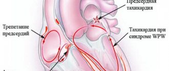
The study does not allow assessing the nature of the rhythm and electrical activity (an ECG is required), the condition of the coronary arteries (coronary angiography is performed), and the blood supply to the myocardium.
Depending on the equipment and assessment methods, the following types of echocardiography are distinguished:
- Standard . Transthoracic option: the sensor is placed on the chest. The study can be one-dimensional, two-dimensional or three-dimensional.
- Doppler . Aimed at assessing the characteristics of blood flow in the cavities of the heart and large vessels.
- Stressful . Ultrasound examination during stress tests.
- Transesophageal . Examination of the heart through the wall of the esophagus.
The gold standard for examination is two-dimensional Doppler-enhanced echocardiography.
Preparation for cardiac echocardiography
Ultrasound of the heart does not require special preparations. On the eve of the procedure, the patient is free to eat as usual and perform normal activities. The only thing that will be asked of him is to give up alcohol, caffeine-containing drinks, and strong tea.
If the patient is constantly taking medications, this must be warned in advance so that the results of the study are not distorted.
For each subsequent ultrasound of the heart, you should take a transcript of the previous one. This will help the doctor see the process over time and draw the right conclusions about your condition.
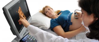
The examination itself takes from 15 to 30 minutes.
The patient, undressed to the waist, is lying on his back or side. A special gel is applied to his chest, ensuring easier sliding of the sensor over the area under study (the patient does not experience discomfort).
The specialist performing echocardiography has access to any areas of the heart muscle - this is achieved by changing the angle of the sensor.
Sometimes standard ultrasound of the heart does not provide complete information about the functioning of the heart, so other types of echocardiography are used. For example, fat on the chest of an obese person can interfere with the passage of ultrasound waves. In this case, transesophageal echocardiography is indicated. As the name suggests, the ultrasound probe is inserted directly into the esophagus, as close to the left atrium as possible.
And to screen cardiac function under stress, the patient may be prescribed stress echocardiography. This study differs from the usual one in that it is performed with a load on the heart, achieved by physical exercise, special medications or under the influence of electrical impulses. It is used primarily to identify myocardial ischemia and the risk of complications of coronary artery disease, as well as for certain heart defects to confirm the need for surgery.
In what cases is additional examination necessary?
Sometimes the results obtained using ultrasound do not allow an accurate diagnosis. Additional examination is prescribed for patients with the following disorders:
- suspicion of pulmonary hypertension arises when signs such as slow opening of the aortic valve, its closure during the systole phase, pathologically increased ejection of the right cardiac ventricle, deviation from the norm in the thickness of the ventricular wall;
- An open type arterial defect may be indicated by an increase in the wall of the atrium and ventricles, and the movement of blood from the aorta to the pulmonary artery. If such signs are detected, the patient needs to undergo additional examination methods;
- a defect in the septum separating the ventricles is indicated by thickening of the walls of the organ, developmental delay in the patient, blood entering from the left ventricle into the right;
- violation of the integrity of the valves and their branches often indicates the development of endocarditis of infectious origin;
- with a decrease in the number of heart contractions, a decrease in the ejection fraction and an increase in the volume of the organ chambers, a suspicion of an inflammatory process of the myocardium arises;
- The occurrence of exudative pericarditis is indicated by an excessive amount of fluid in the heart sac.
Echocardiography is a method for detecting many heart diseases
Myocardial infarction is often indicated by slow contraction of areas of the myocardium. Thickening of the walls of the left ventricle and atrium, weak compression of the mitral valve leaflets indicates its prolapse.
How is cardiac ultrasound interpreted?
- Competent ultrasound doctors with many years of experience
- Expert device with high resolution
- All types of ultrasound (ultrasound of the pelvic and abdominal organs, ultrasound of blood vessels, ultrasound of the heart, ultrasound of the joints)
Conclusion during the study.
Burning to disc
Sign up for an ultrasound
Prices for ultrasound
Echo-CG interpretation occurs by comparing normal indicators with the indicators of the study. What are these indicators:
- Myocardial mass
- Volume and dimensions of the left and right ventricles
- Wall thickness of the left and right ventricle
- Aortic diameter
- Thickness of the interventricular septum
It seems simple, but in reality everything turns out to be much more complicated. For competent decoding, it is necessary to know the individual characteristics of the patient, his medical history and many other factors. Therefore, to interpret echocardiography, contact a specialist!
The essence of echocardiography
Ultrasound of the heart is the process of studying all the main parameters and structures of this organ using ultrasound.
When exposed to electrical energy, the echocardiograph transducer emits high-frequency sound that travels through the structures of the heart, is reflected from them, captured by the same transducer and transmitted to the computer. He, in turn, analyzes the received data and displays it on the monitor in the form of a two- or three-dimensional image.
In recent years, echocardiography has been increasingly used for preventive purposes, which makes it possible to identify cardiac abnormalities at an early stage.
What does an ultrasound of the heart show:
- heart size;
- integrity, structure and thickness of its walls;
- sizes of the cavities of the atria and ventricles;
- contractility of the heart muscle;
- operation and structure of valves;
- condition of the pulmonary artery and aorta;
- pulmonary artery pressure level (to diagnose pulmonary hypertension, which can occur with pulmonary embolism, for example, when blood clots from the veins of the legs enter the pulmonary artery);
- direction and speed of cardiac blood flow;
- condition of the outer shell, pericardium.
1 ECHO-KG in "MedicCity"
2 Echocardiography at MedicCity
3 Ultrasound of the heart at MedicCity
Ultrasound of the heart in Moscow
The most competent interpretation of a heart ultrasound may not bring results if a poor-quality diagnosis was carried out: this can happen if you were examined using outdated equipment, and the protocol was drawn up by an inexperienced, unqualified doctor.
Don't risk your health! Contact a trusted clinic! The Elena Malysheva Diagnostic Center is your confidence in the quality of diagnostics! Experienced doctors with many years of experience, an expert-class ultrasound scanner and affordable prices are at your service!
Sign up for an interpretation of cardiac ultrasound at the Elena Malysheva Diagnostic Center near the Baumanskaya metro station (see map) by phone: 8 (495) 127-03-71 or leave a request on the website.
Baumanskaya metro station (see map) 8
leave a request on the website
Norms of indicators in newborns
Echocardiography is often performed among newborns. This method allows you to identify deviations in the functioning of the organ and its defects. Interpretation of echocardiography in children is a complex process, which is dealt with by a sinologist.
Normal ultrasound examination parameters in babies after birth are determined using a special table.
In this case, the end-diastolic size of the left ventricle in boys should be in the range from 17 to 22 mm, and in girls – from 16 to 21 mm. An indicator such as the end-systolic volume of the left ventricle in children of both sexes ranges from 11 to 15 mm. The diameter of the right ventricle in boys should not exceed the boundaries of 6 to 14 mm, in girls - from 5 to 13 mm. The size of the left atrium in boys should be from 12 to 17 mm, in girls - from 11 to 16 mm.
Ultrasound of the heart in newborns helps identify various defects
An important indicator is the condition of the septum between the left and right ventricles. It should not be hypertrophied, otherwise it indicates the presence of a defect.
Another vital indicator is the cardiac ejection fraction. This concept refers to the volume of blood that the left ventricle pushes out. It should be between 65 and 75%. While the speed of blood movement through the pulmonary valve is 1.4–1.6 mm/s.
With age, these indicators change. After 14 years, the child’s standards correspond to adults.
Important! An ultrasound of the baby's heart is sometimes performed in utero. This diagnostic method allows you to identify various defects at an early stage of development and select the necessary treatment.
Symptoms of reduced ejection fraction
All symptoms that may suggest a decrease in the contractile function of the heart are caused by CHF.
Therefore, the symptoms of this disease come first. However, according to the observations of practicing ultrasound diagnostic doctors, the following is often observed: in patients with severe signs of CHF, the ejection fraction remains within the normal range, while in people with no obvious symptoms, the ejection fraction is significantly reduced. Therefore, despite the absence of symptoms, patients with cardiac pathology must undergo echocardioscopy at least once a year.
So, symptoms that suggest a violation of myocardial contractility include:
- Attacks of shortness of breath at rest or during physical exertion, as well as when lying down, especially at night,
- The load that provokes the occurrence of shortness of breath attacks can be different - from significant, for example, walking long distances (more than 500-1000m), to minimal household activity, when it is difficult for the patient to perform the simplest manipulations - cooking, tying shoelaces, walking to the next room etc,
- Weakness, fatigue, dizziness, sometimes loss of consciousness - all this indicates that the skeletal muscles and brain receive little blood,
- Swelling on the face, legs and feet, and in severe cases - in the internal cavities of the body and throughout the body (anasarca) due to impaired blood circulation through the vessels of the subcutaneous fat, in which fluid retention occurs,
- Pain in the right half of the abdomen, an increase in abdominal volume due to fluid retention in the abdominal cavity (ascites) - occurs due to venous stagnation in the hepatic vessels, and long-term stagnation can lead to cardiac cirrhosis of the liver.
In the absence of proper treatment for systolic myocardial dysfunction, such symptoms progress, increase and become increasingly difficult for the patient to tolerate, therefore, if even one of them occurs, you should consult a general practitioner or cardiologist.
Normal ultrasound of the heart
Here are some of the indicators that are taken into account when assessing results:
- Myocardial mass: men - 130-180 g; women - 90-140 g.
- Left ventricle size: At rest - 5.7 cm in men, 4.6 cm in women)
- During contraction – 4.3 cm in men, 3.1 cm in women)
Interpreting the results of a cardiac ultrasound is a serious process that requires the doctor to have fundamental knowledge of human anatomy. Only a specialist can competently assess deviations and understand whether it is worth sounding the alarm. You may need additional examinations.
Take care of your health! Trust the decryption to professionals!
If you have any questions, ask our specialist!
Ask a Question
Is cardiac echocardiography safe?
During this study, there is no radiation or other load on the organ. Therefore, if necessary, it can be prescribed even several times a week.
This study is characterized by the absence of complications and side effects.
EchoCG does not harm either the expectant mother or the fetus during pregnancy.
Limitations for the procedure may include inflammatory diseases of the skin of the chest, chest deformities and some other reasons.
An important diagnostic method
Ultrasound examination of the heart
Echocardiographic examination of the cardiovascular system is a very important and also quite accessible diagnostic method. In some cases, the method is the “gold standard”, allowing one or another diagnosis to be verified. In addition, the method makes it possible to identify hidden heart failure that does not manifest itself during intense physical activity. Echocardiographic findings (normal values) may vary somewhat depending on the source. We present the guidelines proposed by the American Association of Echocardiography and the European Association of Cardiovascular Imaging in 2015.
Decoding EchoCG
After the examination, the doctor draws up a conclusion. First, a visual picture with the presumed diagnosis is described. The second part of the study protocol indicates the patient’s individual indicators and their compliance with standards.
Decoding the data obtained is not a final diagnosis, since the study can be done not by a cardiologist, but by an ultrasound diagnostic specialist.
It is the cardiologist, based on the collected medical history, examination results, interpretation of tests and data from all prescribed studies, who can draw accurate conclusions about your condition and prescribe the necessary treatment!