We offer you a comprehensive examination of the heart and cardiovascular system. If a disease is detected, we will offer you the help of a therapist, cardiologist, etc. In addition to conducting the study, our specialist will interpret the ECG, ABPM results, etc.
- Methods for studying the cardiac vascular system
- Search for the cause of the disturbance in the functioning of the cardiovascular system
Watch our video about diagnostics of the heart, blood vessels and the function of external respiration
. The heart performs the main pumping function in our body, its task is to ensure adequate supply of blood to the tissues, and with it oxygen and nutrients. With heart diseases (coronary artery disease, atherosclerosis, carditis, etc.), not only the heart itself suffers, but also other organs, including the brain.
In our clinic, research of the cardiovascular system takes place in two directions:
- Study of the structure and function of the heart muscle;
- Vascular examination;
- Search for the cause of the disturbance in the functioning of the cardiovascular system.
MRI procedure
MRI is magnetic resonance imaging, that is, layer-by-layer scanning of the heart. The operation of the tomograph is based on the use of the magnetic resonance effect. During an MRI scan, the machine creates a powerful magnetic field. Cell atoms, when exposed to an RF signal in a magnetic field, begin to vibrate, and this resonance is picked up by a tomograph. The obtained information is translated by an MRI device into three-dimensional images, from which the doctor can determine the presence of the smallest pathologies of the heart muscle and coronary vessels in the patient. The MRI procedure itself is quite simple. The patient is placed on a movable table on his back, the body is fixed with a special coil, and the head is fixed with rollers, after which the table is pushed into a cylindrical chamber. The patient's task is to lie still and calm throughout the magnetic resonance examination. The duration of cardiac MRI is from 25 to 30 minutes. During the procedure, the patient may experience discomfort due to various sounds produced by the device. If a person is concerned about this, they should ask the diagnostician for headphones before the test. This will soften the noise. If the patient begins to feel uncomfortable inside the tomograph tube or experience a panic attack, he always has the opportunity to contact the diagnostician, since the scanner is equipped with two-way communication. After an MRI examination, the patient receives a report from a radiologist and an examination recorded on disk in the form of a series of high-quality images. Waiting for the result can take up to one hour, in rare cases 1-2 days. It all depends on the complexity of the scan and diagnosis. To reduce the waiting time for results, the conclusion in most medical centers in St. Petersburg can be obtained by e-mail.
Previous Next
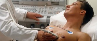
Ultrasound
Ultrasound is an ultrasound examination of the heart. The operating principle of an ultrasound machine is based on the physical properties of ultrasound. Supersound is not perceived by the human ear, since the vibrations of the sound wave in this case are reproduced at a very high frequency. The ultrasound sensor sends ultrasound into the cavity of the object under study, which, when reflected, creates an echo. It is captured by a computer and converted into images. The anatomical accuracy and level of detail on ultrasound is significantly lower than on cardiac MRI because it is difficult for ultrasound to penetrate the bony barrier of the chest. Ultrasound diagnostics of the heart cannot distinguish between changes ten years ago and changes occurring now. It cannot reveal the severity or depth of the ongoing inflammatory or tumor process. Ultrasound can only indirectly indicate that there are some thrombotic changes in the coronary vessels of the heart.
Four flights of stairs
Before taking the test, it is important to do some physical exercise: for example, take a brisk walk. Muscles, including the heart, must be toned. After a short rest, you need to climb the 60-step staircase as quickly as possible. Usually this is 4 standard spans. You cannot break into a run while moving.
When testing, scientists measured the energy expenditure of the subjects before and immediately after the test: in metabolic equivalents (ME), which characterize the volume of oxygen.
- rise in 40–45 seconds. at a cost of 9–10 ME: the best result, an indicator of excellent cardiac health;
- rise in 1.5 minutes. with a consumption of less than 8 ME: an alarming condition in which a detailed medical examination is recommended;
- rise in more than 1.5 minutes: heart disease is present in most cases, an urgent need to see a cardiologist.
Advantages and disadvantages of ultrasound and MRI of the heart
Today, high-quality cardiac MRI requires very expensive equipment - a tomograph with a magnetic field power of at least 3 Tesla. Research using such an ultra-high-field device will be expensive, so the main disadvantage of cardiac MRI is its high price. In addition, MRI of the coronary vessels and heart cannot measure the speed of blood flow and its actual volume. Cardiac ultrasound can better cope with this task.
Previous NextSquat test
Here you don’t even need a ladder; you can check your heart function at home. You will need a little more than 1 m² of free space, a stopwatch or timer. No preparation for the study is needed:
- Measure your pulse per minute while at rest. Remember the result.
- Time yourself for 30 seconds and try to do 20 squats during this time. As you lower, stretch your arms forward, and as you rise, press them to your hips.
- After finishing the exercise, immediately repeat the heart rate measurement.
- Rest for 3 minutes and measure the beat frequency again.
A comparison of three results: at rest, after exercise and after rest shows whether everything is fine with the heart:
- the pulse after squats increased to 25% and recovered to the initial result: excellent;
- the frequency increased by 25–50% after exercise, and after rest remained increased by 20–30%: the result is good, but you need to monitor your health;
- the load caused the heart rate to increase by 50–75%, after rest it remains just as high: there are violations and reasons to consult a doctor;
- The pulse fluctuates after exercise, becoming 76% higher or higher, there was not enough rest for recovery, it quickly returned to normal: diagnostics and medical assistance are urgently required.
When is ultrasound better than cardiac MRI?
Cardiac ultrasound is well suited for diagnosing both children, from infancy, and adults. An ultrasound examination will clearly show the functional features of the heart:
- efficiency of the heart muscle;
- thickness of the heart walls;
- functional state of four chambers and valves;
- the size of the heart cavities and the pressure in them;
- speed of intracardiac blood flow.
Cardiac ultrasound is better suited than MRI for examining young children, since it does not require immobilization of the patient through anesthesia so that the baby can lie quietly. Ultrasound is an inexpensive way to initially screen the heart if you need to evaluate the condition of the organ due to some primary symptoms of illness or if you want to do an ultrasound of the heart as a preventive measure for cardiovascular diseases. Cardiac MRI is an expert diagnostic method. It is best used when a pathology is identified and it is necessary to verify or clarify the diagnosis.
Search for the cause of the disturbance in the functioning of the cardiovascular system
- Vascular examination If the cause of heart muscle suffering is atherosclerosis, we will offer you to conduct an ultrasound examination (ultrasound) of the vessels of the brain and neck, ultrasound examination of the vessels of the arms and legs because with atherosclerotic damage to the vessels of the heart, there is a high probability that some other vessels are involved in the process.
- Biochemical blood test (lipid profile study, i.e. the ratio of harmful and good cholesterol);
- Immunological and microbiological studies (bacteriological cultures, PCR, determination of antibodies to infections). These studies may be useful for selecting targeted treatments for carditis.
- An immunogram can be useful to find the reason why the causative agent of carditis was able to enter the body. Typically, the immune system prevents any foreign agent (bacteria, virus, etc.) from entering the body. Our task is to find the dysfunctional link in the immune system and prevent the development of carditis in the future.
When there is an imbalance in various forms of cholesterol, atherosclerosis develops.
Atherosclerotic cholesterol plaques block the lumen of blood vessels, impair blood circulation, and can cause myocardial infarction and heart pain. We recommend regular heart examinations, namely bicycle ergometry, Echo-CG (ultrasound of the heart), ABPM and others. This will help identify problems with the cardiovascular system before obvious symptoms appear. Bicycle ergometry is an indispensable study to check how the myocardium (heart muscle reacts) to physical activity or stress; ultrasound of the heart shows the contractility of the myocardium. Daily blood pressure monitoring (ABPM) will help identify trends in blood pressure changes and help in identifying factors that contribute to a rise in blood pressure.
When is MRI better than cardiac ultrasound?
The level of anatomical detail of the heart area on MRI is higher than on ultrasound. During the scan, the doctor receives up to 1000 images of the mediastinal area. The scanning step of a 3 tesla device can be only 0.8 mm. This means that the doctor can look in detail at the condition of the heart itself and the neighboring coronary vessels. In this case, both planar and three-dimensional data will be provided. MRI best visualizes pathologies and diseases such as:
- inflammatory processes in tissues;
- tumor formations;
- blood clots, vascular malformations, occlusions, aneurysms;
- congenital anomalies of the heart structure;
- heart muscle defects;
- pericardial condition.
Author: Telegina Natalya Dmitrievna
Therapist with 25 years of experience
>
Nikolay Amosov test
The creator of the artificial heart valve has proposed an even simpler ladder test that does not require outside help. You can do it on the way home.
- Before you start, you need to measure your pulse. At rest, its norms are: 60–80 beats per minute in women and 50–85 beats in men.
- Slowly walk up the stairs of the 4th floor.
- Measure your pulse again. If it doesn't exceed 100 hits, great. At 100–120 blows: satisfactory, 120–140 blows: the result is alarming. If it is more than 140, accompanied by severe shortness of breath, you must stop the test and see a doctor.
Next stage: climbing stairs for 4 minutes:
- managed to climb 15 floors: excellent;
- no more than 12 floors: good;
- 10–11 floors: something to worry about;
- 9 floors or less: time to see a doctor.
If there are no high-rise buildings nearby, you can modify the test: when you reach the top floor, go back down. In this case, descending 3 floors is equal in load to ascending 1.
Possible complications after the procedure
Like any other surgical intervention and violation of the integrity of the vessel, coronary angiography can have a number of complications, even if it is performed by an experienced specialist. Possible complications include:
- heart attack;
- rupture of an artery or heart;
- heart attack or stroke due to a blood clot breaking off from the wall of a vessel;
- arrhythmia;
- bleeding;
- allergies.
Of course, complications do not always arise and are often limited to small hematomas or swelling at the puncture site. However, the possibility of serious consequences cannot be excluded, so before the procedure you need to carefully study the contraindications to it.
Contraindications for the study
Contraindications for cardiac vascular examination with contrast are:
- various respiratory diseases that are accompanied by elevated body temperature;
- anemia;
- open bleeding of various locations;
- low potassium levels in the blood;
- poor blood clotting;
- diabetes;
- obesity or underweight;
- renal, heart failure;
- lung damage.
Computed tomography of cardiac vessels
Computed tomography of the blood vessels of the heart (CT angiography) is performed using a computer tomograph, which contains an X-ray tube inside. Rotating, it provides a three-dimensional image of the vascular bed being examined, helping the doctor identify various vascular diseases. Before the procedure, a radiopaque contrast agent is injected into the patient’s vein, after which he lies down on the tomograph table. It is not advisable to move during the procedure, as this may distort the data. CT is significantly inferior in information content and reliability to coronary angiography.
Interpretation of coronary angiography
How coronary angiography of the heart vessels is performed and what it is is clear. The main thing is not just to take a picture, but to interpret it correctly, that is, decipher it. For the most informative and accurate result, several specialists are involved in decoding: a radiologist, a cardiologist, a vascular surgeon and others.
The thickness of the vessels, their length and width are assessed from the images. Compactions and darkening are revealed. An alarming signal is a violation of the boundaries of blood vessels - blurriness and indistinctness. The pathology of the vessels will also be indicated by their incorrect location.
Diagnosis of CVD
The level of modern diagnostics makes it possible to identify most diseases of the cardiovascular system at an early stage, when the pathological process is reversible and responds well to therapy.
Methods for studying heart disease are divided into physical, laboratory and instrumental.
Physical examination methods performed by the doctor at the first appointment include:
- examination of the skin and mucous membranes;
- palpation - palpation;
- percussion - tapping;
- auscultation - listening.
Despite their simplicity, physical examination methods allow the doctor to make a preliminary diagnosis and outline the range of necessary laboratory and instrumental tests.
- During the examination, changes in the color of the skin (pallor, cyanosis, yellowness) are noted; the presence or absence of pastiness or swelling of the extremities, pulsation of the cervical arteries.
- Using percussion, the boundaries of the heart are determined, which, in case of myocardial pathologies, go beyond normal limits.
- Using a phonendoscope, auscultation is performed - listening to heart sounds and noises and assessing their sonority and rhythm.
- Blood pressure is measured.
Currently, these methods are relegated to the background, since there are modern, faster and more effective ways to detect pathologies of the cardiovascular system.
Who can benefit from CT coronary angiography?
To examine in detail the condition of the heart vessels, assess blood circulation in them and determine the location of blockages, CT coronary angiography is used.
Vladislav Vasilievich Babenko, a radiologist at the advisory and diagnostic department of the Expert Institute, told us in detail about this study.
– Vladislav Vasilievich, what is CT coronary angiography?
– This is a non-invasive and effective screening method for examining the heart vessels using computed tomography (CT). A more medically correct name for coronary angiography is coronary angiography or coronary angiography.
Read more about the computed tomography method in our article
During CT coronary angiography, the coronary calcium index and the risk of acute coronary events are determined. This technique is carried out using an iodine-containing contrast agent and is a radiation research method.
The procedure is recommended for patients with a low and intermediate risk of pre-test probability of stable coronary artery disease.
– When is coronary angiography prescribed?
– The patient is referred for coronary angiography in the following cases:
- for differential diagnosis in the presence of unclear pain in the chest (with a questionable or negative stress test);
— for uninformative stress tests or as an alternative to stress tests if there are contraindications to them;
— to decide whether further in-depth examination and treatment is necessary (for example, performing invasive coronary angiography, stenting or coronary artery bypass grafting);
— for preventive examinations (screening) of people without symptoms of coronary heart disease in order to identify patients with atherosclerosis of the coronary arteries;
- to control the progression of atherosclerosis of the heart vessels;
- to determine the effectiveness of the treatment.
Read materials on the topic:
Coronary heart disease: diagnosis and treatment
Serious question: what happens to the heart during an angina attack?
How to prevent myocardial infarction?
– What does CT coronary angiography show?
– This method allows you to visualize the vessels of the heart, assess the condition of atherosclerotic plaques, determine the variant anatomy (structural features) of the vascular bed and exclude developmental anomalies.
“We must remember that atherosclerosis, which develops against the background of increased cholesterol concentrations, is a chronic pathology that requires long-term, many-year cooperation between the doctor and the patient.” Quote from the material “How to distinguish good and bad cholesterol?”
– Do you need special preparation for the examination?
- Yes. The patient must bring the results of a blood creatinine test from a week ago and ECG results with interpretation. In order to achieve stabilization of the heart rate (HR) before the examination, special medications are prescribed. Heart rate should be in the range of 50-65 beats per minute. If the heart rate is higher than the permissible value, CT coronary angiography is not advisable, since the study will be uninformative.
Before the procedure, all patients undergo a consultation with a therapist or cardiologist.
– How is CT coronary angiography performed?
– At the first stage (before the administration of a contrast agent), the coronary calcium index is calculated and the patient is familiarized with the upcoming examination, and instructions are given. Next, preliminary medicinal preparation of the patient and stabilization of heart rate are performed. The third stage is the introduction of an intravenous catheter into the right ulnar vein. The fourth is the research itself. The patient is placed in the machine and a contrast scan is performed. And the last, fifth stage is monitoring the patient to exclude side effects from the administration of a contrast agent and providing assistance if they occur.
– What are the contraindications to CT coronary angiography?
– Relative contraindications include:
- allergic reactions to the administration of iodine-containing contrast agent;
- the patient’s inability to lie still for a long time;
— the patient’s inability to follow the operator’s and doctor’s instructions;
- uncorrectable heart rate (HR);
- decreased kidney function (glomerular filtration rate);
- thyrotoxicosis (increased levels of thyroid hormones) and thyrotoxic crisis;
— the patient’s body weight is more than 120 kg;
— pregnancy regardless of term (including potential pregnancy);
- children's age, a clear justification (vital indications) for conducting the study is necessary.
“There are two concepts: hyperthyroidism and thyrotoxicosis. Sometimes they are combined, in particular in foreign sources. But there is a difference." Quote from the material “If the thyroid gland is “raging”: what is hyperthyroidism?”
It should be added that if this study is vitally necessary, it is performed in the presence of contraindications in a hospital setting.
– Tell us about other types of coronary angiography. What is the difference between them?
– In addition to CT coronary angiography, invasive coronary angiography (sometimes called traditional coronary angiography) is used. This procedure is performed in a hospital setting. Surgical access is through the femoral artery, in contrast to CT coronary angiography, which uses intravenous access. The contrast agent is administered in a larger volume than during CT coronary angiography.
– Which is better: CT coronary angiography or traditional coronary angiography?
Each method has its own advantages and disadvantages. Traditional invasive coronary angiography has higher resolution and a higher percentage of specificity in assessing stenosis (narrowing) of blood vessels, but is more likely to cause adverse reactions. With invasive coronary angiography, it is possible to measure the fraction of reserve blood flow (this is the “gold standard” in the assessment of cardiac stenosis), and also immediately install a stent if necessary. But this procedure is usually carried out within the framework of compulsory medical insurance only for health reasons. If the patient does not have vital indications, then this study will be very expensive: in comparison, CT coronary angiography is approximately 10 times cheaper. The advantage of CT coronary angiography is its speed and the ability to perform the study on an outpatient basis.
– Vladislav Vasilievich, is CT coronary angiography different from CT of the heart, and if so, how?
– It differs in its methodology, as well as in the fact that cardiac CT has other research purposes and patients with different pathologies.
The patient may be referred for a cardiac CT scan, for example, to evaluate the left atrium and left atrial appendage. For what? In order to confirm or exclude the presence of a blood clot, assess the condition of the mouths of the pulmonary veins, their diameters, the presence or absence of stenoses. That is, these are patients who have arrhythmia and need to find out the cause. Depending on what was revealed on a CT scan of the left atrium, further surgical or conservative treatment will be performed.
The editors recommend:
What will a heart ultrasound tell you?
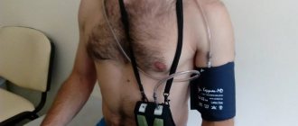
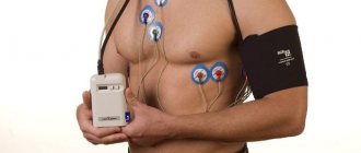
Holter (24-hour) ECG monitoring – complete instructions for the patient
When is daily blood pressure monitoring prescribed?
Interested in other articles on CT scanning? Read more materials in our section
For reference:
Babenko Vladislav Vasilievich
In 2015 he graduated from the Faculty of Medicine of Voronezh State Medical University. N.N. Burdenko.
2015 – 2021 – residency in the specialty “Radiology”.
Currently, he is a radiologist at the advisory and diagnostic department of the Expert Institute.