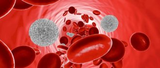
Leukocytes are a group of blood cells that are characterized by the absence of color, the presence of a nucleus and the ability to move. The name is translated from Greek as “white cells”. The group of leukocytes is heterogeneous. It includes several varieties that differ in origin, development, appearance, structure, size, core shape, and functions. White blood cells are produced in the lymph nodes and bone marrow. Their main task is to protect the body from external and internal “enemies”. Leukocytes are found in the blood and in various organs and tissues: in the tonsils, intestines, spleen, liver, lungs, under the skin and mucous membranes. They can migrate to all parts of the body.
Types of leukocytes
White cells are divided into two groups:
- Granular leukocytes - granulocytes. They contain large, irregularly shaped nuclei, consisting of segments, the more of which the older the granulocyte is. This group includes neutrophils, basophils and eosinophils, which are distinguished by their perception of dyes. Granulocytes are polymorphonuclear leukocytes. You can learn more about granulocytes from this article.
- Non-granular - agranulocytes. These include lymphocytes and monocytes, which contain one simple oval-shaped nucleus and do not have a characteristic granularity.
What are leukocytes
Several types of formed elements float in the blood, which support the health of the whole organism. White cells that have a nucleus inside are called leukocytes. Their peculiarity is the ability to penetrate the capillary wall and enter the intercellular space. It is there that they find foreign particles and absorb them, normalizing the vital activity of the cells of the human body.
Leukocytes include several types of cells that differ slightly in origin and appearance. The most popular division is based on morphological characteristics.
| Morphology | View | Normal quantity |
| Granular (having granules in the cytoplasm) | Neutrophils | 70% |
| Eosinophils | 1-5% | |
| Basophils | Up to 1% | |
| Non-granular (no granules) | Lymphocytes | 21-35% |
| Monocytes | 4-8% |
The ratio of these cells is the same in all healthy people and is expressed by the leukocyte formula. By changing the number of any type of cells, doctors draw conclusions about the nature of the pathological process.
It is leukocytes that maintain human health at the proper level. Most infections that enter the human body are asymptomatic due to a timely immune response.
Where are they formed and how long do they live?
The bulk of white cells, namely granulocytes, are produced by red bone marrow from stem cells. From the maternal (stem) cell, a precursor cell is formed, then passes into a leukopoetin-sensitive cell, which, under the influence of a specific hormone, develops along the leukocyte (white) series: myeloblasts - promyelocytes - myelocytes - metamyelocytes (young forms) - rods - segmented. Immature forms are found in the bone marrow, mature forms enter the bloodstream. Granulocytes live for about 10 days.
The lymph nodes produce lymphocytes and a significant proportion of monocytes. Some agranulocytes from the lymphatic system enter the blood, which transports them to the organs. Lymphocytes live a long time - from several days to several months and years. The lifespan of monocytes ranges from several hours to 2-4 days.
General characteristics
Leukocytes in the blood are a group of cells that are responsible for the human body’s resistance to various pathogenic bacteria, viruses, helminths, parasites and other pathological microorganisms.
They also fight not only infectious agents, but also any foreign object:
- malignant or benign neoplasms of any location;
- transplanted donor organ;
- a foreign object that may accidentally enter the body.
The place of formation of leukocytes is blood stem cells, which are localized in the red bone marrow. In order to fully carry out their work, they undergo a large number of transformations, during which their structure and functions change.
In addition to blood, they are also found in fluids such as:
- urine;
- cerebrospinal fluid;
- pleural effusion;
- feces;
- gastric juice.
However, their concentration in such cases will be significantly lower, for example, for urine analysis, from 4 to 6 leukocytes are acceptable, and no more than 8 white blood cells should be present in the cerebrospinal fluid.
An increase or decrease in such blood components in any of the above structures most often indicates the occurrence of a disease.
In addition to the main task, the functions of leukocytes include:
- release of specific substances to combat various tumors;
- absorption and digestion of the pathogenic agent;
- relief of hemorrhages;
- acceleration of wound healing.
As stated above, white blood cells have several subtypes.
- The norm of leukocytes in the blood of women by age in the table, about increased and decreased values
Thus, there are the following types of leukocytes:
- neutrophils - aimed at destroying bacterial infection;
- lymphocytes – responsible for the immune system and immune memory;
- monocytes - absorb and digest particles of foreign cells;
- eosinophils - fight allergen carriers;
- basophils - assist other particles in detecting foreign agents, however, they perform all their “duties” outside the bloodstream - in the internal organs.
It follows from this that subtypes of leukocytes perform their own mission.
Types of leukocytes
All types of such substances, in addition to their functions, differ in the following indicators:
- dimensions;
- core shape;
- way of development.
It is also worth noting about the structural features of each type of white blood cell. For example, neutrophils, eosinophils, basophils and monocytes are born from myeloblasts, the precursor of which is myelopoiesis. This happens under the influence of a stimulator cell in the bone marrow.
The lifespan of leukocytes is on average 2-4 days, and they are often destroyed in the liver, spleen and foci of inflammatory processes. The only exceptions are lymphocytes, some of which live in the human body from birth to death.
In neutrophils, eosinophils and basophils, the entire life cycle takes place in the bone marrow, which is why their immature cells are normally completely absent from the blood. Monocytes continue to exist in the spleen, liver and skeletal system, where they degenerate into macrophages and dendrocytes. Lymphocytes have a longer “life” in the spleen, lymph nodes and thymus.
Leukocytes received their common name - white blood cells - because, unlike red blood cells, they are colorless.
From all of the above, it follows that if there are no leukocytes in the blood, the human body simply will not be able to function.
- Increased level of leukocytes in the child’s blood: causes of leukocytosis, consequences and treatment
Structure
The structure of leukocytes of different types is different, and they look different. What they all have in common is the presence of a core and the absence of their own color. The cytoplasm can be granular or homogeneous.
Neutrophils
Neutrophils are polymorphonuclear leukocytes. They are round in shape and have a diameter of about 12 microns. There are two types of granules in the cytoplasm: primary (azurophilic) and secondary (specific). Specific small, lighter and make up about 85% of all granules, contain bactericidal substances, the protein lactofferin. Ausorophilic ones are larger, they contain about 15%, they contain enzymes, myeloperoxidase. In a special dye, the granules are colored lilac, and the cytoplasm is colored pink. The granularity is fine, consists of glycogen, lipids, amino acids, RNA, enzymes, due to which the breakdown and synthesis of substances occurs. In young forms, the nucleus is bean-shaped, in rod-nuclear forms it is in the form of a stick or a horseshoe. In mature cells - segmented - it has constrictions and looks divided into segments, which can be from 3 to 5. The nucleus, which can have processes (appendages), contains a lot of chromatin.
Eosinophils
These granulocytes reach a diameter of 12 microns and have a monomorphic coarse granularity. The cytoplasm contains oval and spherical granules. The granularity is stained pink with acidic dyes, the cytoplasm becomes blue. There are two types of granules: primary (azurophilic) and secondary, or specific, filling almost the entire cytoplasm. The center of the granules contains a crystalloid, which contains the main protein, enzymes, peroxidase, histaminase, eosinophil cationic protein, phospholipase, zinc, collagenase, cathepsin. The eosinophil nucleus consists of two segments.
Basophils
This type of leukocyte with polymorphic granularity has dimensions from 8 to 10 microns. Granules of different sizes are stained with a basic dye in a dark blue-violet color, the cytoplasm is stained pink. The grain contains glycogen, RNA, histamine, heparin, and enzymes. The cytoplasm contains organelles: ribosomes, endoplasmic reticulum, glycogen, mitochondria, Golgi apparatus. The core most often consists of two segments.
Lymphocytes
By size they can be divided into three types: large (from 15 to 18 microns), medium (about 13 microns), small (6-9 microns). The latter are in the blood most of all. Lymphocytes are oval or round in shape. The nucleus is large, occupies almost the entire cell and is painted blue. A small amount of cytoplasm contains RNA, glycogen, enzymes, nucleic acids, and adenosine triphosphate.
Monocytes
These are the largest white cells, which can reach a diameter of 20 microns or more. The cytoplasm contains vacuoles, lysosomes, polyribosomes, ribosomes, mitochondria, and the Golgi apparatus. The nucleus of monocytes is large, irregular, bean-shaped or oval, may have bulges and indentations, and is colored reddish-violet. The cytoplasm acquires a gray-blue or gray-blue color under the influence of the dye. It contains enzymes, saccharides, and RNA.
Content
Leukocytes in the blood of healthy men and women are contained in the following ratio:
- segmented neutrophils – from 47 to 72%;
- band neutrophils – from 1 to 6%;
- eosinophils – from 1 to 4%;
- basophils – about 0.5%;
- lymphocytes – from 19 to 37%;
- monocytes – from 3 to 11%.
You can learn about the white blood cell count in pregnant women from this article.
The absolute level of leukocytes in the blood in men and women normally has the following values:
- band neutrophils – 0.04-0.3X10⁹ per liter;
- segmented neutrophils – 2-5.5X10⁹ per liter;
- young neutrophils – absent;
- basophils – 0.065X10⁹ per liter;
- eosinophils – 0.02-0.3X10⁹ per liter;
- lymphocytes – 1.2-3X10⁹ per liter;
- monocytes – 0.09-0.6X10⁹ per liter.
You can read about the number of blood leukocytes in children here.
Functions
The general functions of leukocytes are as follows:
- Protective – consists in the formation of specific and nonspecific immunity. The main mechanism is phagocytosis (capture of a pathogenic microorganism by a cell and deprivation of its life).
- Transport - lies in the ability of white cells to adsorb amino acids, enzymes and other substances found in plasma and transport them to the right places.
- Hemostatic - involved in blood clotting.
- Sanitary – the ability, with the help of enzymes contained in leukocytes, to dissolve tissues that have died due to injury.
- Synthetic – the ability of some proteins to synthesize bioactive substances (heparin, histamine and others).
Each type of leukocyte has its own functions, including specific ones.
Neutrophils
The main role is to protect the body from infectious agents. These cells capture bacteria into their cytoplasm and digest them. In addition, they can produce antimicrobial substances. When an infection enters the body, they rush to the site of penetration, accumulate there in large quantities, absorb microorganisms and die themselves, turning into pus.
Eosinophils
When infected with worms, these cells penetrate the intestines, are destroyed and release toxic substances that kill the helminths. In allergies, eosinophils remove excess histamine.
Basophils
These leukocytes take part in the formation of all allergic reactions. They are called first aid for bites of poisonous insects and snakes.
Lymphocytes
They constantly patrol the body in order to detect foreign microorganisms and out-of-control cells of their own body, which can mutate, then quickly divide and form tumors. Among them there are informants - macrophages, which constantly move throughout the body, collect suspicious objects and deliver them to lymphocytes. Lymphocytes are divided into three types:
- T-lymphocytes are responsible for cellular immunity, come into contact with harmful agents and destroy them;
- B lymphocytes identify foreign microorganisms and produce antibodies against them;
- NK cells. These are real killers that maintain normal cellular composition. Their function is to recognize defective and cancer cells and destroy them.
How to count
To count leukocytes, an optical device is used - the Goryaev camera
White cell (WBC) levels are determined during a clinical blood test. Leukocyte counting is carried out using automatic counters or in a Goryaev chamber, an optical device named after its developer, a professor at Kazan University. This device is highly accurate. It consists of thick glass with a rectangular recess (the chamber itself), where a microscopic mesh is applied, and a thin cover glass.
The calculation is as follows:
- Acetic acid (3-5%) is tinted with methylene blue and poured into a test tube. Blood is drawn into a capillary pipette and carefully added to the prepared reagent, after which it is mixed thoroughly.
- The coverslip and chamber are wiped dry with gauze. The coverslip is rubbed against the chamber to create colored rings, fill the chamber with blood and wait for a minute until cell movement stops. Count the number of leukocytes in one hundred large squares. Calculated using the formula X = (a x 250 x 20): 100, where “a” is the number of leukocytes in 100 squares of the chamber, “x” is the number of leukocytes in one μl of blood. The result obtained from the formula is multiplied by 50.
Signs of leukocytosis
It is often impossible to determine the presence of leukocytosis based on certain symptoms. In most cases, the pathology occurs unnoticed by the patient. Clinical manifestations are observed when the level is significantly exceeded. This condition can only be provoked by very serious pathologies, the signs of which will not go unnoticed by the patient. The main features include:
- Unreasonable weakness and fatigue
- Decreased ability to work
- Frequent dizziness
- Brief fainting
- Decreased appetite and resulting weight loss
- Increased work of sweat glands
- Labored breathing
Against the background of leukocytosis, the patient's lymph nodes may become enlarged. During diagnostic procedures, an enlargement of the liver or spleen is sometimes detected. Some patients experience deteriorating vision and discomfort in the abdominal area, characterized as tingling.
Due to changes in qualitative and quantitative blood parameters, bruising, increased sensitivity of the mucous membranes, and bleeding gums are noted. A slight excess of the norm of leukocytes is not accompanied by noticeable manifestations, however, the patient may experience symptoms of diseases that led to leukocytosis.