Diagnostic measures
The presence of SE in itself does not indicate the presence of any heart disease. The diagnosis is made based on:
- patient complaints;
- general examination with listening and measurement of heart rate (HR);
- data on the patient’s lifestyle, bad habits, past illnesses and surgical interventions, heredity; laboratory blood test (general, biochemical, thyroid and adrenal hormones).
If necessary, an ECG, Holter monitoring, ultrasound examination of the heart, stress tests with ECG recording before and after exercise are prescribed.
Differential diagnosis of SE is carried out using an ECG and electrophysiological study of the heart (EPS), which record intracardiac potentials.
First aid for SE:
- calm the person down
- take off your outerwear (if the attack did not happen outside during the cold season) or unbutton your collar,
- give me some water to drink,
- plant in a cool, quiet place.
Diagnostic methods that determine both the fact of extrasystole and the fact that it is supraventricular are divided into general and special. They are listed in the table.
| General methods | Special methods |
| Finger pulse examination | Daily Holter monitoring |
| Listening to the heart (auscultation through a phonendoscope) | Cardiac stress tests |
| Electrocardiography (ECG) | Ultrasound of the heart |
| Electrocardiostimulation |
Tactics for managing patients with supraventricular tachyarrhythmias
Oksana Mikhailovna Drapkina , professor, doctor of medical sciences:
– Next lecture – Professor Olga Nikolaevna Miller, “Tactics for managing patients with supraventricular tachyarrhythmias.”
Olga Nikolaevna Miller , Doctor of Medical Sciences, Professor:
– Good afternoon, dear colleagues! We're really going to talk about supraventricular tachyarrhythmias today. And first of all, I want to present this clinical electrocardiographic classification, according to which, in fact, we, all cardiologists-arrhythmologists, have been working since 2006. It has been changed somewhat, indeed, since 2006, and you see that the so-called sinoatrial reentry tachycardia (or re-entry tachycardia) is distinguished. The group of cardiac arrhythmias of atrial origin has decreased slightly - you see, only two types of cardiac arrhythmias are left in brackets: focal and multifocal, or, as we also call it, chaotic atrial tachycardia. A very large group of atrioventricular tachycardias, which includes AV nodal reciprocal tachycardia, associated with the presence of additional conduction pathways. And, undoubtedly, what is very widely discussed today are two heart rhythm disorders, in particular of supraventricular origin: atrial fibrillation and atrial flutter. We will not discuss them today, but, indeed, the whole world is discussing this problem, and not only “how to treat?”, “how to stop?”, “how to prevent?”, but also very widely discusses antithrombotic therapy.
So we'll start with stopping and, of course, talking about preventing these supraventricular tachyarrhythmias. And first: if we have a patient with reciprocal tachycardia, that is, if it is supraventricular tachycardia caused by the re-entry mechanism, then there is no problem stopping it using transesophageal pacing. Of course, endocardial can also be used, but this is mainly carried out in arrhythmology departments during surgical interventions, for example, during radiofrequency ablation surgery. Basically, in all hospitals, at least those who know these techniques, perform transesophageal cupping. You can use it as reducing, competing, increasing or ultra-fast cardiac pacing. And in these two fragments you see how sinus rhythm is restored with a volley of impulses, where there is an effect on the AV nodal tachycardia on the top film, and below is orthodromic tachycardia, again a volley of impulses, restoration of sinus rhythm. And you see perfectly well that in the sinus rhythm there are signs of pre-excitation, pre-excitation, that is, a short PQ interval, a delta wave and a somewhat widened QRS complex.
Let's see: if it is not possible to stop tachycardia, how should we treat it if we really made the diagnosis correctly? So, sinoatrial reciprocal tachycardia. It is clear that it is caused by the re-entry mechanism, and therefore the first thing you can do is to use vagal maneuvers. You can use anything: squatting, inducing a gag reflex, Valsalva maneuver, that is, a breath-hold test. The best thing, of course, is carotid sinus massage. I remind you that the Aschner maneuver or Aschner test is prohibited today; this pressure on the eyeballs is not recommended for restoring sinus rhythm.
You can use ATP. I won’t talk about Adenosine, because Adenosine has never been on the pharmaceutical market of the Russian Federation, but ATP can be administered in a dose of 10-20 milligrams. It is administered very quickly, as a bolus, as it is written. But bolus is sometimes a loose concept. Let me just remind you that ATP is indeed injected very quickly, within 1-2 seconds, and without dilution.
You can use Verapamil. Its dose can be 5 or 10 milligrams, it is administered no faster than 2 minutes, so as not to cause severe hypotension during the administration of this drug.
The following medications may be used: Digoxin, beta blockers, Amiodarone are listed here. Digoxin has an advantage in older people. Of course, we understand that Digoxin does not belong to antiarrhythmic drugs if we take into account the Williams classification, but, nevertheless, sometimes Digoxin helps in stopping supraventricular tachyarrhythmias. I am not talking, I repeat once again, about atrial fibrillation and atrial flutter.
If your patient with sinoatrial reciprocal tachycardia has hemodynamic disturbances, then, of course, electrical cardioversion is performed. And I would like to draw your attention to the fact that the discharge power during electrical cardioversion should be small. At least 50 joules is enough to restore sinus rhythm.
And, of course, as I have already shown, electrical cardiac stimulation, which in 100% of cases stops reciprocal tachycardia.
The second question is how to warn? And you see the first phrase that there have been no controlled studies on the prevention of sinus reentrant tachycardia. In general, of course, if antiarrhythmic therapy is hemodynamically poorly tolerated or ineffective, catheter ablation should be performed. But here surgeons must work very delicately, very carefully, because if they act powerfully on the sinoatrial zone, they can damage the sinus node, and then the second point of surgical intervention is the implantation of a pacemaker.
You can use medications, that is, tablets, to prevent episodes of sinus reciprocal tachycardia. This is Verapamil, a beta blocker, just please titrate the dose to the maximum tolerated. In older patients, you can use Digoxin, and also, but only as a last resort, you can use Amiodarone or Sotalol. I repeat once again - in tablets. Why amiodarone last? Because Amiodarone, we have talked about this many times, has a very high organotoxic effect.
The second thing that was presented on the electrocardiographic classification of supraventricular tachyarrhythmia is focal atrial tachycardia. And we must also understand that this is a focus that is located in the conduction system or in the atrial myocardium. Focal atrial tachycardia is not relieved by ATP, is not relieved by vagal maneuvers, and is not treatable by pacing.
If your patient has stable hemodynamics, then in this situation, please reduce the heart rate. What can you use? You can use Verapamil or Diltiazem, as well as beta-blockers.
It has been shown in some studies that drugs of the first A class have a good relief effect, but we are talking about relief. Of the first class A drugs on the pharmaceutical market of the Russian Federation, the only drug is Novocainomide, although you can also use it. The first C class is a good drug, Propafenone, but, unfortunately, we do not have it in injection form. You can try a third class drug - Cordarone.
If your patient has a low ejection fraction or there are clear signs of worsening heart failure, then in no case should you intervene with drugs of the first A, first C class.
And again, in case of hemodynamic disturbances - electrical cardioversion, and the discharge power - I emphasize again - is low, not 360 joules, but only 50-100 joules are enough to restore sinus rhythm.
How to warn? Try to start preventing this atrial tachycardia with beta-blockers, but I repeat once again - do not stop at a fixed dose. Titrate and select this dose of beta-blockers specifically for each patient. If there is no effect, use drugs of the fourth class of antiarrhythmics. And if both groups of drugs do not help, then you can use Amiodarone and Sotalol. Even if these medications do not help and it is not currently possible, for example, to perform radiofrequency ablation of this ectopic lesion, then you can try a combination of medications. In particular, what drugs are welcome? These are drugs of the first A or first C class in combination with beta-blockers or calcium channel blockers, that is, with Verapamil.
As a matter of fact, if we take this algorithm for preventive therapy for focal atrial tachycardia, then a therapist or cardiologist has a lot of options.
But multifocal atrial tachycardia - unfortunately, you see the first line: it says that treatment of the underlying disease is important. Yes, we really understand that the cause of multifocal atrial tachycardia is most often COPD with exacerbations, the same bronchial asthma, multifocal tachycardia can occur in long-term febrile patients, in patients with a severe functional class of chronic heart failure and with severe diabetes mellitus.
And therefore, of course, we will first of all focus on reducing the heart rate. And for this purpose, you can use Verapamil or beta blockers, but it is clear that the latter are contraindicated due to exacerbation of COPD.
It is possible to use Amiodarone, but remember also that when Amiodarone is administered, broncho-obstructive syndrome may develop in patients with COPD.
Cases of successful restoration of rhythm with the help of magnesium sulfate have been described, that is, when you administer a 25% solution of magnesium sulfate in a dose of 2 grams, which is 8-10 milliliters of this drug, and no faster than 3-5 minutes, you can restore sinus rhythm. You can repeat exactly the same dose after 5-10 minutes.
It is written further: drugs of the first C class are effective, in particular Propafenone. But, once again, due to the lack of an injection form, we cannot use this drug today.
But electrical pulse therapy or electrical cardioversion, electrical pacing for multifocal atrial tachycardia is ineffective. There are many ectopic foci, and therefore once again I draw attention to the first line - the treatment of the underlying disease occupies an important place.
AV nodal reciprocal tachycardia. Previously, it was believed that it could develop for no particular reason. Today, if there is no coronary heart disease, sick sinus syndrome, or any pathological changes in the area of the atrioventricular junction, then this is a manifestation of connective tissue dysplasia syndrome. I won't dwell on this.
Let's see how to stop AV nodal tachycardia. Of course, the number one drug is calcium channel blockers, undoubtedly, that is, Verapamil. The effectiveness of Verapamil and this group of drugs is 95%, according to some data even 98%. Beta-blockers and Digoxin are still written in the same line. But remember that the effectiveness of beta-blockers is low, only 50%, and Digoxin exhibits its antiarrhythmic effect in this type of heart rhythm disorder only in the second hour.
If you are treating a patient without structural heart pathology, that is, the ejection fraction is sufficient, there is no problem. You can use Propafenone if it is in injectable form, but in 2013 representatives still promise that it will be on our pharmaceutical market.
In most cases, as is written in all recommendations, Amiodarone and Sotalol are not used. It will not be a mistake, of course, if you introduce the same Amiodarone. No one has ever seen sotalol injected. Of course, it will not be a mistake whether you stop or not stop this AV nodal reciprocal tachycardia.
But further it is written that class IA drugs are used to a limited extent. Why? Because Novocainomide, which exists in injections, unfortunately, has a vagolytic effect on the atrioventricular connection, and having, for example, tachycardia with a frequency of 160 per minute. And by introducing Novocainomide, you can get the same type of tachycardia, but it will already occur at a frequency of, for example, 180-200. Vagolytic action, that is, acceleration of the movement of the impulse in this vicious circle. Or, of course, cardiac pacing will stop reciprocal tachycardia in 100% of cases.
What else would I like to remind you? That beta blockers should not be administered in combination with Verapamil, Diltiazem, or in rapid succession, because this can lead to severe bradycardia and even asystole. And in this regard, I would like to recall the expression of Richard Fogoros, who in his book “Antiarrhythmic Drugs” wrote back in 1999 that the idea of antiarrhythmic drugs as a “softening balm” is not just naive, but even dangerous. For if we adhere to this view of antiarrhythmic drugs as a “softening balm,” then in the case when the arrhythmia does not respond to a certain drug, what does the doctor do? Either he naturally increases the dose of the same drug, or, even worse - I want to emphasize, Richard Fogoros writes correctly - he adds another drug. Not a single ambulance doctor will write a note on the accompanying certificate if he administered the drug and did not stop the tachycardia; if the patient’s hemodynamics are stable, bring him to the hospital, then we will figure it all out and try to stop this type of tachycardia.
What else do you need to remember? If, with AV nodal reciprocal tachycardia, the ejection fraction is low or there are obvious manifestations of heart failure, understanding that Verapamil is 98% effective, then you need to use either Amiodarone or Digoxin. Do not forget that Verapamil does have a negative inotropic effect and in this situation, despite such high efficiency, it is contraindicated.
If you encounter AV nodal reentrant tachycardia, the patient has low blood pressure numbers. Yes, indeed, it is described in the literature that vozopressors can interrupt such tachycardia. Because of which? Due to a reflex increase in vagal tone when trying to raise blood pressure numbers. We are talking, naturally, about Mesotone, a 1% solution that you can administer for arrhythmic collapse or severe hypotension in a dose of 0.1-0.2 milliliters.
How to prevent it? But, strictly speaking, there are no large clinical studies on the prevention of AV nodal reciprocal tachycardia. Abroad, these are mainly surgical interventions. And you see, some drugs were compared, and in fairly high doses. Pay attention to the dose of Digoxin, Verapamil, Propranolol - they had the same effectiveness. As for Propafenone - it is effective in 80% of cases - I will show another graph in general on the effectiveness of drugs.
The next thing is oral administration of Amiodarone and the maintenance dose, you see, was 200-400 milligrams, and for 66 days it prevented paroxysms in all patients. I specifically put three question marks because I found this study. There were a total of 17 patients in the study. Of course, there is no evidence base here. But, nevertheless, I repeat once again that these patients are mainly taken for surgical treatment.
If it is not possible to carry out surgical treatment, then with a reduced ejection fraction, of course, and with a severe functional class of CHF, you need to use Amiodarone.
Some antiarrhythmics that are listed in this table (and you see - the years 1987, 1999 and the last year - 2008) are good at preventing AV nodal reciprocal tachycardia, in particular, we have Propafenone in Propanorm tablets. With AV nodal reciprocal tachycardia, what was discussed now is up to 80%, in no way inferior to either Sotalol or Amiodarone.
Another study by our Russian authors - Professor Bunin and co-authors, 2010 - showed that Propanorm, as a representative of the first C class, is quite good - you see, 75% and 81% - prevents both focal atrial tachycardia and AV nodal reentrant tachycardia , and AV reentrant tachycardia associated with an accessory conduction pathway. The effectiveness of this drug is very good.
The next type of tachycardia that I wanted to talk about very briefly is the so-called orthodromic tachycardia - tachycardia associated with additional conduction pathways. How to stop orthodromic tachycardia, that is, tachycardia with narrow complexes? The first point is occupied by all these drugs. If you don’t reread everything, then I can simply say that any antiarrhythmic drug for narrow complex tachycardia, except for drugs of the first B class: except for the Lidocaine class. That is, drugs of the first A class can be used, drugs of the first C class, beta-blockers, which affect the atrioventricular node. We don’t care where to break this re-entry circle: either act on the accessory pathway or on the atrioventricular connection. These are antiarrhythmics and third class, and, of course, Verapamil drugs.
As for tablet drugs, it is written here: the combination of Diltiazem and Propranolol had an effectiveness of 94%. But if you have never tried to use this combination, the first time yourself, it is necessary under the supervision of your doctor, then just do not recommend this combination of drugs to the patient. Moreover, this combination, you see - 94%, turned out to be even more effective than the use of a drug of the first C class, in particular we are talking about Flecainide.
And, of course, transesophageal pacing, because orthodromic tachycardia is AV reentrant tachycardia. I repeat once again: all reciprocal tachycardias are very easy to stop.
Regarding preventive therapy, you see this percentage of how well antiarrhythmic drugs work. Not a bad drug, you see, Sotalol, Propafenone, Flecainide, and 100% - I am always confused by 100% in general in medicine and especially in arrhythmology - but, nevertheless, the combination is not bad: Propafenone (Propanorm) + beta-blockers - 100% . Again, when you open this study, you will see that there were a total of 27 or 26 patients in the study. Basically, if we have a patient with accessory atrioventricular connections—of course, it's radiofrequency ablation of those accessory pathways.
Another little piece is atrial fibrillation plus additional pathways. We quite often see such cardiograms, very ugly. If you look at this cardiogram, it will very much remind you of monomorphic ventricular tachycardia. But we will see a clear inequality of RR intervals. Yes, the QRS complexes are wide, but there is a clear inequality of RR intervals, because if it were monomorphic ventricular tachycardia, there may be an inequality, but it is delicate, it should not be more than 0.02 seconds, that is, no more than 20 milliseconds . Here we see ordinary atrial fibrillation, but, unfortunately, the discharge of impulses occurs through an additional pathway available to the patient. That's why it's such an ugly film. And if you see a patient with such an electrocardiogram, in addition to the fact that we will now discuss how to stop such tachycardia, immediately tell the patient: “You must definitely go to a surgical clinic.”
So, what to buy? With a wide QRS complex, only three drugs are indicated: Propafenone, Amiodarone and Novocainomide (it is called Procainomide abroad).
Of course, you should avoid prescribing Digoxin, but with WPW syndrome, we all know about Digoxin - under no circumstances. Verapamil is not allowed with wide complexes, Lidocaine is not allowed, as well as beta-blockers. Why? I will return to this film again. Imagine that you, as a doctor, mistook this irregular heart rhythm for ventricular tachycardia. It is good that at the moment Amiodarone takes priority in stopping ventricular tachycardia. But if you still love and give priority to Lidocaine as, from your point of view, a safer drug, imagine that for this atrial fibrillation with conduction through an additional conduction pathway you will administer Lidocaine. You will simply end up with the death of the patient. Therefore, we know about Digoxin and Verapamil, and, of course, we don’t forget about beta blockers. But you may ask: “Why is Lidocaine mentioned here?” It’s not for nothing that I returned to this film to show this feature.
How to prevent it while the patient is on the waiting list, waiting in line for radiofrequency ablation? What is used? A good, excellent drug is Propafenone (Propanorm), you can prescribe Amiodarone or Sotalol. But I repeat once again: due to the high frequency of organotoxic effects from Amiodarone, still try to use it last, especially if the patient is planning for surgery. Sometimes long-term administration of drugs - we are talking about Amiodarone - can interfere with surgeons during electrophysiological studies and surgical intervention.
Why are first class A drugs not used? Because they do affect the accessory pathway, but they do not affect conduction through the normal atrioventricular conduction system. Why is Verapamil not used with beta blockers? They, on the contrary, affect the atrioventricular connection and have virtually no effect on conduction in the accessory pathway. So, Propaphenone (Propanorm), Amiodarone, Sotalol, which kill two birds with one stone: they inhibit the conduction of excitation through the atrioventricular system and affect the accessory pathway.
If a patient has chronic heart failure, but preserved systolic function - I always emphasize this too, that is, we are talking about diastolic dysfunction, that is, if the ejection fraction is preserved - combination antiarrhythmic therapy can be used. Yes, of course, we treat the underlying disease, where we use basic basic medications, including beta-blockers, and if titrated, selected doses of beta-blockers do not help, then you can add Propafenone (Propanorm) to the beta blockers. The combination of Propafenone + Verapamil is possible in the absence of again pronounced manifestations of heart failure. And, if there is no effect from other combinations, the combination of Propafenone + Amiodarone is possible. But it is advisable to begin this combination therapy in a hospital setting.
And almost the last pictures. I always start with the fact that if you have a patient with any kind of supraventricular tachycardia, and if this supraventricular tachycardia causes hemodynamic disturbances, you don’t need to remember any antiarrhythmic drugs - you need to perform electrical cardioversion. The discharge power, I repeat once again, is small. The only thing is that if you are using an older model non-synchronized defibrillator, then for atrial fibrillation the power of the first shock is - note - 200 joules.
And the last picture. As for surgery. Here, however, the title also contains typical atrial flutter and supraventricular tachycardia, and we will pay attention to the yellow and green columns first of all. I wasn’t going to talk about atrial flutter today, so we will evaluate those tachycardias that we just talked about. Not bad efficiency, actually. We are talking about radiofrequency ablation. You see – 90-94%. The mortality rate is very low, and repeat radiofrequency ablations in the yellow and green bars are 8% and 5%.
The most important thing is with AV nodal reciprocal tachycardia, when our surgeons go to the atrioventricular connection - after all, the length of the node is 6 millimeters, the width is only 3 millimeters - it is very difficult to influence the fast and slow conduction channels, so the most important thing is for surgeons not to damage the atrioventricular node. Abroad, if we damage the atrioventricular connection, we get the so-called artificial complete AV block, and then we have to implant a pacemaker - this is considered a complication of this operation.
Extrasystole in pregnant women
During gestation, the load on the heart increases significantly, and the rhythmic activity of the myocardium may be disrupted. During pregnancy, this is not such a rare disease, and it is characterized by additional irregular contractions.
The pathology affects the lower, upper part of the heart or the entire organ; this is one of the types of arrhythmia.
Ventricular extrasystoles occur due to hormonal changes in the body, especially if before pregnancy there were problems with the cardiac system or there was a predisposition to them.
Causes
NVEs develop due to many reasons.
Even a simple sneeze or fear can cause extraordinary contraction of the myocardium. The most common culprits of extrasystoles are various heart diseases: coronary disease, cardiomyopathy, congenital and acquired defects, myocarditis, pericarditis, chronic heart failure, etc. Also, supraventricular extrasystole develops with the following factors, conditions and diseases:
- violation of autonomic regulation (autonomous dysfunction syndrome);
- physical and emotional stress;
- neurotic disorders;
- reflex irritation of the cardiac nerves in diseases of the gastrointestinal tract: duodenal ulcer, cholelithiasis;
- presence of bad habits;
- coffee addiction;
- taking pills: antidepressants, psychostimulants to reduce appetite, vasoconstrictor nasal drops, medications for high blood pressure. Even some antiarrhythmic drugs in some cases cause EVE;
- infectious diseases;
- severe diseases of the respiratory system: bronchial asthma, chronic broncho-obstructive pulmonary disease;
- pathology of endocrine organs: Graves' disease, Hashimoto's thyroiditis, diabetes mellitus;
- excess or deficiency of minerals in the body (calcium, magnesium, sodium);
- chest injuries.
In some cases, the cause of the rhythm disturbance cannot be identified. Then a diagnosis of “NVE of unknown etiology” is made.
Features of the pathology
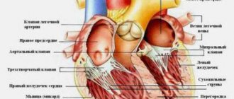
The vital organ consists of the atria and ventricles. Under the influence of automatically occurring shocks, they contract. Their generation occurs at the very apex of the heart, called the sinus node. The impulses pass through all parts of the vital organ, causing synchronous relaxation and tension first of the atria and then of the ventricles.
- Features of conducting and interpreting ECG during pregnancy
During supraventricular extrasystole, the formation of exciting impulses occurs not only in the sinus node, but also outside it. Under the influence of an extraordinary impulse, the heart contracts, which is why the heart muscle does not rest and is not filled with blood at the moment of relaxation.
If the extrasystoles are more than 5-6 per minute, then:
- Redistribution of blood flow.
- Disruption of blood flow to all organs and tissues, development of heart failure.
- Overstrain and exhaustion of the myocardium.
If extrasystoles occur in the sinus node about five times per minute, then this is normal. If an additional lesion appears in the area above the ventricles, pathology is diagnosed.
Although in 90% of cases single extrasystoles are not accompanied by any symptoms, the likelihood of complications developing in the future is high.
Classification and types
There are many types of NLEs, divided according to different characteristics.
Depending on the source of the impulse, atrial extrasystoles and extrasystoles (ES) from the atrioventricular (AV) connection are distinguished. Based on the number, they distinguish between single and double. Three or more ES in a row is already considered an episode of tachycardia (also called a “jog”).
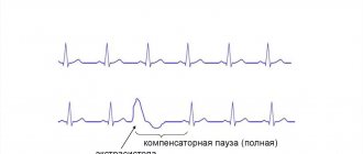
In my patients, I often observe such an ECG phenomenon as allorhythmia - the regular occurrence of extrasystoles. There are the following types:
- bigeminy - the appearance of ES on the cardiogram after each normal contraction of the heart (read more about this phenomenon here)
- trigeminy - after every second complex;
- quadrigeminy - after every third complex.
Depending on the cause, the following types of NVEs are distinguished:
- functional - during physical activity, reflex effects;
- organic - for heart diseases;
- toxic - in case of drug overdose;
- mechanical - for injuries.
Single extrasystoles
The most benign variant of NVE, mainly found in healthy individuals, are single supraventricular extrasystoles. They almost always go unnoticed by humans and do not pose a threat to health.
Treatment
Therapeutic tactics include the following rules for stopping premature contractile activity of the myocardium. First of all, it is necessary to eliminate bad habits, organize a daily routine and rest, normalize the emotional atmosphere, drink coffee and strong tea in moderation.
Prescription of sedatives - valerian, motherwort, lemon balm, mint. It is imperative to adhere to a potassium-containing diet (dried fruits, potatoes, cherries, grapes), and, if necessary, take medications such as Asparkam, Panangin.
Such measures are effective when the arrhythmia is functional in nature and is not a deviation from the norm.
When an extrasystolic disorder negatively affects the patient’s well-being, occurs due to cardiac pathology and risks initiating a paroxysm of tachyarrhythmia, antiarrhythmic therapy is appropriate. For this purpose, the following drugs are used:
- Class 1a (novocainamide).
- Class 2 (beta blockers).
- Class 4 (calcium channel antagonists).
Expert advice
Despite the fact that most often EVEs are relatively harmless, if they occur frequently and are accompanied by symptoms (feelings of freezing, interruptions in heart function, dizziness, feeling of lightheadedness), you should consult a doctor to find out the cause, including examination for cardiac problems. and other diseases. I try to explain to my patients that eliminating the causative factor is of no small importance in the treatment of EVE. Therefore, I give recommendations for lifestyle changes: you need to quit smoking, try to avoid severe stress, and significantly limit the consumption of alcohol and coffee. If a person develops signs of EVE while taking medications, be sure to tell the doctor about it. Reducing the dosage or changing the medication often helps get rid of extrasystoles.
Extrasystole in a child
Deviations in young children and newborns are usually detected during a routine examination on an ECG, since due to their age they cannot describe any of their own sensations.
- Single polymorphic ventricular extrasystole
For babies, the disease does not cause noticeable concern; older children may complain of fatigue, shortness of breath, and rapid heartbeat. In 80% of cases, the disease occurs in adolescence (12–15 years), most often in boys .
A type of such arrhythmia does not cause any particular harm to the child; possible complications are dangerous, especially atrial fibrillation, which causes the cessation of cardiac impulses.
A feature of the disease in children is the occurrence of relapses, when after a long remission the disease reappears.
Why are supraventricular extrasystoles dangerous and what are their consequences?
Extraordinary supraventricular extrasystoles themselves do not pose a threat to human life and often go unnoticed. However, they can provoke the appearance of more severe rhythm disturbances: supraventricular tachycardia, atrial fibrillation and flutter, which lead to a sharp decrease in blood pressure, deterioration of blood supply to the myocardium and an increased risk of blood clots in the heart. A combination is often observed .
Long-term polytopic and blocked ES are considered the most unfavorable.
The consequences of supraventricular extrasystole are determined by the presence of: coronary heart disease, chronic heart failure, etc. The rhythm disturbance itself almost does not cause any complications.
Clinical case
A 33-year-old man came to see me with complaints of rapid heartbeat, periodic sensations of “fading” and interruptions in heart function over the past 3 weeks. He does not take any medications on his own. Doesn't smoke, doesn't drink alcohol. A general examination revealed a high heart rate (105 beats per minute) and increased blood pressure - 140/80 mmHg. Art. During the conversation, I noticed the patient’s uncharacteristic irritability and bulging eyes. When questioning about the presence of diseases in relatives, the man noted that his father suffered from Graves' disease. Holter ECG monitoring was prescribed. Sinus tachycardia, atrial extrasystole of the bigeminy type, and a large number of single extraordinary contractions (967) were detected. A referral was issued to an endocrinologist to check the thyroid gland. On the recommendation of a specialist, an ultrasound examination was performed and blood was taken for hormonal tests. The results obtained: diffuse enlargement of the thyroid gland, decreased TSH levels, increased concentrations of free T4, high titers of antibodies to the TSH receptor. Diffuse toxic goiter was confirmed . Mercazolil therapy was prescribed with subsequent monitoring of hormone levels. To slow down the heartbeat and combat extrasystole, beta-blockers (Bisoprolol) are recommended.