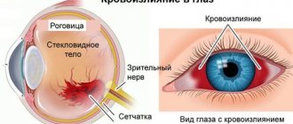
The meaning of the ancient Greek roots from which the term “chorioretinitis” is derived is as follows. The choroid is a network of blood vessels in the posterior wall of the eyeball that nourishes the retinal (retinal) tissue; The ending “-itis” in medical terminology always indicates inflammation. Thus, chorioretinitis is an inflammatory process that extends to both the retina and its vascular system. According to the type of course, chorioretinitis is divided into acute and chronic; by genesis (origin) - congenital and acquired.
Chorioretinitis of the eye
With inflammation occurring in the posterior part of the retina, chorioretinitis develops, which affects the retinal vessels in the posterior part of the eye.
In this area, the vessels form a wide bed, which in turn leads to a slowdown in circulation processes.
Attention! The danger in this case lies in the delay of all infections in this area, and if they penetrate into this area, there is a high probability of damage to the capillaries, and as the disease develops, it spreads to the choroid as a whole.
Chorioretinitis
The diagnosis is based on anamnestic data, the results of instrumental and laboratory research methods. An objective examination does not reveal pathological changes. This is an important criterion that allows you to differentiate chorioretinitis from pathology of the anterior pole of the eyeball. Laboratory diagnostics boils down to:
- Bacteriological culture
. The material for the study is a biopsy of the orbital conjunctiva or conjunctival fluid. The purpose of the method is to identify the pathogen and determine sensitivity to antibacterial therapy. - Enzyme-linked immunosorbent assay (ELISA).
The study of antibody titer (Ig M, Ig G) is used to detect pathogens of chlamydia, herpes simplex, toxoplasmosis, and cytomegalovirus. ELISA allows you to assess the stage of activity of the inflammatory process. - C-reactive protein test
. Detection of protein in the blood makes it possible to exclude or confirm the autoimmune nature of the disease. If the test for C-protein is positive, rheumatic tests are performed.
In order to make a diagnosis and assess the extent of the lesion, the ophthalmologist uses instrumental methods. Using visometry, a decrease in visual acuity of varying degrees of severity is determined with a tendency to the myopic type of refraction. An increase in intraocular pressure (IOP) is observed only in moderate and severe cases. Specific diagnostics include:
- Gonioscopy
. An accumulation of pus is detected in the anterior chamber of the eyeball, which indicates hypopyon or exudate. Hemorrhage into the anterior chamber of the eye leads to hyphema. - Ophthalmoscopy
. During an ophthalmoscopic examination, lesions of a grayish-yellow hue with unclear contours and pinpoint hemorrhages are visualized. Detection of a limited area of white color indicates atrophy. The macula area is pigmented. - Fluorescein angiography of the retina (FA)
. It is possible to visualize signs of retinal vasculitis. When conducting FA with contrast, dark spots are revealed at the site of accumulation of indocyanine green. - Perimetry
. In the peripheral form of the disease, there is a concentric narrowing of the visual field. Focal damage leads to the loss of small areas from the field of view.
Differential diagnosis
Differential diagnosis is carried out with macular degeneration and malignant neoplasms of the choroid. Unlike a tumor, chorioretinitis reveals a perifocal focus of inflammation with blurred ridges. With dystrophic changes in the macula, there are no signs of inflammation and opacification of the vitreous body. If the origin of the disease is traumatic, radiography of the orbit is performed, which makes it possible to identify pathological changes in the retrobulbar tissue and bone walls of the orbit (fracture, displacement of debris).
Symptoms of the disease
Symptoms of chorioretinitis can vary depending on the type and severity of the disease. In most cases, the patient exhibits the following symptoms:
- a sharp decline in the quality and acuity of vision;
- the appearance in a person’s field of vision of such visual interference as “sights” and dots;
- the presence of flashes before the eyes , regardless of whether the eyes are exposed to light sources or whether the person is in complete darkness;
- a sharp drop in the quality of vision with the onset of darkness.
Despite the presence of such subjective symptoms, there are no physiological changes in the anterior part of the eyeball , which can cause difficulties in diagnosis.
Important! With a timely visit to a specialist and a correct history, in most cases it is possible to establish not only the presence of the disease itself, but also to determine its form.
Symptoms
The clinical picture of chorioretinitis significantly depends on the location of the inflammatory focus. Thus, central chorioretinitis is characterized by predominant damage to the macula (“macula”, the most light-sensitive and specialized area of the retina, responsible for the clarity of the central field of vision), equatorial and peripheral. There are also several options based on the nature of the focality: focal, multifocal (disseminated, divided into several separate foci) and diffuse, involving the entire retinal tissue without clear focal boundaries. Acute chorioretinitis can last up to three months, chronic chorioretinitis has a tendency to frequent relapses.
The content of subjective complaints is also determined by the localization of the inflammatory process. Thus, peripheral chorioretinitis often does not manifest itself at the level of subjective sensations at all - and in this case it is diagnosed by chance, during a consultation for another reason or during a medical examination. Macular chorioretinitis, on the contrary, manifests itself with multiple and varied visual disturbances: “fog before the eyes”, scotomas (blind or dark areas in the visual field), photopsia (illusory sparks or flashes of light), a noticeable decrease in the acuity and quality of vision. “Night blindness” is quite typical, as well as a distorted perception of the size and shape of objects observed by the patient.
Symptoms of this kind, no matter what the final diagnosis turns out to be, require immediate attention to an ophthalmologist.
Diagnostic methods
In order to accurately diagnose chorioretinitis, a specialist must conduct a series of examinations, which include the following procedures:
- Biomicroscopy - allows you to identify pathologies and transformations occurring in the vitreous body.
- Initial examination for visual acuity impairment. In this case, correction using traditional optical methods is ineffective.
- Ultrasound of the eyeball - performed to determine the degree of clouding of the lens of the eye.
- Computer perimetry . Designed to detect the presence of “blind spots”, as well as dark spots in front of the eye, due to which the sensitivity of the retina may decrease.
- Electroretinography is a procedure to determine the functional abilities of the retina.
- Refractometry. A control procedure, the indicators of which, in the presence of a disease, should not change in one direction or another.
- Coherence optical tomography of the retina - to identify the causes of the disease.
- Fluorescein angiography. This event is necessary to identify changes in the vascular system of the fundus.
- Research in transmitted light.
Need to know! Also, if chorioretinitis is suspected, the patient undergoes general urine and blood tests and is examined for hepatitis, HIV, herpes, toxoplasmosis and cytomegalovirus.
It is necessary to additionally do fluorography and the Mantoux reaction.
If the disease is not ophthalmological in nature, additional examinations may be necessary from other specialists (venereologist, dentist, phthisiatrician, pediatrician, immunologist, ENT).
Our doctors who will solve your vision problems:
Fomenko Natalia Ivanovna
Chief physician of the clinic, ophthalmologist of the highest category, ophthalmic surgeon. Surgical treatment of cataracts, glaucoma and other eye diseases.
Yakovleva Yulia Valerievna Refractive surgeon, specialist in laser vision correction (LASIK, Femto-LASIK) for myopia, farsightedness and astigmatism.
You can find out the cost of a particular procedure or make an appointment at the Moscow Eye Clinic by calling in Moscow 8 (499) 322-36-36 (daily from 9:00 to 21:00) or using the ONLINE REGISTRATION FORM.
Causes of the disease
Among the factors that can provoke pathology, the main ones are the following:
- penetration of infections into the organs of vision ;
- the presence of herpes virus and HIV in the body;
- autoimmune pathologies;
- eye injuries (regardless of their origin);
- complications resulting from myopia ;
- allergic reactions;
- exposure to radioactive radiation .
Pathology can also occur as one of the complications of influenza.
Causes
The main factors under the influence of which the retinal-vascular complex can become inflamed include:
- local and systemic infections caused by bacterial or viral pathogens (toxoplasmosis, herpes, and many others); the most common local primary sources of chorioretinitis are untreated infectious foci in adjacent organs - the nasopharynx and oral cavity;
- radiation (radiation) injury;
- intoxication, incl. products of decay of one’s own tissues (in particular, long-term non-resorbable blood after intraocular hemorrhage);
- allergic reactions;
- autoimmune disorders and diseases;
- immune deficiency (AIDS, exhaustion of various origins, recovery period after a major operation, etc.);
- ophthalmotrauma.
Treatment
The main methods of treating chorioretinitis are the following procedures:
- Immunotherapy with immunosuppressants (immunostimulants can be used as an alternative).
- Laser coagulation of the retina.
- Hemodesis (detoxification procedures).
- Injections directly into the eyeball .
- A course of taking anti-inflammatory drugs .
- Antibacterial drugs that should stop the root cause of the disease (antibiotics of the penicillin group are mainly used for this).
- Antiviral drugs.
- Treatment with physiotherapeutic methods (the most effective is electrophoresis ).
Note! The use of traditional medicine methods for chorioretinitis is recognized as ineffective and is unacceptable either as the main treatment or as a means of helping to relieve symptoms.
Moreover, such methods can only aggravate the situation, provoking the development of inflammatory processes.
Chorioretinitis: causes
Among the causes of chorioretinitis, the following conditions can be identified, which are mainly associated with general diseases of the body:
- Infectious diseases (tuberculosis, syphilis, herpes, cytomegalovirus infection, toxoplasmosis), including local ones (ENT organs, oral cavity)
- Exposure to radiation
- Toxins (with prolonged hemophthalmia, destroyed blood cells can be toxic)
- Allergy
- Autoimmune disorders
- Sarcoidosis
- Immunodeficiencies (in HIV carriers, weakened people, during recovery after long-term treatment)
- Penetrating eye injuries.
Possible complications
If not treated in a timely manner using inappropriate medications, chorioretinitis can lead to the following complications:
- retinal hemorrhages with the possibility of subsequent relapse;
- neovascular membrane formation;
- total blindness;
- retinal detachment of varying severity;
- retinal vascular thrombosis
- formation of atrophic foci .
With diffuse chorioretinitis, which involves large foci of inflammation, connective tissue may form at the bottom of the eyeball, the appearance of which reduces visual acuity.
Symptoms of chorioretinitis
Regardless of the type of chorioretinitis diagnosed, all patients exhibit the following symptoms:
- The quality of vision is greatly reduced (this happens abruptly and for no particular reason);
- the patient sees a large number of black dots before his eyes;
- a person sees as if a plastic bag was covered from above the eye;
- disorientation in space (a person thinks that he is moving in one direction, but is actually heading in the opposite direction);
- decreased visual acuity with the onset of darkness (sometimes it is even impossible to distinguish one object from another);
- blind spots appear in a person’s field of vision;
- the visibility of colors is impaired (the patient cannot distinguish between them);
- the patient cannot understand what shape and size the object is;
- more acute stages are characterized by the fact that most objects are seen by a person as one large black spot.
Classification
Classification according to etiology:
- infectious;
- allergic;
- systemic;
- post-traumatic;
- combined.
Infectious chorioretinitis of the eye is caused by the activity of microorganisms - fungi, viruses, bacteria. Allergic pathology occurs as a result of intolerance to any substance or ingredient. Systemic pathology develops against the background of internal diseases of a systemic nature. Post-traumatic pathology is a consequence of trauma to the visual apparatus. Combined chorioretinitis is a tandem of several causes.
Classification according to the localization of inflammation:
- central serous chorioretinitis;
- peripapillary;
- equatorial;
- peripheral.
Central chorioretinitis affects the macula - the yellow spot of the retina, peripapillary is located next to the optic nerve, equatorial - located around the circumference of the eyeball, peripheral is located next to the dentate line of the eye.
Classification according to the number of foci of inflammation:
- one area is focal;
- several areas - multifocal;
- several foci of inflammation are combined into one - diffuse.
Classification according to the nature of the course:
- active form;
- inactive;
- subactive.
The active form is characterized by deterioration in the quality of visualization and rapid eye fatigue. In the inactive form of the disease, there is a deterioration in the quality of visualization in the absence of symptoms of the inflammatory process; the pathology is detected during hardware diagnostics. The subactive form is detected during the diagnosis of other diseases, since it does not manifest itself in anything.
Chorioretinitis can also be acute or chronic. There are two ways of infection: endogenous and exogenous. The endogenous path - inflammation forms directly in the structure of the eye, the exogenous - passes from neighboring tissues.
The endogenous form of the pathology occurs due to mechanical injury to the eyeball, while the exogenous form is a consequence of systemic or autoimmune pathologies.
Central serous chorioretinitis
Central serous chorioretinitis leads to retinal separation. Blood plasma accumulates between it and the adjacent membranes of the eyeball. The pathology is caused by the fragility of the capillaries and their high permeability. Dangerously severe complications, requires treatment by an ophthalmologist. Self-treatment with folk remedies or pharmacy products is unacceptable. In chronic and subacute cases of the disease, laser surgery is required.
Toxoplasmosis
Toxoplasmosis chorioretinitis can appear with reduced immunity, and an infant can also become infected with it during the prenatal period of its development. With this form of the disease, the central nervous system, internal organs, and visual organs are affected. Retinal detachment, retinal hemorrhage, and the growth of additional capillaries may occur.
Tuberculous
Tuberculous chorioretinitis in its initial form proceeds completely unnoticed and is discovered randomly during an examination by an ophthalmologist. With secondary tuberculosis, the clinical picture changes and occurs with vivid symptoms. This pathology is dangerous due to serious complications: it leads to cataracts, damage to the optic nerve, and retinal detachment.
Syphilitic
The syphilitic form of the disease appears either in the prenatal period or in adulthood. The disease progresses brightly: hemorrhage occurs in the retina, atrophic changes in tissues form. If the pathology develops in adults, it is characterized by a diffuse form of changes.
Treatment of chorioretinitis
Surgery is considered one of the treatment options for eye chorioretinitis, because only with its help it is possible to completely get rid of the inflamed area and fight the disease that caused the infection.
In order for the operation to be successful, the patient is preliminarily prescribed the following treatment:
- anti-inflammatory drugs (non-steroidal). More often these are drops in the eyes, which must be applied after 3-4 hours;
- antibiotics - help destroy the original source of infection; it is also advisable to take after surgery to reduce the risk of developing infection in the operated area of the eye;
- hormonal drugs (can be prescribed in the form of installations or hydrocortisone injections). The injection is made directly into the lower part of the eye. In this case, it is additionally necessary to use hydrocortisone ointment;
- mydriatics (help prevent fluid accumulation in the eye and reduce the risks of developing congestive processes and glaucoma);
- biostimulants and reparants (help restore and regenerate affected areas of visual structures);
- physiotherapy (recommended for acute and subacute course of the disease, as well as for chronic types of chorioretinitis). It helps speed up the recovery process after surgery.
It is also necessary to prescribe eye drops that have the ability to dilate the pupil.
If chorioretinitis was a consequence of an injury, then surgical intervention is necessary.
In the absence of timely treatment, the result is atrophy of the retina and choroid, which leads to complete blindness.
{banner_horizontalnyy3}
Folk remedies
The use of tinctures and decoctions can speed up the healing process. It is recommended to use vasodilators and vitamins. This will complement the conservative course of treatment, but will not replace it.
An infusion of hazel bark is a vasodilator. The crushed bark is steamed with boiling water, wrapped and allowed to rest for about an hour. 10 g of crushed bark is consumed per glass of water. The product is taken orally in a teaspoon several times a day.
To strengthen the immune status, take vitamins. Riboflavin, folic acid, and retinol have a good effect. You need to eat fresh vegetables and fruits: squeeze juice out of them, stew them. If the pharmacy does not have riboflavin, it can be found in fermented milk products.
Prevention is regular examination by an ophthalmologist, timely treatment of ENT diseases and elimination of infection.
Prevention of chorioretinitis
To reduce the risk of developing chorioretinitis, each person must adhere to several rules of prevention, which are as follows:
- treat infectious diseases in a timely manner (especially acute respiratory viral infections);
- in case of a sharp decrease in vision, immediately consult an ophthalmologist;
- do not stay in the cold for a long time;
- compliance with personal hygiene rules (especially hand washing);
- undergo routine medical examinations annually.
In particular, you need to visit the dentist regularly. Advanced caries can provoke inflammatory and purulent processes in the oral cavity, which can subsequently spread to the visual structures.
The prognosis of chorioretinitis with timely diagnosis and adequate treatment is favorable in most cases.