A blood test is the most common type of laboratory test, used for almost all diseases. The accuracy of the results depends not only on the patient’s preparation for the procedure, but also on the blood sampling technique. Taking blood from a vein in the traditional way, that is, using a conventional syringe, is associated with the following difficulties:
- duration of the procedure;
- blood reacting with the environment;
- blood passing through the needle twice;
- blood clotting in the needle;
- destruction of red blood cells;
- difficulties in maintaining the ratio of the amount of blood and reagent;
- blood getting on the lab technician’s hands;
- damage to tubes, blood spillage.
In this regard, distortion of results is possible, which means errors in diagnosis and treatment, not to mention the risk of infection of medical staff. Today, in laboratory practice, vacuum systems – vacutainers – are increasingly used for blood collection. This method is convenient, safe and provides more accurate test results.
Advantages of the new method
Vacuum systems have a number of significant advantages over the conventional method of collecting venous blood. Among them:
- safety and comfort for both the patient and the laboratory assistant;
- less severe pain;
- vacuum tubes are sealed and do not break;
- procedure time – about 10 seconds;
- exact observance of the ratio of blood volume and reagent;
- in a short period of time you can collect material into several tubes, without the need to insert the needle into the vein again;
- reliable and simple marking of test tubes: for each type of analysis - a test tube with a lid of a certain color, which eliminates errors when choosing it;
- ease of transportation and centrifugation;
- the system is completely closed, there is no air access to the blood;
- no need to pour the resulting material into other containers;
- no direct contact with blood;
- no need to open the tube cap when working with analyzers;
- the possibility of an individual approach to patients: the kit includes a set of different needles that are used depending on the condition of the veins;
- simplicity of design and use;
- savings on reagents and tube disposal.
Benefits of using negative pressure systems
All the benefits of negative pressure systems come from their design. Their use allows:
- completely eliminate contact of medical personnel with the patient’s blood during sampling;
- standardize the process of blood collection and sample preparation, create a simple algorithm of actions;
- reduce the number of operations spent on preparing a sample for testing in the laboratory;
- Primary tubes included in negative pressure systems can be used directly in many automated analyzers. This saves money on purchasing secondary plastic tubes and time on transferring samples into them;
- make the transportation and centrifugation of biomaterials safer, since the tubes are sealed and made of unbreakable materials;
- facilitate the identification and labeling of samples by type of examination, thanks to color coding of the caps of negative pressure systems;
- reduce laboratory material costs for the purchase and processing of additional secondary tubes;
- simplify staff training methods;
- reduce the occupational risk of infection;
- reduce the time spent collecting venous blood using the method discussed in the article.
Multi-colored test tubes made from high-strength modern materials ensure the safety of medical staff working with blood
What is the system?
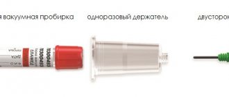
The closed system includes a vacuum tube with a lid, a double-sided needle, and a needle holder. Depending on the purpose, they are filled with appropriate reagents. The complete set of the system may vary depending on the type of analysis, the condition of the patient’s veins, and the experience of the medical worker.
The vacuum system includes double-sided needles that differ in length and diameter
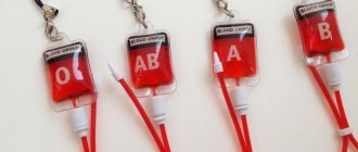
Vacuum transparent test tubes are made of plastic. The lids consist of a plastic body and a rubber stopper. They ensure tightness and sterility and maintain a vacuum state for up to two years. To distinguish test tubes for different purposes, the plastic body has a certain color, depending on the composition of the filler: red, green, blue, purple, black, gray. There is an international standard for color coding of reagents, which all colors used in vacuum systems must comply with.
- Tubes with a red cap contain a clotting activator or have no filler. They are intended for biochemical analysis, bacteriological, immunochemical, and for determining blood group.
- The EDTA tube has a purple cap. Designed for general analysis, gene diagnostics, immunochemistry.
- The blue cap indicates that the test tube contains sodium citrate. Used to test coagulation.
- A test tube with a green cap contains heparin. With its help, biochemical and immunochemical analysis is carried out.
- The black cap indicates that the reagent is sodium citrate, and it is intended for determining ESR.
- A test tube with a gray cap contains a glucose stabilizer and an anticoagulant, used to determine sugar levels.
To draw blood, depending on the patient’s veins, needles of different types and sizes (length and diameter) are used. Standard double-sided needles are usually used. One side of the needle is inserted into the patient's vein, the other side pierces the elastic stopper of the tube. In addition, butterfly needles and standard luer-lock needles can be used. In this case, a luer adapter is required. A butterfly needle equipped with protrusions is used for thin and hard-to-reach veins.
There are three types of holders: standard, extended and automatically resetting the needle.
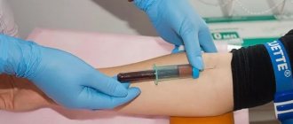
The vacutainer has the same operating principle as a regular syringe. Blood is drawn by creating a vacuum in the test tube, resulting in a pressure difference that acts as a piston.
Vacuum systems make venous blood collection comfortable and safe for physicians and patients
Venous blood collection
See Necessary equipment for collecting venous blood
Preparing the patient for the blood donation procedure
- Preparation may vary depending on the analysis. For each specific analysis, if necessary, information can be obtained from the laboratory.
- Blood sampling occurs in the morning before 12.00, preferably between 8.00-10.00.
- Before the blood collection procedure, the patient must remain awake for at least 1 hour.
- On the day before the test, the patient can drink and eat as usual, but should only limit the consumption of alcohol, coffee and fatty foods.
- At least 10-14 hours should pass between the last meal and fluid intake and the blood draw. If necessary, you can drink no more than one glass of water without any additives.
- Before drawing blood you should avoid: Excessive physical and emotional stress
- Taking medications (if possible)
- Alcohol consumption
- Smoking
It is important to know
- The vacuum tube should be filled strictly to the specified volume to ensure the correct ratio of blood and additive - the blood in the tube should not be less than the specified volume (dilution effect) or more (danger of clotting).
- When drawing blood, you should hold the tube in such a position that the blood flows along the wall of the tube. If blood splashes onto the bottom of the tube and forms foam, it may promote hemolysis.
- Test tubes containing an anticoagulant must be turned upside down immediately after filling so that the blood mixes with the anticoagulant - at least 4 times when taking blood clotting tests, in all other cases 8 times. The air bubble should move from one end of the tube to the other. The test tube must not be shaken or shaken!
- If blood does not flow into the tube or the flow stops before the tube is filled, the cause may be a failed venipuncture or the vessel wall being sucked into the needle hole.
- Solution: change the position of the needle - insert the needle a little deeper, pull it back, change the angle at which the venipuncture was performed. If venipuncture is unsuccessful, another blood vessel should be punctured. Repeated puncture of the same vessel should be avoided.
- If there is enough blood in the yellow cap tube (serum tube) for testing, but the vacuum tube is not filled to the required volume, hemolysis may occur due to the remaining vacuum in the tube. To avoid this, quickly remove the stopper from the test tube to allow air to enter the test tube and close with a new stopper. The remaining tubes cannot be used if they are insufficiently filled.
- When drawing blood, do not rub or pat the intended puncture site. Excessively strong hand pumping can cause hemolysis and hemoconcentration.
- After disinfecting the skin, it should be allowed to dry so that particles of the disinfectant, which promote hemolysis, do not enter the test tube through the needle.
- Apply a tourniquet approximately 10-12 cm up from the puncture site. The tension of the tourniquet should be such that a finger can be placed between the tourniquet and the skin.
- Immediately loosen the tourniquet, making sure that blood flows into the tube.
- When drawing blood, it is important that the time the tourniquet compresses the blood vessels is minimal. Keeping the tourniquet in place for up to 1 minute does not particularly affect routine studies performed on blood serum.
- As a rule, blood is drawn in the elbow from the central vessel. For the patient, the elbow is less painful to pierce with a needle because the blood vessel is close to the skin. Alternative sites for venipuncture may include the inner forearm, wrist, and dorsum of the hand.
- Venipuncture/blood sampling is not performed: from an extensive scar surface, from a wound surface (burn wound), from an edematous surface, from a surface with a hematoma, from a cannulated vein, from a limb (arm) with a fistula for dialysis.
Venous blood collection procedure
1. Identify the patient. 2. Complete/check directions. 3. Find out whether the patient followed the prescribed diet and whether he is allergic to substances contained in the disinfectant for cleansing the skin at the venipuncture site. 4. Provide the patient with a comfortable and suitable position for drawing blood - the patient’s arm should be extended so that the arm forms a straight line from the shoulder to the wrist. 5. Wear gloves. 6. Prepare the necessary test tubes - shake them lightly to remove possible droplets of additives from the stopper. The test tubes will be installed in the required order. 7. Select a puncture needle of the required diameter and make sure that the needle is stable at the attachment point. Make sure that the protective cap does not interfere with vein puncture. 8. Select the puncture site. Ask the patient to clench his hand into a fist so that the veins are better visible. 9. Clean the site of the intended puncture with a disinfectant and allow the skin to dry. 10. If necessary, apply a tourniquet to your arm. 11. Remove the protective cap from the needle. Check whether the needle hole is free. 12. Perform a vein puncture - to do this, fix the vein with your finger slightly lower from the puncture site and slightly stretch the skin so that the vein does not move; Use a needle to pierce the vein wall at an angle of 15-30 degrees, the hole of the needle should be directed upward. 13. When drawing blood, make sure that the stopper of the tube is located higher and the bottom lower, to avoid substances from the test tube getting into the needle. 14. Holding the needle attachment point firmly with one hand, place the vacuum tube as close as possible to the needle attachment point so that the tip of the needle, covered with a latex cap, passes through the tube stopper. 15. The test tube should be held so that the blood flows down the wall of the test tube. 16. Immediately loosen the tourniquet, making sure that blood flows into the test tube. 17. When the test tube is filled with the required amount of blood and blood stops flowing into the test tube, remove the test tube from the mount and, if necessary, place the next test tube there. 18. Immediately after filling, the test tube with additives should be turned upside down and back again 4-5 times with calm movements so that the blood is mixed with the additives. 19. Place a dry cloth at the puncture site and remove the needle from the vein. 20. After use, immediately close the needle with a protective cap. To do this, you need to push the protective cap onto the needle with your finger. 21. Press the puncture site with a napkin for 3-5 minutes, the patient’s arm should be straightened. If necessary, apply a bandage to the puncture site. 22. Label the test tubes. 23. With calm movements, turn the test tube upside down and back again 4-5 times. Place the test tubes on a stand in a vertical position. 24. Dispose of the used needle and needle holder in an appropriate waste container.
How is it carried out?
- The needle is opened immediately before blood is drawn.
- The cap is removed from the needle on the rubber membrane side, the needle is inserted into the holder and turned until it stops.
- The holder is in the right hand, while the needle cannula is held with the index finger. Vacuum test tube - in the left hand.
- The skin and vein are pierced with a needle. You need to pay attention to the cannula located between the holder and the needle. If the needle is in a vein, blood will appear in the cannula.
- The test tube is inserted all the way into the holder from the inside. At the same time, an elastic membrane is pierced in its lid. Blood begins to flow into the test tube, thanks to the vacuum created in it.
- The required amount of material is collected, the test tube is removed from the holder.
- If it is necessary to take blood several times, insert the following tube into the holder, observing the order: biochemistry, prothrombin analysis, general analysis.
- When the blood sampling is completed, the needle is removed from the vein, and the injection site is clamped with a cotton swab soaked in alcohol.
You can tell the purpose of the test tube by its appearance
How to prepare for the procedure?
The blood collection algorithm requires special preparation for the event. The reliability of the analysis results is influenced by the following factors:
- time of collection of biological fluid;
- food intake, the nature of foods in the diet;
- drinking alcoholic beverages, smoking;
- taking medications;
- physiotherapy;
- intense physical activity;
- stressful situations;
- instrumental diagnostic methods (MRI, ultrasound, x-ray);
- cyclical changes in a woman’s body (mensis).
Before taking blood from a vein, you should adhere to general rules that will increase the efficiency of the study and minimize the risk of obtaining false results.
- Blood is donated on an empty stomach in the morning (8.00 – 11.00). You can drink water without carbon dioxide.
- On the eve of the examination, it is not recommended to overeat, eat salty, spicy, or fatty foods.
- The day before the test, avoid drinking alcohol.
- It is necessary to submit biomaterial before undergoing instrumental examination and physiotherapeutic treatment.
- Coordinate the discontinuation of medications with your doctor.
- One hour before the examination, you should not smoke; it is necessary to exclude stressful situations and physical overexertion.
Repeated blood tests to monitor indicators over time should be carried out under the same conditions (time, diet) and in the same laboratory, since the blood sampling algorithm, study methods and reference values (norms) may differ significantly in different medical institutions.
Classification
Vacuum test tubes differ in the following characteristics:
- size (length and diameter);
- volume;
- purpose (hematological, biochemical analysis, blood clotting test, etc.);
- reagent filling the test tube;
- sample volume;
- color;
- cover type.
By filling, the test tubes are distinguished as follows:
- containing sodium citrate – intended for coagulological reactions;
- containing sodium citrate - to determine ESR;
- with EDTA K-2;
- with EDTA K-3;
- containing a blood clotting activator and gel;
- no filler.
Description of the vacutainer
The BD Vacutainer is a disposable device designed specifically for collecting blood from a vein. The device also has another name: vacutainer. The set consists of:
- test tubes with a tight screw cap;
- sterile disinfected needle;
- adapter holder.
To collect, transport and preserve biological material, a container is used, inside of which there are various reagents and a vacuum. Excipients are necessary to obtain the desired material, namely serum, plasma and whole blood. The lid body is plastic and unbreakable, and the stopper is made of rubber. Air is not able to penetrate inside; the biomaterial is reliably protected. There are standard holders, elongated ones, and those that automatically reset the needle. The tubes have markings that indicate what each one is for.
Price
The price of vacuum tubes depends on the material of manufacture, volume and filling. Their approximate cost is:
- with sodium citrate - from 7.5 to 11 rubles per piece, depending on the volume;
- with EDTA K-2 and EDTA K-3 – from 7.5 to 10.5;
- with a coagulation activator – from 7.5 to 8.5;
- with coagulation activator and gel – from 10.5 to 13.5.
A standard double-sided vacuum needle costs about 6 rubles, the same price for a needle with a transparent cannula and a butterfly needle with a luer adapter. The cost of a needle holder is about 4 rubles per piece.
Main advantages
Unlike a disposable syringe, a vacutainer has many advantages, the most important thing is that the system guarantees the accuracy of the study.
In this case, there is no need to transfuse biological material, so its contact with hands or air is excluded. The danger of infection to the laboratory assistant is eliminated. The analysis is carried out directly from the device. There are different types of vacutainers that are used for various laboratory tests. All test tubes are numbered, so it is impossible to replace or mix them up. The vacuum timer system is made of plastic that does not break. The procedure takes 10-15 seconds. The patient's veins are not injured. Attention! If you need to carry out more than one examination at once, then you just need to change the container.
Blood collection scheme
Taking blood using a vacuum system is almost no different from the procedure performed with a simple syringe. Comfort and safety is explained by the fact that when the biomaterial flows into the test tube, a volumetric vacuum is formed. It allows you to take the required amount of exclusively sterile biomaterial. When working with the device, it is important to adhere to the correct scheme. First, the nurse unseals the vacutainer and removes the cap from the disposable needle.
Possible mistakes
When a medical professional takes venous blood for analysis, he may unwittingly make some mistakes. Sometimes the biomaterial does not go into the test tube after it has been connected to the needle holder. Often the reason is that the needle did not enter the vein. You need to change its position; you don’t need to pull it out completely. Sometimes this happens because the end of the needle is stuck into the vein wall. Needs to be corrected carefully. If the puncture is made through the entire vein, then blood will also not flow into the test tube. Here, too, the situation is being carefully corrected.
In all these situations, the container is not disconnected from the adapter (holder). If less than the required amount of biomaterial enters the vacuum tube, the reason may be due to air penetration into the device. You will need to do the job a second time using a new vacutainer. Low pressure may develop in the vein, causing it to collapse. You should disconnect the tube from the holder and wait until the vein is filled with blood.