Analysis of urine
Cancer of the urinary system manifests itself as blood in the urine.
Urine may also contain ketone bodies, which indicate tissue breakdown. However, these symptoms also accompany diseases not related to oncology, for example, they indicate the presence of stones in the bladder or kidneys, or diabetes mellitus. For diagnosing other types of cancer, urine analysis is not informative. It cannot be used to judge the presence of cancer, but deviations from the norm indicate health problems. If the deviations are serious and confirmed by the results of other basic tests, then this is a reason to conduct special tests to determine cancer.
The exception is multiple myeloma, in which specific Bence Jones protein is determined in the urine.
For the study, morning urine is collected in a sterile container, which can be purchased at a pharmacy. You need to take a shower first.
What does ESR show?
ESR is not a specific indicator for any particular disease, that is, it is impossible to establish a specific diagnosis based on its increase.
This test is considered useful for identifying hidden forms of various diseases and determining the activity of chronic inflammatory conditions. ESR can also serve as an indicator of the effectiveness of therapy.
However, measuring ESR is in no way used to diagnose cancer.
Stool analysis
Blood may also be present in the stool, and it is almost impossible to notice it visually. Laboratory analysis will help determine its presence.
The presence of blood in the stool is a sign of intestinal cancer (most often colon), but it is also a symptom of many benign gastrointestinal diseases. Polyps in the intestines can bleed. Moreover, it should be remembered that polyps tend to degenerate into a malignant tumor. In any case, the presence of blood in the stool is a reason to undergo a more in-depth diagnosis and take tests to detect cancer.
Feces are also collected in a sterile container in the morning.
What does ESR depend on?
If you take a flask, pour blood into it and then add an anti-clotting agent inside, the liquid will separate over time. Pure plasma will remain on top, and at the bottom there will be sediment, mainly consisting of red blood cells. This occurs under the influence of gravity due to the difference in weight.
The normal ESR level for an adult woman is in the range from 2 to 15 mm/h, for a man - from 2 to 12 mm/h. In a newborn child, regardless of gender, this figure is around 1-2 mm/h.
What blood test shows cancer?
Many patients are convinced that it is possible to detect cancer using a blood test. In fact, there are several types of this diagnostic procedure, starting with a general analysis and ending with an analysis for tumor markers. The following types of cancer diagnosis using blood tests with varying degrees of information content are distinguished:
- general analysis;
- biochemical analysis;
- blood clotting test;
- immunological blood test (for tumor markers).
Even if cancer has not yet manifested itself with painful symptoms, negative changes are already occurring in the body, which can be recorded by a blood test. When a malignant tumor grows, it destroys healthy cells that help the body grow and releases toxic substances. These changes are noticeable even with a general blood test, but they can also be a sign of dozens of diseases not related to cancer.
The most informative is considered to be an analysis of tumor markers - specific substances that are released into the blood as a result of the vital activity of tumor cells. However, given that tumor markers are contained in the body of any person, and their number increases during inflammation, this analysis does not 100% prove the presence of cancer. It only becomes a reason to undergo more reliable tests to determine oncology.
Timely help is the key to successful recovery
By consulting a doctor in time, you can successfully resolve the problem and get rid of the disease. Oncology specialists are ready to help you. Doctors note that children’s bodies tolerate chemotherapy better and it is easier for them to recover after all procedures, which increases the chances of recovery.
The center's oncologists will advise, provide timely assistance, and select the correct course of treatment. The medical staff will find an approach to the child and help him quickly rehabilitate after the course of treatment.
Among the main advantages of oncological treatment it is necessary to highlight:
- world standards of treatment;
- increased life expectancy after diagnosis;
- social adaptation;
- rehabilitation.
When the first signs appear, you cannot do without the help of a qualified doctor. He will help determine the stage of cancer, the size of the tumor, advise parents, prescribe an examination, and conduct treatment. The entire course of treatment takes place under the strict supervision of doctors; the center has all the necessary conditions for this. You can make an appointment with a pediatric oncologist by calling the phone number listed on the website. You can also order a call back or use the form on the website by sending a request to a center specialist.
Will a general blood test show cancer?
This analysis does not provide complete information about the presence of a tumor in the body. However, this is one of the basic tests that helps detect cancer at an early stage, when it does not yet manifest symptoms. Therefore, if you decide which tests to take to check for cancer, then you need to start with it.
The following changes in the structure of the blood may indicate malignant processes in the body:
- decrease in the number of lymphocytes;
- increase or decrease in the number of leukocytes;
- decrease in hemoglobin;
- low platelets;
- increased erythrocyte sedimentation rate (ESR);
- increase in the number of neutrophils;
- presence of immature blood cells.
If a patient, in the presence of one or several of the listed signs at the same time, experiences weakness, quickly gets tired, loses appetite and weight, it is necessary to undergo a more detailed examination.
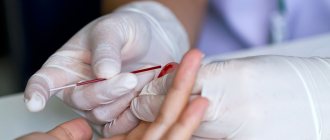
Blood is donated on an empty stomach or at least 4 hours after eating. The sampling is carried out using a finger.
How to calculate the individual ESR norm in elderly patients?
The easiest way is to use Miller's formula:
For example, the permissible ESR limit for a 60-year-old woman is: (60 years + 10): 2 = 35 mm/hour
When changes are detected in a clinical blood test, the first thing the patient does is go to a general practitioner. A useful point is that ESR is included in the Clinical Blood Test, which means that the doctor simultaneously sees the level of leukocytes, platelets, and hemoglobin. When making a diagnosis, the doctor first chooses between three groups: infections, immune diseases and conditions, and malignant diseases. The doctor interviews and examines the patient, after which, based on symptoms, examination and diagnostic data, he determines further tactics.
If the reason for the increase in ESR has not been identified, the analysis should be repeated after 1-3 months. Normalization of the indicator is observed in almost 80% of cases.
Blood chemistry
The method identifies abnormalities that may be a sign of cancer. It should be taken into account that the same changes are characteristic of many non-oncological diseases, so the results cannot be interpreted unambiguously.
The doctor analyzes the following indicators:
- Total protein. Cancer cells feed on protein, and if the patient has no appetite, then its volume is significantly reduced. In some cancers, the volume of protein, on the contrary, increases.
- Urea, creatinine. Their increase is a sign of poor kidney function or intoxication, in which protein in the body is actively breaking down.
- Sugar. Many malignant tumors (sarcoma, cancer of the lung, liver, uterus, breast) are accompanied by signs of diabetes mellitus with changes in blood sugar levels, since the body does not produce insulin well.
- Bilirubin. An increase in its volume may be a symptom of malignant liver damage.
- Enzymes ALT, AST. Increased volume is evidence of a possible liver tumor.
- Alkaline phosphatase. Another enzyme, an increase in which may be a sign of malignant changes in bones and bone tissue, gall bladder, liver, ovaries, and uterus.
- Cholesterol. With a significant decrease in volume, liver cancer or metastases to this organ may be suspected.
Blood is drawn from a vein. It must be taken on an empty stomach.
Symptoms of oncology in children
Common symptoms of the first signs of cancer in children include:
- fatigue without good reason;
- irritability (the child constantly cries, is capricious for no reason);
- the skin becomes pale (a feeling of intoxication occurs);
- poor sleep, development of insomnia;
- increase in body temperature (it will stabilize itself, then rise sharply and fall again);
- nausea and vomiting;
- enlarged lymph nodes.
The child will be afraid of everything, stop communicating with strangers, and become withdrawn. Each type of disease has its own symptoms, let’s look at the main ones.
Leukemia
If the level of certain cells in the blood decreases, this leads to pathological growths of the bone marrow. Symptoms:
- poor appetite;
- irritability;
- the skin becomes pale;
- weight loss;
- dyspnea;
- vomit.
This problem occurs in up to 35% of children. Some patients experience loss of coordination, symptoms are accompanied by bleeding and enlarged lymph nodes. Redness and hematomas may appear on the skin.
Brain tumor
The cerebellum and brainstem are the first to be at risk. Symptoms:
- headache;
- loss of coordination;
- insomnia;
- visual impairment;
- hearing impairment.
The child’s handwriting changes, especially headaches, which torment the baby in the morning. Movements such as rubbing the head against the wall may appear, as the baby tries to get rid of the discomfort. Severe headaches occur when tilting your head back and forth. The disease is also accompanied by convulsions, hallucinations, and vomiting in the morning.
Nephroblastoma
Kidney damage is typical for children under 3 years of age. Symptoms:
- hard breathing;
- increased body temperature;
- loss of appetite;
- the appearance of pain.
Parents can feel changes in the area of the celiac cavity themselves; when the first signs appear, it is recommended to consult a doctor. Another sign is diarrhea, which is initially assessed by parents as a common stomach upset.
Neuroblastoma
Signs are typical for both newborns and children under 9 years of age. Symptoms:
- increase in abdominal volume;
- pain in the abdominal area;
- bone damage.
Pain sensations are concentrated only in one place - in the abdomen; the disease does not spread to other organs and areas. The child may have swollen limbs; during palpation, the doctor can determine the source of the disease; there is a malfunction in the gastrointestinal tract and bladder.
Retinoblastoma
This is a malignant tumor that affects the eye area. Symptoms:
- loss of vision;
- pain in the eye area;
- redness of the eyes (the disease is accompanied by frequent hemorrhages);
- bulging eyes.
Symptoms are typical for children under 6-7 years of age. The last stage of the disease is expressed in complete loss of vision. Children develop strabismus and develop the impression of a “cat’s eye” – a glow in the pupil that parents and doctors cannot notice.
Osteosarcoma
Bone damage, the disease is typical for adolescent children. Symptoms:
- pain;
- slight swelling;
- insomnia.
The pain is especially intensified at night, the early stage of the disease does not last long, pain symptoms and swelling at the site disappear on their own, but then appear in a more acute form.
Ewing's sarcoma
Children under 16 years of age can get this disease. In the affected area are the scapula and collarbone. Symptoms:
- pain in the affected area;
- increased body temperature;
- sweating (especially at night);
- enlarged lymph nodes.
The child will become irritable, aggressive, get tired quickly, and exhibit causeless mood swings.
Khodzhikin's lymphoma
The disease is typical for adolescence. The main symptoms include:
- increased body temperature;
- excessive sweating;
- weakness;
- fatigue.
The child's lymph nodes will enlarge; this syndrome is expressed painlessly, may disappear over time, and then reappear.
Immunological blood test: tumor markers
If we talk about what tests show oncology, then this examination is quite informative and allows you to determine the presence of cancer. It is also used to detect relapses after treatment.
Tumor markers are special types of protein, enzymes, or protein breakdown products. They are released either by malignant tissue or by healthy tissue in response to cancer cells. Now the existence of more than 200 species has been scientifically proven.
Tumor markers are also present in small quantities in the body of a healthy person; their volume increases moderately, for example, with a cold, as well as in women during pregnancy, and in men with prostate adenoma. However, the appearance of certain specific types in large quantities is characteristic of certain tumors. For example, tumor markers CEA and CA-15-3 can signal breast cancer, and CA 125 and HE-4 can signal ovarian cancer. To obtain the most objective result, it is recommended to be tested for several tumor markers.
By increasing the level of a particular tumor marker, it is possible to determine which organ or system is affected by the tumor. Also, this analysis can show that a person is at risk of developing cancer. For example, in men, an increase in the PSA tumor marker becomes a precursor to prostate cancer.
An immunological test is taken on an empty stomach, blood is taken from a vein. Tumor markers are also determined by urine analysis.
Prevention of cancer
Unfortunately, at this point in time it is not possible to completely prevent the development of cancer. But following some preventive rules can reduce the risk of disease to a minimum:
- Complete cessation of smoking and alcohol abuse;
- Diet;
- Sports activities;
- Maintaining a daily routine;
- Reducing time spent in direct sunlight;
- Annual follow-up with a doctor, especially for those at risk.
Cytological examination
This is the most informative type of laboratory examination, which accurately determines the presence or absence of malignant cells.
The analysis consists of taking a tiny section of tissue in which the presence of a cancerous tumor is suspected, with further examination under a microscope. Modern endoscopic technologies make it possible to collect biomaterial from any organ - skin, liver, lungs, bone marrow, lymph nodes.
Cytology is the study of cellular structure and function. The cells of a cancerous tumor differ significantly from the cells of healthy tissues, so laboratory testing can accurately determine the malignancy of the neoplasm.
The following biomaterials are used for cytological examination:
- imprints from the skin, mucous membranes;
- liquids in the form of urine, sputum;
- swabs from internal organs obtained during endoscopy;
- tissue samples obtained by puncture with a thin needle.
This diagnostic method is used for preventive examinations, clarifying the diagnosis, planning and monitoring treatment, and identifying relapses. It is simple, safe for the patient, and results can be obtained within 24 hours.
Reasons for increasing ESR
Physiological conditions
An increase in ESR does not always indicate a pathological process. Some physiological conditions also cause an increase in ESR. For example, the cause of such an increase in ESR may be food intake, insufficient fluid intake, or intense physical activity. An increase in ESR also occurs during pregnancy. The indicator increases with each trimester, reaching a maximum at childbirth. Numerous studies have noted that ESR gradually increases with age (every 5 years by 0.8 mm/hour). Therefore, in almost all elderly people, an ESR of up to 40-50 mm/hour is observed in the blood. Approximately 10-15% of absolutely healthy people experience an increase in ESR.
Infections
Infectious diseases are recognized as the most common cause of increased ESR in the blood. The pathogenetic mechanism is that the resulting inflammatory proteins (fibrinogen, C-reactive protein) and immunoglobulins (antibodies) to foreign microorganisms, which have a positive charge, are adsorbed on the surface of red blood cells, reducing their negative charge. This weakens the force of mutual repulsion of red blood cells, which leads to their agglutination, aggregation ("sticking together"), the formation of "coin columns", which is why they settle faster than normal.
- Acute infections
. An increase in ESR occurs somewhat later than the onset of clinical symptoms of the pathology, the appearance of leukocytosis in the blood and fever, and correlates with the severity of the infection. With bacterial and fungal infections (tonsillitis, salmonellosis, candidiasis), the ESR is much higher than with viral infections (influenza, measles, rubella). It reaches a maximum after the reverse development of pathological processes, persists for some time after recovery, then gradually decreases. - Chronic infections
. Chronic infections of the urinary system and oral cavity are recognized as a common cause of persistently increased ESR. Quite often, an increase in ESR may be the only manifestation of such sluggish infectious inflammatory processes as tuberculosis, helminthic infestations, and chronic viral hepatitis C.
Aseptic inflammation
The cause of high ESR is also pathological conditions accompanied by tissue damage and decay. These are infarctions of various organs (myocardium, lung, kidneys), surgical interventions, non-infectious inflammation of the gastrointestinal tract (pancreatitis, cholecystitis). Under the influence of tissue breakdown products, proteins of the acute phase of inflammation are produced, mainly fibrinogen, which binds to the membrane of erythrocytes, which causes their aggregation. An increase in ESR does not occur immediately, but approximately on the 2-3rd day; it increases rapidly at the end of the 1st week, when the level of leukocytes in the blood begins to decrease. This phenomenon is especially typical of myocardial infarction and is called the “crossover sign.”
Immune inflammation
The change in the negative charge of erythrocytes in nosologies characterized by immunopathological reactions is caused by the deposition of immune complexes and gamma globulins on the membrane of red blood cells. An increase in ESR develops gradually, reflects the activity of the inflammatory process, and normalizes during remission. ESR serves as an indicator of the effectiveness of pathogenetic treatment. It is noteworthy that its increase occurs much earlier than the onset of symptoms of these diseases (joint pain, skin rashes, etc.).
Of this group of diseases, the most common causes of increased ESR in children are considered to be acute rheumatic fever, in adults - rheumatoid arthritis, in the elderly - polymyalgia rheumatica.
- Joint diseases
: ankylosing spondylitis (ankylosing spondylitis), reactive arthritis. - Diffuse connective tissue diseases (collagenoses)
: systemic lupus erythematosus, Sjogren's syndrome, systemic scleroderma, dermatomyositis. - Systemic vasculitis:
giant cell arteritis, granulomatosis with polyangiitis, polyarteritis nodosa. - Inflammatory bowel diseases
: Crohn's disease, ulcerative colitis. - Other autoimmune pathologies
: glomerulonephritis, autoimmune hepatitis, thyroiditis.
Hemoblastoses
The cause of a pronounced increase in ESR (up to 100 mm/hour and above) is tumors of the B-lymphocyte system (paraproteinemic hemoblastosis). These include multiple myeloma, Waldenström's macroglobulinemia, and heavy chain disease. These pathologies are characterized by the secretion of a large number of paraproteins (abnormal proteins), causing hyperviscosity of the blood and changing the membrane potential of red blood cells. Moreover, an increase in ESR often develops several years before the onset of the first symptoms (itching, sore throat, bleeding). An increase in ESR, although less sharp, occurs in patients with other oncohematological pathologies (leukemia, lymphoma).
Oncological diseases
Sometimes the cause of an increase in ESR in the blood is solid (non-hematopoietic) tumors. The increase in ESR is explained by two mechanisms: an increase in the level of fibrinogen, tumor markers and the breakdown of malignant formation. The degree of increase in ESR is determined not by the histological structure of the tumor, but by its size and damage to surrounding tissues. Often the appearance of elevated ESR in the blood precedes clinical symptoms.
Rare causes
- Metabolic disorders
: amyloidosis, familial hypercholesterolemia, generalized xanthomatosis. - Endocrine pathologies
: thyrotoxicosis, hypothyroidism. - Heavy metal poisoning
: intoxication with arsenic, lead. - Use of medications
: dextrans, estrogen-containing drugs (oral contraceptives).
Instrumental diagnostics
If a cancer is suspected or a malignant neoplasm is detected, the patient must undergo more detailed examinations to determine the location of the tumor, its volume, the extent of damage to other organs and systems (the presence of metastases), and also to develop an effective treatment program. For this purpose, a complex of instrumental examinations is used. It includes various types of diagnostics, depending on the suspicion of a particular disease.
Modern clinics offer the following types of instrumental examinations:
- magnetic resonance imaging (with or without contrast agent);
- computed tomography (with and without the use of X-ray contrast agent);
- plain radiography in frontal and lateral projection;
- contrast radiography (irrigography, hysterosalpingography);
- ultrasound examination with Dopplerography;
- endoscopic examination (fibrogastroscopy, colonoscopy, bronchoscopy);
- radionuclide diagnostics (scintigraphy and positron emission tomography combined with computed tomography).
These types of examinations make it possible to detect cancer with high accuracy.
Diagnosis of cancer in children at early stages of development
Early diagnosis of cancer in children helps to promptly pay attention to the disease, provide medical care and get rid of the disease. The main role is assigned to parents; they must monitor the baby’s health, respond to all changes in a timely manner, and contact specialists for any need. You also need to listen to the child’s complaints and not let things take their course. There are cases when the first symptoms are characteristic of other diseases, for example, nausea, vomiting, diarrhea, headache.
Parents do not fully appreciate the severity, accepting the symptoms as a mild disorder of the body. Do not self-medicate, show your child to a doctor. The specialist will conduct an examination and, if signs of cancer appear, will prescribe an examination. It could be:
- inspection;
- palpation of the place where the baby hurts;
- X-ray;
- ultrasonography;
- MRI;
- CT;
- taking tests.
The doctor’s task is to determine the cause of the symptoms, to understand why the child complains of pain and other changes. If pain bothers you in the abdominal area, endoscopy of the stomach, esophagus, and duodenum is prescribed. To better study the condition of the internal organs, scintigraphy with radioisotopes is performed, thanks to which it is possible to study the organs and see all changes in a two-dimensional position.
A blood test is required to help determine whether there are changes in the composition of the cellular ratio. If surgery is required, in most cases young patients are prescribed a biopsy, and this can also be a separate additional type of examination.
Two methods of performing a laboratory blood test for ESR
In Russia, two approaches to conducting this analysis are widespread. They differ in the degree of accuracy, the type of biological material being analyzed and the procedure for collecting it.
1.According to Panchenkov
To perform this test, capillary blood is usually taken from a finger. It is mixed with a special anticoagulant that prevents clotting in a ratio of 1 to 4. Then it is placed in a Panchenkov stand - a glass tube with a stand and a scale with 100 divisions. After an hour, when the blood is divided into plasma and heavier formed elements (erythrocytes, platelets and leukocytes), a measurement is taken.
This method is extremely common in the post-Soviet space.
2.According to Westergren
According to the method, blood is first taken from a vein. Then the analyzed material is mixed with an anticoagulant and placed in a special tube 300 mm high. Moreover, it is filled to ⅔. measurements are taken within an hour. It turns out that the result is assessed on a scale with 200 divisions - its accuracy is higher.
Westergren analysis complies with international standards.
Note! In 5% of the total population of the world, ESR indicators from birth differ from the standard. And this is not accompanied by any pathologies.


