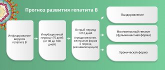
When is a general blood test prescribed?
Donating blood for a general analysis is a familiar procedure to everyone, used both for primary diagnosis and for preventive purposes. Infections, inflammatory reactions or any other pathologies affect the concentration of certain elements in the biomaterial. Changes in each individual or several indicators are very specific and characteristic of a different group of diseases.
A clinical examination is necessarily included in a routine preventive examination and precedes any vaccination. The donation of biological material is also prescribed before the start of drug treatment for a number of diseases, since some drugs affect the composition and, therefore, the properties of blood. For example, the use of anticoagulants is contraindicated when platelet concentrations are low due to the risk of internal bleeding.
Platelets: friends or enemies?
Today we will talk about cells, life without which, without exaggeration, would be impossible.
Galina Petrovna Episheva, a therapist at the Expert Clinic Kursk, talks about platelets.
— Galina Petrovna, what are platelets?
— This is one of the types of blood cells (along with red blood cells and leukocytes). Sometimes they are also called blood platelets. The diameter of the platelet is small - only 2-3 microns. It has no core. The platelet contains a large number of granules containing substances of various chemical compositions.
Platelets are produced by red bone marrow, their predecessors are large bone marrow cells - megakaryocytes.
— What is the role of platelets in the body?
— Today the following functions of platelets are known:
- they protect the walls of blood vessels from mechanical damage;
- prevent blood loss;
- nourish blood vessels;
- participate in the regeneration (restoration) of damaged tissues.
There are also reports that platelets have some antiparasitic effects.
— What is the platelet rate in humans?
— The platelet count is measured in thousands per 1 μl (microliter) of blood (the measurement in liters is also used). The norm is determined depending on gender:
- for men – 200-400 thousand/µl (or, otherwise, 200-400x109/l);
- for women – 180-320 thousand/µl. During menstruation, the rate may decrease, ranging from 75 to 220 thousand/μl. A decrease can also be observed during pregnancy - approximately 100-310 thousand / μl.
In children, the platelet count changes according to age.
Please note that the normal platelet count in the blood may vary depending on the specific laboratory in which the analysis is performed.
— If platelets are increased or decreased, what does this mean? What can lead to an increase or decrease in their number?
— The reasons for the increase in platelets may be:
- stress;
- physical overload;
- use of certain medications (increased platelet count as a side effect). For example, these could be corticosteroids, adrenaline;
- injuries (fracture, cuts, burns);
- erythrocytosis;
- some types of leukemia (leukemia);
- a number of infections;
- enteritis;
- pneumonia;
- acute meningitis;
- anemia;
- autoimmune diseases (rheumatoid arthritis, sarcoidosis, vasculitis);
- cirrhosis of the liver;
- splenectomy.
Reasons for a decrease in platelet count may include:
- pregnancy;
- idiopathic thrombocytopenic purpura (Werlhof's disease);
- use of a number of medications (decreased platelet count as a side effect). For example, these could be antidepressants, antibiotics;
- oncopathology, including tumors of the hematopoietic system;
- chemotherapy;
- hypothyroidism and hyperthyroidism;
- bleeding;
- severe injuries;
- long menstruation;
- liver diseases (in particular hepatitis);
- surgical interventions;
- avitaminosis;
- poisoning with alcohol, heavy metals;
- HIV infection.
— Does it happen that the number of platelets is normal, but their functions are impaired?
- Yes. Platelet dysfunction can result from many causes. I will list some:
- mutation of genes responsible for platelet membrane glycoproteins. This happens, for example, with thrombasthenia - Bernard-Soulier disease;
- abnormalities of platelet granules, in particular, deficiency of dense granules containing ATP, calcium, serotonin;
- changes in the function of cytosolic enzymes, other platelet proteins and a number of others.
— How to identify problems with platelets, in particular, changes in their number and function?
— A number of studies are being carried out. Among them:
- complete blood count with platelet determination;
- coagulogram (blood clotting test);
- study of platelet aggregation.
You can read more about a general blood test in our article
— For what pathological processes and conditions is a platelet test prescribed?
— The list of them is extensive. This:
- injuries;
- thrombosis and thromboembolism;
- blood loss;
- anemia and erythrocytosis;
- leukemia, lymphogranulomatosis, other oncological pathologies;
- poisoning;
- enteritis;
- liver diseases;
- hypo- and hyperthyroidism;
- hypersplenism (increased spleen function);
- prolonged menstruation;
- alcoholism;
- chemotherapy.
— What complaints and signs can be a reason to study the number and functions of platelets?
— The appearance of hemorrhages/bruises (“bruises”) on the skin and/or mucous membranes is typical, and not necessarily associated with even a minor injury. In other words, they can arise spontaneously. The patient may also experience bleeding gums and possible nosebleeds. In women, the duration of menstruation increases.
— How to properly prepare for a platelet test?
— If we are talking about determining the number of platelets in a general blood test, then this is:
- observing a 12-hour fast before donating blood;
- exclusion of fatty foods and alcohol on the eve of the study;
- 2 hours before taking blood, it is advisable to limit physical and emotional stress, stress; you should not smoke for half an hour.
— Which doctor should you contact if you have symptoms indicating a possible platelet problem?
- See a therapist, general practitioner or specialist - an otolaryngologist (for example, for nosebleeds), a gynecologist (for prolonged menstruation). Based on the results of the survey, examination and tests, a consultation with a hematologist or other specialist may be recommended.
You can make an appointment with specialists here
ATTENTION: the service is not available in all cities
Interviewed by Enver Aliyev
The editors recommend:
Leukocytes. What will a blood test tell you?
What does a biochemical blood test show?
Immunity is not on the side of Rosa Luxemburg and Clara Zetkin. Why doesn't nature recognize equality?
For reference:
Episheva Galina Petrovna
She graduated from the Faculty of General Medicine of Kursk State Medical University in 1990.
From 1990 to 1991 she completed an internship in therapy. Doctor of the highest category.
Currently a general practitioner at the Expert Clinic Kursk. Receives at the address: Karl Liebknecht St., 7.
How is blood taken and is preparation needed for the procedure?
Donating blood is a simple and virtually painless procedure that even infants can easily tolerate. Biological material for general examination is taken mainly from a finger, but to check against an extended list of parameters and in some other cases, doctors prescribe venous puncture.
To prevent infection and take clean material, the nurse wipes the pad of one of the fingers of the left hand with a swab soaked in alcohol. Immediately after this, a tiny incision about 3 mm deep is made with a special instrument, a scarifier. The blood protruding from the incision is collected with a collection pipette and poured into a tall tube. A small portion of the liquid is smeared onto a glass slide.
If a doctor prescribes a venous puncture, a rubber tourniquet is placed on the patient's forearm to slow down the blood flow below the dressing. Then the puncture site on the inner surface of the elbow is disinfected with alcohol, after which a hollow needle is inserted into the vein. Through it, blood from the vessel fills a special test tube, and a small amount of biomaterial is applied to a glass slide.
No special preparations are required for the procedure. The only thing doctors can ask is not to have breakfast before collecting biomaterial, since eating food can distort the results obtained.
In some cases, the analysis has to be done several times over a set time period, including to track the effectiveness of the prescribed treatment. To obtain the most accurate data for comparison, repeat blood donation is carried out approximately in the same period of time as the first time.
Platelet count (according to Fonio)
Platelets are involved in the entire process of blood coagulation, starting from the formation of a primary thrombus in the area of damage to blood vessels and ending with participation in the processes of regulating vascular permeability and tone.
The calculation of the most accurate number of platelets is carried out using the Goryaev camera and the Fonio method. The Fonio platelet count is quite accurate. The Fonio method is most convenient for automatic counting.
Units
In a stained smear, 1000 red blood cells and all platelets found are counted. The result is a number expressed in ppm. To obtain the absolute number of platelets, a clinical laboratory diagnostics physician multiplies this value by the number of red blood cells present in 1 μl, and then divides by 1000.
What biomaterial can be used for research?
Venous, capillary blood.
How to properly prepare for research?
- Eliminate alcohol and medications from your diet (in consultation with your doctor) the day before donating blood.
- Do not eat for 8 hours before the test; you can drink clean still water.
- Avoid physical and emotional stress and do not smoke for 30 minutes before the test.
General information about the study
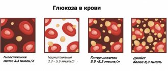
An important condition for the correct functioning of the coagulation system is the presence of a certain number of mature platelets in the bloodstream. Deviations in any direction can have adverse consequences. The normal platelet count in the blood differs depending on age and gender. If their level is reduced, there is a tendency to bleeding, blood clotting worsens, and is difficult to stop. If the platelet count exceeds the norm, the blood becomes thick, and there is a risk of blood clots and blockage of blood vessels.
Platelets are formed in the bone marrow from giant cells called megakaryocytes. In fact, they are fragments of these cells and are small, nuclear-free, colorless oval or round shaped plates surrounded by a membrane, including granules. The diameter of platelets ranges from 2 to 4 microns. They are usually called blood cells, like leukocytes and red blood cells, although they are not. These are postcellular structures, or Bizzazero plaques (named after the Italian scientist who made a great contribution to their study). About 2/3 of all platelets are found in the blood, the rest are found in the spleen.
Functions of blood platelets
When the vascular wall is damaged, blood platelets are immediately activated. They take on a spherical shape and outgrowths appear on them, making them look like a star. Platelets in their active form are capable of sticking to each other (aggregation) and sticking to the vessel wall (adhesion). They secrete an enzyme into the blood plasma, under the influence of which soluble fibrinogen is converted into insoluble fibrin, which entangles the formed elements of the blood with its threads, like nets. The resulting thrombus closes the defect in the damaged vessel. Platelets take part in the dissolution of the fibrin clot and maintain spasm of damaged vessels. Another important function is to provide nutrients to the cells that line the inner surface of blood vessels.
What do the results mean?
Platelets
| Age | Reference values |
| Less than 10 days | 99 - 421 *10^9/l |
| 10 days – 1 month | 150 – 400 *10^9/l |
| 1-6 months | 180 – 400 *10^9/l |
| 6 months – 1 year | 160 - 390 *10^9/l |
| 1-5 years | 150 – 400 *10^9/l |
| 5-10 years | 180 - 450 *10^9/l |
| 10-15 years | 150 – 450 *10^9/l |
| More than 15 years | 180 - 320 *10^9/l |
Thrombocytopenia
Causes:
- thrombocytopenic purpura/hemolytic-uremic syndrome;
- DIC syndrome (disseminated intravascular coagulation);
- drug thrombocytopenia (co-trimoxazole, procainamide, thiazide diuretics, heparin);
- hypersplenism;
- idiopathic thrombocytopenic purpura.
It should be remembered that in pregnant women, platelets can normally decrease to 75-150×109/l.
Thrombocytosis
Causes:
- Primary thrombocytosis (malignant disease of the myeloid lineage of the bone marrow, including essential thrombocytosis and chronic myeloid leukemia);
- Secondary thrombocytosis after removal of the spleen, during an infectious process, iron deficiency anemia, hemolysis, trauma and malignant diseases (reactive thrombocytosis).
- An increase in Hb, MCV, or total leukocyte count suggests primary thrombocytosis.
General blood test indicators
The widespread dissemination of the procedure made it necessary to standardize its data. The standard analysis contains information about:
- hemoglobin (g/l);
- erythrocytes (x106/µl);
- blood color index;
- reticulocytes (%);
- platelets (x103/µl);
- thrombocrit;
- leukocytes (x103/µl);
- leukocyte formula (neutrophils, eosinophils, basophilic granulocytes, lymphocytes, monocytes);
- plasma cells (U).
Hemoglobin (Hb) levels are extremely important for breathing. This substance is able to temporarily combine with oxygen molecules and transport it, delivering it to every cell of the body. Having released oxygen, the protein binds with hydrogen ions and carbon dioxide, carrying the latter back to the lungs. Hemoglobin levels depend on gender and age, but especially fluctuate in childhood (from 145-225 g/l in the first days after birth to 95-135 g/l at 3-6 months, followed by an increase to normal levels in adults). The indigenous people of the highlands have a naturally higher level of hemoglobin in the bloodstream compared to other people.
Red blood cells (RBCs) are the most abundant in blood. It is these bodies that contain hemoglobin, performing the same functions as iron-containing protein. The main role of red blood cells is to transport hemoglobin through the bloodstream and protect the body from the toxic effect of free protein.
The role of the color index (MCHC) is to determine the degree of saturation of red blood cells with hemoglobin.
The percentage of reticulocytes—immature red blood cells—in the bloodstream varies between children and adults. In newborns, their number can reach up to 10%, gradually decreasing and stopping at an older age by 0.5-2%. These cells are also capable of carrying oxygen, but their main task is to compensate for the deficiency of red blood cells when the indicator drops.
The platelet count (PLT) measures the blood's ability to clot. The platelet content in the bloodstream is normally the same in men and women (150-400 thousand/µl), but is slightly different in newborns (180-490 thousand/µl) and older children (160-390 thousand/µl). Platelets ensure the rapid formation of a protective plug that closes the rupture of the vessel.
In addition to the quantitative indicator, the CBC results display the percentage of platelets in the bloodstream - thrombocrit (PST).
An important indicator of health is the erythrocyte sedimentation rate (ESR), which reflects the ratio of plasma proteins.
The task of white blood cells (WBC) is to protect against foreign pathogens (viruses, bacteria, allergens), as well as to eliminate cell debris from the body after they die. The number of white blood cells in children is higher than in adults. In babies under one year old, their number reaches 6.0-17.5 thousand/µl, but with age the level of leukocytes drops, normally amounting to 4.5-11.0 x10³/µl in adults.
The body produces 5 main types of leukocytes, the percentage of which is indicated by the leukocyte formula. The majority of white blood cells are neutrophils - microphages responsible for the absorption of foreign particles. After phagocytosis, they die, releasing antibiotic proteins into the blood. Eosinophils are also capable of microphagocytosis, but provide antiparasitic protection through the release of cytotoxic components.
Monocytes are the main macrophages responsible for the fight against viruses, microbes, parasites, and tumors due to the secretion of cytotoxins, interleukins, interferon, and antitumor factors.
Lymphocytes are the main immune cells that provide detection, storage of antigens, and production of antibodies against them. They also regulate the intensity of the immune response.
Basophils are involved in the formation of an allergic reaction, and also contribute to enhancing the immune response at the site of inflammation.
A separate form of lymphocytes, normally not detected in a blood test, are plasma cells. They appear as a response to the penetration of a pathogen and the onset of an inflammatory reaction, from B lymphocytes and are involved in the production of immunoglobulins.
The short form of the general analysis does not provide the values of ESR and leukocyte formula. But to establish a diagnosis, knowledge of the physiological norms of blood components is not enough. It is important to correctly evaluate the obtained values and correlate them with existing symptoms, since a deviation from the normal number of different types of cells is just a nonspecific sign indicating the presence of a pathological process.
Blood in the human body performs important functions: it carries food products that come in during the digestion of food; takes oxygen from the lungs, gives it to tissues and carries carbon dioxide from them to the lungs; maintains a constant body temperature, participates in immunity mechanisms. Blood consists of a liquid part - plasma and formed elements. Formed elements make up 45% of the total blood volume. These “components” of ours will be discussed.
The most numerous inhabitants of the blood are red blood cells. Their number is in the millions. An erythrocyte is a tiny, nuclear-free cell that, in 100-110 days of its life, runs a path of almost 150 km through the vessels, ending its life in the spleen and liver. Each red blood cell has a yellowish-red color due to the hemoglobin pigment it contains. A feature of hemoglobin is its ability to quickly become saturated with oxygen and release it to tissues. But the role of hemoglobin is not limited to oxygen transport. It also actively frees tissues from carbon dioxide formed during metabolism.
In the blood, along with “red-skinned” red blood cells, their “pale-faced” (or rather, colorless) neighbors, leukocytes, coexist. They are larger than red blood cells, contain a nucleus and have the ability to “devour” microbes.
The most numerous population of leukocytes are granulocytes (eosinophils, basophils, neutrophils). Another community of leukocytes consists of monocytes and lymphocytes. A special place is occupied by the family of blood cells such as platelets. Their main function is to participate in blood coagulation, the formation of a clot-thrombus, which “locks” the lumen of a damaged blood vessel.
In medical practice, the most common practice is to perform a standard general clinical blood test, which includes determining the concentration of hemoglobin, the number of erythrocytes and leukocytes in 1 liter of blood, calculating the leukocyte formula, calculating the color index and determining the ESR.
In most modern laboratories, basic blood parameters are determined using hematological analyzers. The designations in the analysis form for the average person are most often “Chinese letters”. "Deciphering" the test result is the doctor's priority, but by looking at your test, you can also have a general idea of your health.
Basic blood parameters on hematology analyzers
Let's consider some deviations from the accepted norm.
Hemoglobin
The level of hemoglobin in the blood can increase not only in patients, but also in healthy people living in high mountain areas (a small amount of oxygen in the air leads to a compensatory increase in the production of hemoglobin carriers - red blood cells), workers in hot shops (excessive sweating causes blood thickening and an increase in the level hemoglobin). Also, the level of hemoglobin can increase in pathological conditions not directly related to the blood system: dehydration with excessive vomiting, diarrhea, extensive burns. Blood diseases that cause an increase in this indicator are associated with increased production of red blood cells in the bone marrow. More often than an increase there is a decrease in hemoglobin content. The reasons for this phenomenon can be different: iron deficiency in food, impaired absorption of iron in diseases of the stomach and intestines, lack of vitamin B12 and folic acid in the body, blood loss (including frequent hemorrhoidal bleeding).
Red blood cells
Deviations from the accepted norm in relation to red blood cells are considered to be changes in their number, diameter, shape, color, as well as the appearance of intracellular formations in them, characteristic of certain diseases.
An increase in the number of red blood cells most often occurs in response to oxygen starvation of tissues (pulmonary diseases, heart defects), as well as blood thickening associated with loss of fluid by the body (burns, diarrhea, taking diuretics).
A decrease in the content of red blood cells is observed with blood loss, a decrease in the rate of formation in the bone marrow, or their accelerated destruction.
Color index
A diagnostically significant indicator related to hemoglobin concentration and the number of red blood cells is the determination of the degree of saturation of the red blood cell with hemoglobin - calculation of the color index. Based on this test, it is possible to differentiate blood diseases (anemia) associated with insufficient red blood cells in the blood. Thus, with anemia as a result of bleeding, decreased production of red blood cells or accelerated destruction of red blood cells, the color index often remains within normal limits; in some diseases (iron deficiency, lead poisoning, pregnancy) - decreases; in some cases (vitamin B12 deficiency, folic acid; stomach diseases) it increases.
Erythrocyte sedimentation rate (ESR)
An increase in this indicator is a highly sensitive test indicating an actively ongoing inflammatory process. An increase in ESR is typical for infectious and inflammatory diseases, sepsis, joint diseases, liver damage, kidney damage, etc.
A decrease in ESR is observed with an increased number of red blood cells in the blood.
Leukocytes
Leukocytes are a kind of indicator that reflects the characteristics of the state of the human body.
An increase in their number is observed in many conditions associated with inflammatory diseases of various organ systems, as well as in patients suffering from acute and chronic leukemia.
A decrease in the number of leukocytes is observed in elderly people, with radiation sickness, and viral diseases.
For a more informative examination, in addition to determining the total number of leukocytes, they use the calculation of the leukocyte formula, which is understood as the percentage of different types of leukocytes.
Neutrophils
A decrease in the number of neutrophilic leukocytes can occur during chemotherapy in cancer patients, certain types of anemia (aplastic, megaloblastic), autoimmune diseases (systemic lupus erythematosus, rheumatoid arthritis), and viral diseases.
Increased numbers of neutrophils are typical for acute infectious diseases, intoxications, cancer, and leukemia.
Lymphocytes
A decrease in the number of lymphocytes is observed in some pathologies of the immune system, anemia, AIDS, radiation sickness, etc.
An increase in the number of lymphocytes is observed in chronic lymphocytic leukemia, chronic joint diseases, and viral infections.
Eosinophils and basophils
An increase in the number of eosinophils and basophils in the blood is a symptom characteristic of allergic reactions (including bronchial asthma), parasitic diseases, and decreased thyroid function.
Monocytes
An increase in the number of monocytes occurs in diseases such as leukemia, tuberculosis, syphilis, viral, fungal, protozoal infections, as well as in systemic connective tissue diseases (arthritis, periarteritis nodosa, systemic lupus erythematosus).
A decrease in the number of monocytes is observed in some rare types of leukemia and aplastic anemia.
In conclusion, I would like to note that certain deviations from the accepted norm should be assessed in conjunction with data from other tests and the patient’s well-being. That is why a doctor should decipher the blood test and interpret the information received.
Trust the professionals and be healthy!
Laboratory doctor, category I N. Obedkova
Print Email
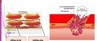
Structure
Rice. 1.
Micrograph of non-activated platelets []
Platelets (from the Greek θρομβοζ - 'clot' and κυτοζ - 'cell') are specialized non-nuclear blood cells, having the shape of a disk with a diameter of about 3 microns and a thickness of about 0.5 microns (Fig. 1). They are formed during the fragmentation of large bone marrow cells—megakaryocytes—and circulate in the bloodstream at a concentration of 200–400 thousand cells per 1 μl of blood. Platelets live in the bloodstream for an average of 5–9 days, and then are destroyed in the spleen and liver.
The structure of a platelet is quite complex. Externally, it is limited by a bilipid layer of the membrane, numerous invaginations of which (an open tubular system) provide a reserve surface for changing shape (Fig. 2). It supports it and at the same time allows for significant changes in the cytoskeleton (framework) of the cell. Inside there are the endoplasmic reticulum (a repository of calcium ions necessary for signaling and the platelet performing its functions) and mitochondria (organelles that ensure respiration). The cytosol contains granules containing substances that spill out into the extracellular space upon cell activation (transition to a new state). Dense granules contain nucleotides (ATP, ADP, GTP, GDP), serotonin, calcium ions in high concentrations, α-granules contain various proteins (including blood clotting factors), and lysosomes contain some enzymes (collagenase, elastase and etc.).
Rice.
2. Scheme of the platelet structure []
After platelet activation, a negatively charged lipid, phosphatidylserine, appears on the outer surface of its membrane. Some coagulation factors bind to it with the help of calcium ions, forming special complexes. They greatly accelerate the reactions leading to gelation of blood plasma at the site of injury (this process is called plasma hemostasis). In other words, phosphatidylserine provides a procoagulant function of platelets that promotes plasma hemostasis.
Why is the lifespan of these blood cells so short-lived (red blood cells, for example, live three to four months), because normally, in the absence of serious damage to blood vessels, they practically do not work? Why do they look like disks? Why does a platelet need mitochondria if its energy expenditure is extremely modest? Why did nature need to accelerate plasma coagulation reactions on cell membranes? Why do α-granules contain coagulation proteins, which are also found in blood plasma? These are just some of the questions that do not yet have clear answers.