Not always indicators of the content of erythrocytes and hemoglobin can provide answers to the questions posed. In such cases, determining MCV, RDW and some other indicators, which we will talk about, will help when making a diagnosis.
MCV – mean erythrocyte volume, or mean corpuscular volume (Mean Corpuscular Volume). An indicator characterizing the average size of cells (in volumetric terms). The unit of MCV is femtoliter (fl) or cubic micrometer (µm3).
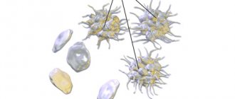
Determined by a hematology analyzer by dividing the cellular volume by the number of blood cells. You should be aware that in some situations it may be unreliable. For example, in the presence of irregularly shaped red blood cells in patients with sickle cell anemia.
On the right in the image is a modified red blood cell in the shape of a sickle
Indications for analysis
MCV blood test is not the only one. To clarify the diagnosis, the therapist may require a retake or refer it for additional tests.
MCV is informative in two cases:
- in order to diagnose one of the types of anemia;
- to determine the type of water-salt imbalance. People are often sent for such an analysis if they have various intestinal infections, acute respiratory diseases and ARVI.
Other, no less serious, reasons for getting tested may be:
- failure of the hormonal system;
- metabolic problems;
- overweight;
- increased blood sugar or diabetes;
- a sharp and causeless decrease in the level of the immune system.
MCV analysis results often help identify such deviations:
- anemia of normochromic type. It is fixed when pathologies appear in the bone marrow, sometimes caused by chronic diseases;
- macrocytic type anemia. It is characterized by an excessive increase in mcv. Red blood cells increase in size due to insufficient amounts of vitamin B and folic acid;
- microcytic anemia. In this case, the disease occurs due to a lack of iron in the blood.
What is the norm and what is considered a deviation
A normal value is considered when the MCV is between 80 and 100 femtoliters. The red blood cell in this case is a normocyte.
If the average volume of a red cell is less than 80 fl, it is a microcyte. Well, if the volume is more than 100, then such an erythrocyte is called a macrocyte.
Accordingly, microcytosis is distinguished when a large number of microcytes are detected in the blood. Low hemoglobin is characteristic. Macrocytosis is characterized by an increased content of macrocytes in the analysis.
Anisocytosis is the presence of red blood cells of different sizes and shapes.
In newborns and children under one year old, the norm is 70-110 femtoliters. With age, this indicator is compared with that of adults
Normal MCV values
| Age, gender | Mean erythrocyte volume, MCV, fl | |
| Children | ||
| 1 day - 14 days | 88,0 — 140,0 | |
| 14 days - 4.3 weeks | 91,0 — 112,0 | |
| 4.3 weeks - 8.6 weeks | 84,0 — 106,0 | |
| 8.6 weeks - 4 months | 76,0 — 97,0 | |
| 4 months – 6 months | 68,0 — 85,0 | |
| 6 months – 9 months | 70,0 — 85,0 | |
| 9 months - 12 months | 71,0 — 84,0 | |
| 12 months - 5 years | 73,0 — 85,0 | |
| 5 years - 10 years | 75,0 — 87,0 | |
| 10 years - 12 years | 76,0 — 90,0 | |
| 12 years - 15 years | Women | 73,0 — 95,0 |
| Men | 77,0 — 94,0 | |
| 15 years - 18 years | Women | 78,0 — 98,0 |
| Men | 79,0 — 95,0 | |
| 18 years - 45 years | Women | 81,0 — 100,0 |
| Men | 80,0 — 99,0 | |
| 45 years - 65 years | Women | 81,0 — 101,0 |
| Men | 81,0 — 101,0 | |
| 65 years - 120 years | Women | 81,0 — 102,0 |
| Men | 83,0 — 103,0 | |
In children under 10 years of age, the index may fluctuate and be inaccurate; later it returns to normal (80-100 fl).
MCHC reduced
A reduced MCHC indicates a condition such as hypochromia, that is, the red cells are not sufficiently saturated with hemoglobin. This condition is associated with pathologies in which the production of iron-containing protein is impaired. Hypochromia can be caused by various types of anemia, hypovitaminosis, lead poisoning, some hereditary congenital diseases, and is also associated with a disorder of iron metabolism in the body. A decrease in the concentration of iron-containing protein in erythrocytes has the following reasons:
- sideroblastic and hypochromic iron deficiency anemia;
- chronic posthemorrhagic anemia;
- hemoglobinopathies, in which hemoglobin synthesis is impaired and some amino acids are replaced in its chains;
- disturbance of water-electrolyte metabolism;
- thalassemia (some types) is a hereditary disease associated with mutations in genes that are responsible for the synthesis of iron-containing protein;
- megaloblastic anemia, in which red cells increase in volume more significantly than they are saturated with iron-containing protein.
When hemoglobin synthesis is impaired, MCHC decreases last. Therefore, a decrease in the concentration of iron-containing protein with normal values of other blood parameters (erythrocyte and hemoglobin content) indicates an erroneous laboratory test result.
MCV is higher than normal
If the results are above normal, this indicates the development of macrocytic anemia. It can be directly related to diseases such as:
- drug intoxication;
- food poisoning;
- problems with the thyroid gland;
- lack of iodine or iron in the body;
- liver dysfunction;
- oncological process of red bone marrow;
- long-term alcoholism;
- disruption of the pancreas.
An increase in mcv can be caused by:
- long-term use of birth control pills that affect hormonal levels;
- addiction to cigarettes and tobacco products;
- prolonged contact with toxic substances (work in hazardous industries);
- taking medications that increase the level of mcv in the blood.
If left untreated, macrocytic anemia can lead to frequent fainting, poor health, and low hemoglobin levels in the blood. Particularly at risk are:
- people who eat poorly, lead a sedentary lifestyle and ignore exercise;
- patients with chronic liver failure;
- people with a genetic predisposition to the disease;
- men over fifty-five years of age who abuse alcohol.
Experts identify some signs by which one can understand that a person’s red blood cell volume is too high:
- unhealthy pale lips;
- abdominal pain for no particular reason, which appears very often;
- the presence of tachycardia (heartbeat too fast), even when the person is at rest;
- skin with a yellowish tint.
If you notice similar symptoms or detect an increased level of mcv in the blood, you must immediately consult a general practitioner for appropriate treatment.
A little about anisocytosis
Anisocytosis is the presence of red blood cells of different sizes in the blood - from microcytes to macrocytes. Quantitatively, the distribution of red blood cells by volume is expressed in a special index, designated RDW.
This is what the distribution width of erythrocytes by volume curve looks like in the analysis
Red blood cell heterogeneity by volume (RDW) shows the deviation from the standard volume, expressed as a percentage.
Anisocytosis can be determined by a blood smear under a microscope, but the exact characteristics of these indicators are obtained using hematological analyzers. Interpretation of the results is carried out by laboratory diagnostic doctors together with MCV and facilitates the diagnosis of anemia.
Normally, the width of the distribution of red blood cells is 11.5-14 percent. You should know that if MCV is reduced with normal RDW, then this is typical for blood transfusion, removed spleen, and thalassemia.
If MCV is elevated with normal RDW, then most likely there is liver pathology. If RDW is elevated and MCV is below normal, beta thalassemia, iron deficiency, or red blood cell sludge may be suspected.
If suddenly both indicators are higher than normal, then we can assume cold agglutination, vitamin B12 deficiency or impaired absorption. This combination is also typical after a course of chemotherapy for cancer.
MCV below normal
Tests showing that the volume of red blood cells is below normal also indicates pathology. Experts name a number of reasons that can lead to such results:
- genetic predisposition;
- insufficient amount of water consumed;
- development of various types of anemia;
- lead intoxication;
- the presence of malignant formations, tumors in the body;
- taking medications that affect test results.
In medical circles, a disease in which the level of red blood cells in the body decreases is commonly called microcytic anemia. The peculiarity of the disease is that red blood cells do not perform their transport function, i.e. they do not deliver oxygen and other useful substances to the body’s cells in the required quantities.
With this pathology, a characteristic clinical picture is observed:
- constant fatigue;
- increased irritability, nervousness;
- decreased concentration and performance;
- absent-mindedness;
- memory impairment.
A decrease in the volume of red blood cells is always observed with various types of blood loss.
Pregnancy and mcv
During pregnancy, due to the body's increased consumption of iron, microcytic anemia can develop. This condition can negatively affect both the health of the unborn child and the well-being of the mother.
Some experts are convinced that mcv indicators are directly related to a person’s psychological state.
Causes of elevated MCV
The average volume of red cells is increased when:
- liver diseases;
- hemolytic anemia;
- development of water-electrolyte imbalance in the form of hypotonic overhydration, which is possible with kidney diseases;
- macrocytic and megaloblastic anemia;
- deficiency of folic acid and vitamin B12;
- myelodysplastic syndrome.
Anemia cannot be ruled out when MCV is not higher than normal. For example:
- after bleeding;
- with hemolytic anemia;
- for some acute poisonings.
Features of the analysis
Today, the mcv test is included in the general blood test or can be performed separately from other indicators. To donate blood, the patient must come to the treatment room, where a laboratory technician or nurse will take blood samples from a finger or vein. Blood sampling is carried out in accordance with all sanitary and epidemiological regulations (SanPiN).
The patient is required to comply with the following rules:
- You need to donate blood on an empty stomach (5-12 hours after your last meal);
- at the time of donation, the woman should not be menstruating;
- feeling normal. It is prohibited to take blood samples if the patient is not feeling well, is in a coma or is in cardiac shock.
Prevention of increased red blood cells
The cause of the increased production of red blood cells is determined by the doctor. To maintain normal red blood cell levels, it is enough to adhere to preventive measures.
To avoid increased blood concentrations, it is recommended to drink more fluid. The water must be purified, so it is better to buy it in bottles or collect it from wells.
Eating fresh vegetables and fruits has a good effect on blood composition. They contain many vitamins and microelements that regulate the content of red blood cells.
You can reduce the concentration of red cells in the blood by eliminating foods containing iron from your diet. You will have to give up red meat, liver, beans, lentils, spinach, cabbage, prunes and raisins. Avoid drinking caffeinated drinks, smoking or taking aspirin. Regular physical activity will help regulate red blood cell levels.
General (clinical) blood test: interpretation and norm
A complete blood count provides essential information about the types and numbers of blood cells, especially red blood cells, white blood cells and platelets. Using a general blood test, the treating doctor can detect the nature of symptoms such as weakness, fatigue, and bruising. A complete blood count is used to diagnose anemia, infection and many other disorders.
A general (clinical) blood test includes the following indicators:
- Absolute white blood cell count ( WBC ). White blood cells protect the body from infection. If an infection develops, these cells attack and destroy bacteria, viruses, or other pathogenic organisms. White blood cells are larger in size than red blood cells, but their number is less. When a person gets sick, the number of white blood cells increases very quickly. Sometimes white blood cell counts are used to detect infection or to evaluate the effectiveness of cancer treatment.
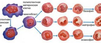
- Determination of the ratio of different types of white blood cells (leukogram). The main types of leukocytes are neutrophils, lymphocytes, monocytes, eosinophils and basophils. Counting immature neutrophils, also called band neutrophils, is part of this test. The number of each cell type can provide important information about the health of the immune system. Too high or too low the number of one type of cell may indicate an infection, an allergy or toxic reaction to a drug or chemical, or various diseases such as leukemia.
- Absolute red blood cell count ( RBC ). Red blood cells carry oxygen from the lungs to the rest of the body. They also carry carbon dioxide back to the lungs for exhalation. When the level of red blood cells is low (anemia), the body does not receive enough oxygen. When levels are high (polycythemia), there is a risk of red blood cell clots that can block tiny blood vessels (capillaries), also making it difficult to carry oxygen.
- Hematocrit ( HCT , precipitated red blood cell volume, PCV ). This test measures the volume of blood per red blood cells. For example, a hematocrit value of 38 means that 38% of the blood is red blood cells. Hematocrit and hemoglobin are the two main tests for detecting anemia or polycythemia.
- Hemoglobin ( Hgb ). Hemoglobin molecules are found in red blood cells; they carry oxygen and give the blood its red color. A hemoglobin test measures the level of hemoglobin in the blood and shows how the body copes with transporting oxygen.
- Erythrocyte index. There are three red blood cell indices: mean erythrocyte volume (MCV), mean erythrocyte hemoglobin content (MCH) and mean erythrocyte hemoglobin concentration (MCHC). All of them are calculated using special equipment, and the numbers are derived from other indicators of a general blood test. MCV reflects the size of red blood cells, MCH is the average hemoglobin content in an individual red blood cell, MCHC is the hemoglobin concentration in an individual red blood cell. These indices are used in diagnosing various types of anemia. The red blood cell distribution width (RDW) index is also calculated, which reflects the difference in size and shape between cells.
- Absolute platelet count. Platelets are the smallest type of blood cell. They play an important role in the blood clotting process. If bleeding occurs, the platelets are activated and bind together, forming a plug that prevents bleeding. If the number of red blood cells is insufficient, there is a risk of uncontrolled bleeding. If there is an excess amount, there is a risk of thrombus formation in a blood vessel. In addition, platelets can be involved in the formation of sclerotic plaques on artery walls (atherosclerosis).
- Mean platelet volume ( MPV ). This indicator reflects the average value of the measured platelet volume. MPV is used in conjunction with platelet count to diagnose certain diseases. If the platelet count is normal, the MPV may be low or high.
Your doctor may also order a blood smear microscopy at the same time as your complete blood count, but this is not part of your routine blood test. For this test, a drop of blood is placed on a plate and diluted with dye. The plate is examined under a microscope, noting the number, size and shape of red blood cells, white blood cells and platelets. Differences in the shape or size of blood cells can indicate various diseases, such as leukemia, malaria or sickle cell disease.
A clinical blood test is carried out for the following purposes:
- Finding the cause of symptoms such as fatigue, weakness, fever, bruising, or weight loss
- Diagnosis of anemia
- Establishing the amount of blood lost during bleeding
- Diagnosis of polycythemia
- Diagnosis of infections
- Diagnosis of blood diseases, such as leukemia
- Monitoring the body's response to drug treatment and radiation therapy.
- Monitoring the blood cell response to abnormal bleeding.
- Screening of indicators before surgery.
- Tracking high or low levels is used to detect various diseases, such as high eosinophil levels that may indicate allergies or asthma.
A complete blood count is performed as part of a routine medical examination. A complete blood count provides valuable information about the overall condition of the body and health.
Preparing for the examination
No special preparation required.
How is the examination carried out?
- The shoulder is tightened with an elastic tourniquet to stop the blood flow. The veins under the tourniquet become larger, making it easier to insert the needle.
- The puncture site is cleaned with alcohol.
- The needle is inserted into the vein. Sometimes more than one injection is required.
- A blood collection tube is attached to the needle.
- When enough blood has been collected, the tourniquet is removed.
- After the needle is removed, a cotton swab is applied to the injection site.
- The area should be pressed and a bandage applied.
If a baby's blood is being tested, a heel prick is done instead of drawing blood from a vein.
Feelings during the procedure
A blood sample is taken from a vein in the arm. The forearm is wrapped with an elastic band, so there may be a feeling of tightness. You may not feel any pain from the needle, but sometimes there will be a slight burning sensation or pain.
Complications
When taking blood from a vein, the chance of complications occurring is very small.
- There may be a slight bruise. By pressing the tampon to the puncture site for a few minutes, you can reduce the chance of its occurrence.
- In rare cases, the vein may become swollen, this is called phlebitis. The problem can be treated with several warm compresses.
- Continuous bleeding may occur in patients with blood disorders. Taking medications such as aspirin, warfarin (such as Coumadin), and other blood thinners increases the chance of bleeding. Tell your doctor before taking a sample if you have problems with blood clotting or are taking blood-thinning medications.
results
A complete blood count provides important information about the types of blood cells - red blood cells, white blood cells, platelets - and their numbers. A complete blood count helps determine the cause of various symptoms such as weakness, nausea, bruising and is used in the diagnosis of various diseases - anemia, infections and many others.
Normal results
IMPORTANT! The normal values, or range of normal values, given here are average values. These vary depending on the laboratory, so where you are being tested will have different readings. The laboratory report should contain information about the reference range used. In addition, overall health and other factors are taken into account when analyzing the results. Therefore, a value that does not fit into the range presented here may be normal for you personally.
The normal range depends on your age, gender, type of blood sample, and how high above sea level the area where you live is. The doctor can use all the indicators of the general blood test when checking the condition of the body. For example, the number of red blood cells, hemoglobin, hematocrit are the main indicators in diagnosing anemia, but erythrocyte indices and smear microscopy also help detect this disease and can show the possibility of its occurrence.
To assess the number and size of leukocytes, the doctor takes into account the results of counting the number of cells and the ratio of their types. To see if there are too many or few cells of a certain type, the doctor compares their number and percentage. There is a norm for each type of leukocyte.
Indicators may change during pregnancy. During each trimester, your doctor should report changes in your normal range.
| Absolute white blood cell count ( WBC ) | |
| Men and non-pregnant women: | 5,000–10,000 WBC per cubic mm (mm3) or 5.0–10.0 x 10⁹ WBC per 1 liter (L) |
| The ratio of different types of leukocytes | |
| Neutrophils: | 50%–62% |
| Band neutrophils: | 3%–6% |
| Lymphocytes: | 25%–40% |
| Monocytes: | 3%–7% |
| Eosinophils: | 0%–3% |
| Basophils: | 0%–1% |
| Absolute red blood cell count ( RBC ) | |
| Men: | 4.5–5.5 million RBC per µl or 4.5–5.5 x 10¹²/l |
| Women: | 4.0–5.0 million RBC per µL or 4.0–5.0 x 10¹²/L |
| Children: | 3.8–6.0 million RBC per µL or 3.8–6.0 x 10¹²/L |
| Newborns: | 4.1–6.1 million RBC per µL or 4.1–6.1 x 10¹²/L |
| Hematocrit (HCT) | |
| Men: | 42%–52% or 0.42–0.52 of the total volume |
| Women: | 36%–48% or 0.36–0.48 of total volume |
| Children: | 29%–59% or 0.29–0.59 of the total volume |
| Newborns: | 44%–64% or 0.44–0.64 of total volume |
| Hemoglobin (Hgb) | |
| Men: | 14–17.4 grams per deciliter (g/dL) or 140–174 grams per liter (g/L) |
| Women: | 12–16 g/dl or 120–160 g/l |
| Children: | 9.5–20.5 g/dl or 95–205 g/l |
| Newborns: | 14.5–24.5 g/dl or 145–245 g/l |
Typically, a normal hemoglobin level is one third of the hematocrit level.
| Red blood cell indices | |
| Mean erythrocyte volume ( MCV ) - Adults: | 84–96 femtoliters (fl) |
| Mean erythrocyte hemoglobin content ( MCH ) - Adults: | 28–34 picograms (pg) per cell |
| Mean erythrocyte hemoglobin concentration ( MCHC ) - Adults: | 32–36 grams per deciliter (g/dL) |
| Red blood cell distribution width by volume ( RDW ) | |
| Norm: | 11.5%–14.5% |
| Absolute platelet count | |
| Adults: | 140,000–400,000 platelets per mm3 or 140–400 x 109/L |
| Children: | 150,000–450,000 platelets per mm3 or 150–450 x 109/L |
| Mean platelet volume (MPV) | |
| Adults: | 7.4–10.4 µm3 or 7.4–10.4 fl |
| Children: | 7.4–10.4 µm3 or 7.4–10.4 fl |
| Blood smear microscopy | |
| Norm: | Normal indicators of the number, shape, color and size of blood cells. |
High performance
Absolute red blood cell count (RBC)
- Conditions and habits that cause high red blood cell counts include smoking, exposure to carbon monoxide, long-term lung disease, kidney disease, some cancers and heart disease, alcoholism, liver disease, a rare bone marrow disease (polycythemia vera), or rare problems related to with hemoglobin, which are associated with the transfer of oxygen.
- Diseases related to the water component of the body can also cause high red blood cell counts. These include dehydration, diarrhea, vomiting, increased sweating, and taking diuretics. Lack of fluid in the body causes red blood cell counts to appear high. This is sometimes called pseudopolycythemia.
Absolute white blood cell count (WBC)
- Causes of high white blood cell counts include infection, inflammation, tissue damage (for example, due to a heart attack), kidney failure, lupus, tuberculosis, rheumatoid arthritis, malnutrition, leukemia, and cancer.
- Taking costosteroids and certain medications, underactive adrenal glands, problems with the thyroid gland, and removal of the spleen can also cause an increase in white blood cell levels.
Absolute platelet count
- High platelet levels can occur due to bleeding, iron deficiency, cancer and bone marrow problems.
Low performance
Absolute red blood cell count (RBC)
- With anemia, the level of red blood cells decreases. Anemia can be caused by heavy menstrual bleeding, stomach ulcers, colorectal cancer, inflammatory bowel disease, tumors, Addison's disease, thalassemia, lead poisoning, sickle cell disease, or a reaction to certain chemicals and medications. One reason for low red blood cell counts may be removal of the spleen.
- Not getting enough folic acid or vitamin B12 can also cause anemia, such as pernicious anemia, which causes problems absorbing vitamin B12.
- Red blood cell indices and smear microscopy can help find the cause of anemia.
Absolute white blood cell count (WBC)
- Low white blood cell counts may be caused by chemotherapy and reaction to drug treatment, aplastic anemia, viral infections, malaria, alcoholism, AIDS, lupus, Cushing's syndrome.
- An enlarged spleen can reduce white blood cell counts.
Absolute platelet count
- Low platelet levels can be caused by pregnancy, immunopathological thrombocytopenic purpura and other diseases that affect the process of generating platelets or destroy them.
- An enlarged spleen can reduce platelet counts.
What can affect the results of the examination?
Reasons why the analysis is not possible or the results will be incorrect:
- When drawing blood, your arm was tied with an elastic tourniquet for a long time.
- Taking drugs that cause low platelet counts, such as certain antibiotics, steroids, thiazide diuretics, chemotherapy drugs, quinidine, meprobamate.
- Very high levels of white blood cells or fats (triglycerides) can cause falsely high hemoglobin levels.
- With an enlarged spleen, the level of platelets (thrombopenia) or white blood cells may be reduced; this condition can also cause some types of cancer.
- During pregnancy, the level of red blood cells is often reduced, and less often, the level of white blood cells is increased.
On a note:
- The absolute white blood cell count can reach up to 2000 WBC per microliter due to exercise, stress or smoking.
- Children tend to have higher levels of white blood cells than adults.
- Often, a red blood cell count test includes:
- Erythrocyte sedimentation rate (ESR). The ESR test measures how quickly red blood cells settle to the bottom of a tube. During inflammatory processes occurring in the body (for example, in the presence of infection or cancer), red blood cells settle more slowly than under normal conditions. Using ESR analysis, inflammatory diseases can be diagnosed if the blood test results are normal.
- Analysis of reticulocyte content. This is an analysis of the number of young red blood cells in a blood sample. As a rule, the content of reticulocytes is lower compared to the content of mature red blood cells. However, recent bleeding or destruction of mature red blood cells may cause increased reticulocyte production. This test is done to detect certain types of anemia and screen for the body's response to treatment.
- Hematocrit measurements may vary depending on the assay method and equipment used.