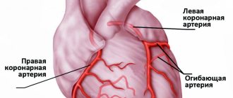
Blood clots can form not only in veins, but also in the cavities of the heart and arteries.
The formation of blood clots is a natural reaction of the body. They help restore damaged blood vessels. A thrombus is a blood clot containing fibrin (a protein necessary for blood clotting). But blood clots in the cavities of the heart are a completely different matter.
The most dangerous thing is that there are no symptoms that would suggest the presence of intracardiac blood clots. A thrombus can be detected in the cavities of the heart only through diagnostics (echocardiography, etc.). A person may experience any symptoms (shortness of breath or suffocation) only with pulmonary embolism.
Thrombi can be in the left or right cavities of the heart, located in the atria or ventricles (most often these are parietal thrombi).
What are blood clots?
- Floating. These are partially attached blood clots. The danger is that the floating part of the clot may come off;
- Parietal, when the clot is completely attached to the wall of the heart chamber;
- Clogging (occlusive). The most dangerous! These clots block blood flow.
Briefly about the treatment method
Removing blood clots (thrombectomy) can be a separate type of treatment, but more often it is a component of it. Vessel thrombosis can form as a result of its narrowing or expansion; a thrombus fragment can be brought from other arteries or the heart. Thrombotic masses in the lumen of blood vessels block blood flow and cause circulatory failure, which can lead to gangrene of an organ or limb. Modern vascular surgery has a number of technologies that make it possible to remove thrombotic masses from vessels.
Advantages of treatment at the ISC
Our clinic uses both traditional technologies and minimally invasive modern methods to remove blood clots.
- Fogarty catheter thrombectomy is a traditional method of removing blood clots from an artery using a balloon probe. The principle is to pass the probe in a folded state through the thrombus, followed by inflating the balloon and pulling it out along with the thrombotic masses.
- Rotarex is a technology for removing chronic blood clots from the arteries. The principle is based on the splitting of blood clots by a special probe head rotating at high speeds and its simultaneous suction by a special spiral mechanism (presented in the film). The technology opens up unlimited possibilities for treating severe vascular pathology without incisions.
- Aspirex is a similar technology for removing fresh blood clots from arteries and veins. Suitable for the thinnest vessels, does not damage venous valves. A special spiral suction atraumatically removes all blood clots from the vessel if the thrombosis period does not exceed 2 weeks.
Statistics
Cardiovascular diseases remain the main cause of high mortality in industrialized countries and Russia. Despite the obvious success in the treatment of acute myocardial infarction (MI) and unstable angina (UA), as the main forms of acute coronary syndrome (ACS), they account for more than 50% of all deaths. Among patients admitted to the emergency departments of Russian hospitals with pain in the chest area, that is, with suspected MI or UA, only 15-25% are found to have ACS. The rest are either discharged from the hospital or transferred to other departments. Therefore, one of the central tasks is the fastest possible selection of patients with ACS in need of urgent special medical procedures. However, as the experience of a number of clinics in Europe and the USA shows, due to an “inaccurate” diagnosis, some of these patients undergo intensive and quite expensive treatment, which, among other things, can negatively affect their health. The other side of the coin is the inaccurate diagnosis of MI and, as a consequence, the inclusion in the group of non-core patients from 4 to 9% of patients with missed MI. In the USA and many European countries, this situation is one of the main reasons for lawsuits accusing doctors of professional incompetence. That is, the key problems in the management of ACS patients are quick and accurate diagnosis, selection of patients in the early stages of hospitalization, assessment of the degree of risk and prognosis of the disease, and the appointment of an adequate system of treatment procedures.
Indications and contraindications for the treatment method
- Acute arterial embolism - blockage of an artery by a blood clot brought from other places. The most common procedure is open embolectomy with a Fogarty probe through an approach below or above the blockage.
- Arterial thrombosis against the background of atherosclerotic plaques (atherothrombosis). It is dangerous to use a Fogarty probe in this case, as the artery wall may collapse. Rotarex allows you to remove thrombotic masses from an artery affected by atherosclerosis without damaging the vascular wall. In addition, soft atherosclerotic plaques are cut off. After removing the thrombus, angioplasty and stenting of the affected arteries are necessary.
- Removal of blood clots from vascular prostheses. Can be performed from open approaches using various modifications of Fogarty catheters. Most often, blood clots are removed from large vascular prostheses (aorto-femoral) as part of repeated operations.
- Removal of blood clots from previously installed stents. Stent thrombosis often requires repeated interventions. It is not possible to use a Fogarty balloon to remove blood clots from stented areas of arteries, as it will rupture against the stent or damage the arterial wall. For such thromboses, it is much more effective and safer to use Rotarex technology.
Standing and sitting work, long flights
Those at risk include cooks, salespeople, hairdressers, surgeons, traffic police officers, accountants, programmers, secretaries, as well as those who, as part of their duties, have to fly a lot on airplanes, stand in traffic jams, sit in a car, when people are forced to stay in a car for a long time. static, sitting position.
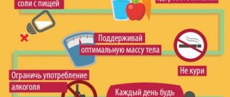
How to warn. When going on a long journey, sitting or standing idle in the office for hours, make it a rule to change your body position every 30 minutes or just walk. To prevent blood clots, you can also wear compression stockings (support stockings, stockings or tights).
Article on the topic
Thick means dangerous. What you need to know about blood clots
Preparing for treatment
In the case of acute thrombosis, surgery to remove blood clots should be performed according to urgent indications in order to save the limb. The urgency of the operation depends on the degree of circulatory failure and the duration of the disease. Preparation for such an intervention is minimal.
- General blood and urine analysis
- Biochemical blood test (urea, creatinine, electrolytes)
- ECG
- Echocardiography - to identify the causes of thrombosis
- Ultrasound of the arteries of the affected limb and possible aneurysms.
- Installation of a urinary catheter
- Installation of an intravenous line catheter
- Cleansing enema
- Shaving surgical access sites
How the treatment method works
Removal of blood clots using a Fogarty probe
An open thrombectomy using a Fogarty probe is performed through the incision. The incision is usually made below the site of thrombotic blockage of the artery, since the removal of blood clots is easier through the blood flow. If the patient has thrombosis of the aorta or iliac arteries in the abdomen, then the incision is made in the groin area; if thrombosis is in the popliteal artery, then the access is made on the lower leg or foot. After isolating and assessing the condition of the artery, a 3-5 mm incision is made on it, into which the Fogarty balloon probe is inserted in a compressed state. For better elasticity, a metal conductor is installed in the probe, which is removed after passing the probe above the blood clot. A syringe is attached to the probe, through which the balloon is inflated. After this, the surgeon removes the inflated balloon, which pushes the thrombotic masses towards the cut artery. Pressure of the blood above the clot helps the process of removing the clot. After removing the clot, the procedure is repeated to ensure complete removal. Vascular clamps are placed on the artery and the hole in it is sutured with a vascular suture. After this, the wound is sutured layer by layer.
This simple operation helps well with acute blockage of intact arteries by thromboembolism (a blood clot brought from the heart or from larger arteries), however, if thrombosis of arteries affected by atherosclerosis occurs, then thrombectomy with a Fogarty probe becomes impossible or even dangerous, since the probe easily breaks on sharp edges atherosclerotic plaques or can get caught on the plaque and tear it. Therefore, for such cases, we use other methods of removing blood clots.
Removing blood clots with a Rotarex Straube probe
The Rotarex Straube probe allows you to clear the artery of blood clots without resorting to a surgical incision, through a puncture of the artery. At the same time, this probe does not damage the walls of the artery, carefully removing thrombotic masses and freeing the lumen of the vessel. The principle of operation is to crush the clot with a rotating probe head and suck its particles into the lumen of the probe and remove them from the lumen of the vessel into a special bag. The use of Rotarex Straube is possible even in cases of chronic thrombosis due to atherosclerotic and diabetic arterial lesions. The Rotarex Straube probe clears blood clots from the vessel, but does not remove atherosclerotic plaques, therefore, to restore the lumen of the vessels, angioplasty and stenting of narrowed arteries of the leg are performed if necessary. The Rotarex Straube endovascular thrombectomy procedure is performed without incisions, only through a puncture of the artery wall, so the main method of pain relief is local anesthesia. Probe thrombectomy is indispensable when removing blood clots from intestinal arteries with mesenteric thrombosis. Considering the severe general condition of patients with this pathology and the difficulties with access to the mesenteric arteries, thrombectomy with a probe from the initial parts of the superior mesenteric artery is the preferred method of restoring blood circulation in the intestine.
Risk factors for thrombosis
Internal:
- arterial hypertension [7];
- pregnancy, childbirth, postpartum period [3];
- biochemical changes in blood [2,3,5,7];
- vasculitis [2];
- age over 40 years [5];
- congenital thrombophilia, thrombosis, varicose veins of the lower extremities [3,5];
- congestive heart failure [5, 6];
- malignant neoplasms, radiotherapy and chemotherapy [3];
- strokes [3, 6];
- myeloproliferation [2, 5, 7];
- nephrotic syndrome and renal failure [5, 6, 7];
- obesity (BMI over 30) [3];
- myocardial infarction [6];
- diabetes mellitus [6, 7];
- systemic lupus erythematosus [2];
- chronic pulmonary diseases [3];
- enterocolitis [5].
External:
- heroin addiction [2];
- hormonal therapy [3,5];
- dehydration due to vomiting, diarrhea, increased sweating, direct lack of fluid [6];
- immobilization [3];
- travel by plane, bus or in a seated car [3];
- infectious diseases, including COVID-19 [1, 3, 5, 9];
- catheterization of central and peripheral veins [2, 5];
- smoking [6, 7];
- sedentary lifestyle [3];
- operations [3];
- fractures of large bones, other injuries [3];
- taking oral contraceptives [5];
- taking Diazepam, Amiodarone, Vancomycin [2];
- sclerotherapy and thermal ablation [2];
- condition after joint replacement [3];
- holding an awkward position [3].
Possible complications during treatment
The following complications are possible when using a Fogarty probe:
- Rupture of the inner wall of the artery with its dissection and secondary thrombosis
- Tearing off the probe head and leaving it in the lumen of the artery
- Perforation of an altered artery wall with a probe with internal bleeding
- Complications associated with access (hematoma, lymph accumulation, wound suppuration)
Such complications are quite rare. Their frequency decreases as clinical experience with thrombectomies accumulates. In our clinic, we have not observed such complications over the past five years.
Complications of Rotarex thrombectomy:
- Dissection of the arterial wall due to rough passage of the probe
- Transfer of pieces of blood clots to small arteries below the site of thrombosis
- Formation of hematoma along the artery
Such complications are usually identified during the operation and are usually corrected immediately.
Prognosis after treatment method
Arterial thrombosis is a complication of various diseases. This means that removing a blood clot (thrombectomy) is an intervention that eliminates complications. Thus, the prognosis after treatment depends on eliminating the causes of thrombosis. If this is thrombosis associated with narrowing of the arteries, then restoration of the lumen by angioplasty and stenting eliminates the causes of thrombosis and provides a good long-term prognosis. If thrombosis or thromboembolism is associated with diseases of other arteries or the heart, then treatment of these diseases is necessary to prevent repeated episodes of blood clot transfer.
What are blood clots and why do they appear?
Thrombi are clots that form from connections between blood cells. Platelets, sticking together in chains, form lumps that are attached to the walls of blood vessels. In some situations, blood clots form due to disorders of the hematopoietic system, in others - as a result of damage to the inner wall of the vessel.
Large growths inside the vein prevent blood flow from passing through the obstructed area. As a result, congestion forms in the veins, leading to varicose veins. When a blood clot blocks the lumen, a heart attack occurs—the death of tissue that has not received oxygen through the bloodstream.
The formation of blood clots is influenced by several factors from a person’s daily life:
- Sedentary lifestyle. Lack of active mobility leads to stagnation of blood in the lower legs. Therefore, constantly sitting at a computer, as well as choosing an escalator instead of stairs, give the blood a reason to stagnate and form clots.
- Insufficient fluid intake. The quality of blood directly depends on what a person eats. With insufficient fluid intake, the blood becomes thick, which means it cannot fully perform its functions and puts a greater strain on the heart. It is easy to pump liquid blood through the system, but thick blood is much more difficult.
- Taking medications that affect the hematopoietic system. In medical practice, drugs are often used to treat certain diseases, one of the side effects of which is blood thickening. Therefore, such medications must be taken simultaneously with anticoagulants that prevent the formation of blood clots.
You can protect yourself from blood clots by regularly exposing your body to physical activity, drinking enough fluids, and including more plant-based foods rich in fiber in your diet.
Observation program after treatment method
After removing blood clots and eliminating the causes of their occurrence, the patient needs to be monitored by a vascular surgeon. While in the hospital, the surgeon evaluates circulatory compensation and blood flow velocity using Doppler ultrasound.
Upon discharge, the patient is prescribed antithrombotic therapy, depending on the causes of thrombosis. These can be drugs such as Plavix, warfarin, aspirin.
A month after discharge, it is necessary to conduct a control duplex ultrasound scan of the arteries to assess the condition of the restored segments and circulatory compensation. It is also necessary to evaluate possible sources of thromboembolism (cardiac cavities, aorta) using echocardiography.
Subsequently, the patient must repeat ultrasound examinations and undergo blood clotting tests (APTT, TV, platelet aggregation) twice a year.
Diagnostics
Making a diagnosis requires a thorough examination of the patient. The doctor identifies pronounced symptoms and the approximate location of the blood clot, and for clarification prescribes:
- Ultrasound of veins;
- Dopplerography to assess the intensity of blood flow;
- Phlebography to examine deep veins;
- MRI to determine the exact location of the thrombus;
- Angiography (for pulmonary embolism);
- Lung perfusion scintigraphy to determine areas of the lung where air enters, but blood flow is impaired;
For differential diagnosis the following are also used:
- X-ray, excluding injuries, pneumothorax, pleurisy, neoplasms.
- Analysis of the level of d-dimers, the level of which increases in PE in 90% of cases.
- Ultrasound of the heart to differentiate thromboembolism from infarction, pericarditis, and acute heart failure.