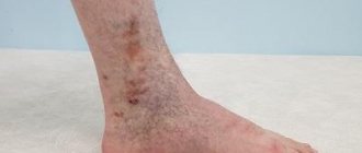
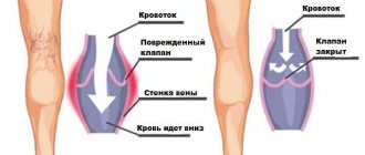
Venous reflux of the lower extremities is an abnormal blood flow in the veins. The occurrence of reflux is caused by incorrect or defective operation of vascular valves. Valves allow blood to flow in only one direction.
Normally, blood moves from bottom to top: from the lower extremities to the heart. Blood is also directed from superficial vessels to deep veins. It often happens that under the influence of various external and internal factors, veins expand, which leads to a decrease in the tone of the vascular walls and improper functioning of the valves. As a result, pathological blood flow appears: blood begins to move from top to bottom and from deep to superficial veins.
Prevalence of the disease
In the world, those diagnosed with chronic venous insufficiency in different countries range from 15 to 25%, depending on the development of medicine in the country. In Russia, symptoms of CVI are observed in every second person aged 20–50 years, of which up to 15% have an established diagnosis, and 4% suffer from a severe form of the disease with complications in the form of trophic ulcers.
Chronic venous insufficiency is caused by upright posture, as a result of excessive stress on the legs. The impetus for its development can be congenital and acquired pathological conditions, injuries and prolonged physical activity. CVI is not always accompanied by visible changes in the veins; in most cases (mild form) it is a feeling of heaviness in the legs, swelling in the evenings, increased fatigue, pain when forced to stand for long periods of time.
Types of vein reflux
Depending on how the blood is distributed, there are 2 types of reflux:
- Vertical.
It can develop in deep and superficial vessels. With this type of reflux, the blood moves in a downward direction. In deep veins it occurs during postthrombophlebitic syndrome and provokes severe venous insufficiency.
It is very difficult to identify this disease in the early stages. Even complex therapy does not guarantee complete recovery, and therefore patients with this disease need to be regularly checked by a doctor.
- Horizontal.
In this case, blood flows from deep veins to superficial ones. Horizontal reflux is considered the main cause of varicose veins. Diagnosed in the initial stages of development.
Causes of CVI
Most often, venous insufficiency occurs with varicose veins, as well as against the background of such diseases and pathological conditions as:
- congenital pathology of the venous system;
- congenital aplasia and hypoaplasia of the deep veins;
- congenital arteriovenous fistulas;
- congenital osteohypertrophic nevus with varicose veins;
- suffered acute thrombosis of the main veins;
- Klippel-Trenaunay syndrome;
- previous phlebothrombosis.
In recent years, a new cause for the development of chronic venous insufficiency has emerged, which is increasingly being identified - phlebopathy. This name refers to the state of venous stagnation in the absence of clinical signs of pathology. In rare cases, CVI begins to develop after a trauma (bruise, rupture, deep burns or hypothermia).
Diagnostic methods in the department of vascular surgery
Several research options are used for diagnosis:
· Doppler ultrasound: assessment of blood flow in the veins without assessment of the structure of the veins.
· Doppler ultrasound angioscanning (color mapping of blood flow) is the best diagnostic method for a comprehensive assessment of the condition of the venous system.
· Phleboscintigraphy: a study by injecting into a vein a drug that is labeled with a radioactive isotope that has a short half-life.
· Emission CT, performed to evaluate venous pathology.
· Phlebography: X-ray examination using a contrast agent.
The mechanism of the onset and development of the disease
Under the influence of gravity, the blood in the vessels descends to the lower extremities, the body has to make efforts to lift it. Venous valves prevent blood from flowing downwards, which are actively helped in this by physical activity, muscle contraction and bending of the knees. The combination of these factors ensures normal blood flow.
Maintaining a constant resistance to gravity is possible due to physiological changes in the lumen of blood vessels when changing body position, the operation of the valve apparatus and the tone (elasticity) of the venous wall. If one of these component mechanisms is disrupted, pathological processes begin to affect the entire system as a whole. Loss of elasticity of the section of the vein below the valve, and its expansion leads to valvular incompetence, the inability to maintain blood flow for subsequent rise. Stagnation of fluid leads to increased pressure to move blood upward. But, over time, increased pressure increases the volume of the part of the vein that has lost its elasticity.
Venous reflux (reverse flow of blood from top to bottom) can join the pathological process. The liquid begins to stagnate and put pressure on the walls of the vessel. As a result, blood plasma leaks into the surrounding tissue, causing swelling. The situation develops similarly with initial valvular insufficiency.
Simultaneously with circulatory failure, the lymphatic system is also overloaded. Trophic disorders contribute to the formation of trophic ulcers. Trophic ulcers are long-term non-healing wounds (6 months or more) affecting the skin and tissues. They form on the lower leg, are surrounded by an area of inflammation, and have a high risk of infection.
Treatment methods
Therapy for CVI should solve several problems: eliminate varicose pathology and the mechanisms that cause it, that is, eliminate pathological blood discharges. Treatment can be conservative (drug or non-drug) and surgical.
In medicinal treatment tactics, phlebotonics , which increase the tone of the walls of the veins, their elasticity, affecting the microcirculation of blood in the veins. Phleboprotectors are also prescribed - drugs that have preventive, anti-inflammatory and anti-destructive properties. Drug treatment of CVI is generally aimed at relieving venous inflammation, improving the tone of vascular walls, eliminating edema, normalizing microcirculation, as well as lymphatic drainage.
Non-drug therapy involves wearing compression garments to reduce chronic swelling. Doctors also prescribe physical therapy and physiotherapeutic measures.
Surgical intervention is indicated if conservative methods are ineffective, CVI progresses, and trophic ulcers develop. Surgeries not only remove the manifestations of the disease, they prevent the occurrence of CVI in the future.
Operational practices are very diverse nowadays. Open surgical interventions are used, when a skin incision is made in the area of the pathological vein, or modern minimally invasive surgical interventions are performed without incisions of the skin and soft tissues.
The most common types of operations include:
· Stripping: a minimally invasive method in which a probe is used to remove a vein (or part of it) and small incisions are made only at the ends of the vessel.
· Microphlebectomy: a minimally invasive method of eliminating a pathological vessel through punctures of the skin with a special hook, thanks to which the diseased vessel can be pulled out, bandaged and removed.
· Transluminal phlebectomy: with this method of minimally invasive intervention, the doctor sees the venous vessel, since the probe has a light bulb at its end.
· Ligation: ligation of the affected vein, usually in addition to phlebectomy or stripping.
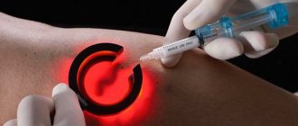
· Sclerotherapy: thanks to the action of a solution injected into a vein, a scar appears at the site of the pathological vessel.
· Laser surgery: the internal venous walls are irradiated with a laser, then the vein is glued together. There is no cut on the skin, no punctures in it.
· Ablation: the vein is destroyed by heating the electrodes of the catheter placed in the vessel.
· Endoscopic surgery: a flexible microcamera is inserted into the vein, and the doctor visually controls the removal of the vessel.
· To remove large veins, more extensive operations with incisions are performed.
· Shunting: in severe CVI, the venous vessels are connected with a special shunt so that blood flows bypassing the diseased vein.
All treatment methods in the vascular surgery department are selected individually.
Risk factors for CVI
A number of factors significantly increase the risk of developing chronic venous insufficiency, these include:
- Genetic predisposition, including pathologies of connective tissue and weakness of the vascular wall.
- Taking hormone-containing drugs, including hormonal contraceptives.
- Low physical activity, sedentary lifestyle, constant heavy lifting, excess weight.
- Chronic constipation.
CVI is more often diagnosed in women, since the formation of venous insufficiency is influenced by a high concentration of estrogens (female steroid sex hormones). The period of pregnancy and childbirth and the use of hormonal contraceptives have a negative impact. With age, the likelihood of developing the disease increases in both sexes due to prolonged exposure to adverse factors.
Causes of vein reflux
The main and most common cause of the appearance and development of venous reflux is a hereditary predisposition. Also factors that provoke the development of reflux of the veins of the lower extremities are:
- obesity;
- taking hormonal contraceptives;
- pregnancy and childbirth cause increased stress on the venous system;
- the onset of menopause. leads to significant hormonal changes in a woman’s body;
- prolonged stay in one position: standing, sitting, lying down;
- lack of physical activity.
Classification of CVI
- 1st degree:
is expressed in a feeling of heaviness, aching pain, passing, minor swelling and night cramps.
- 2nd degree:
persistent swelling, hyperpigmentation (darkening of the skin of the affected limb), lipodermatosclerosis (dystrophic changes in fatty tissue), dry and weeping eczema (serous inflammation of the dermis).
- 3rd degree:
To the above signs of stage 2, trophic ulcers, current or already healed, are added.
- There is also 0 degree
when the patient does not observe changes, there are no complaints, however, the pathological processes have already started. The treatment method for patients with zero degree CVI differs from the treatment of patients at other stages.
In international practice, chronic venous insufficiency is divided into 6 degrees according to clinical manifestations, including zero (CEAP system). The disease also has an etiological classification based on its causes:
- EC is a congenital pathology.
- ES is an acquired form as a result of thrombosis, varicose veins, or trauma.
- EP is an unclear cause of venous insufficiency.
There is also an anatomical classification based on location, taking into account pathophysiology, disability scale and a number of other factors.
Cost of treatment of venous insufficiency
You can find out prices for treatment of venous insufficiency, the cost of admission and consultation in Moscow, as well as make an appointment by phone or leave a request on the website. For diagnosis and treatment of venous insufficiency, contact medical in Moscow and Vidnoye
You may also be interested in:
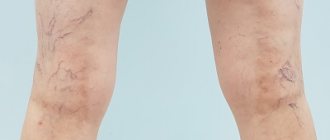
Removing spider veins on the legs
Laser treatment of varicose veins
Venous insufficiency
Symptoms of chronic venous insufficiency
At an early stage, chronic venous insufficiency has a broad clinical picture, expressed in one symptom, followed by a chaotic addition of other manifestations. As a rule, they appear or intensify after long periods of sitting or standing, as well as during periods of increased physical activity:
- heaviness in the legs;
- fatigue, buzzing;
- aching pain;
- transient swelling;
- numbness, temporary loss of sensation;
- cramps that occur at night.
If you regularly experience at least one of these symptoms, you should consult a doctor. Subsequently, with the development of pathology, pigmentation of the skin of the lower leg will occur, the skin will lose elasticity and become dry.
As it progresses (stages 2 and 3), symptoms such as:
- persistent swelling;
- increased pain during exercise;
- formation of ulcers and eczema;
- phlebeurysm.
Lack of blood circulation negatively affects not only the affected area, but also the entire body as a whole. Dizziness and fainting, high mental and physical fatigue may occur. Signs of heart failure are also noted: heart rhythm disturbances, pale skin, shortness of breath.
Prevention
It is necessary to fight excess weight, eat right, avoid prolonged static loads, move a lot, wear shoes with low heels, play sports (swimming, cycling, skiing).
If it is impossible to remove the provoking factors (heredity or work characterized by prolonged static loads), doctors recommend resorting to preventive compression hosiery, and for pregnant women - therapeutic ones.
Preventive measures help eliminate risk factors for CVI.
Diagnosis and treatment of CVI
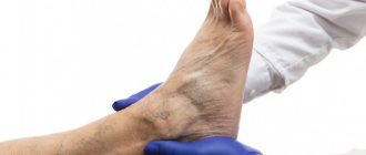
At an appointment with a phlebologist at the Ambulatory Surgery Center, the patient is diagnosed based on the medical history, the patient’s complaints, and the results of the study. The main method of instrumental examination is duplex ultrasound scanning. In our clinic, functional diagnostic doctors during the study not only confirm the presence of CVI, but also help surgeons determine the extent of damage to the venous system and the choice of tactics for further action. In some cases, the attending physician may prescribe a duplex angioscan or X-ray contrast study (phlebography).
Treatment of chronic venous insufficiency is a set of measures, the purpose of which is to restore the functionality of the venous and lymphatic systems, eliminate the pathologies that arise from them and prevent relapses. The method of therapy is selected individually for each patient, taking into account the degree of pathological changes, medical history, age and concomitant diseases.
Conservative treatment
A course of treatment is prescribed, lasting on average 2 - 3 months, then after a certain period it is resumed. The course usually includes:
- medicines (phlebotrobic drugs);
- local use of antiseptic ointments;
- corticosteroid drugs;
- elastic compression (bandages and compression hosiery);
- treatment of associated secondary infections;
- elevated position of the legs when lying down.
In some cases, antibiotics and diuretics are prescribed to reduce swelling. The greatest role in the routine treatment of CVI is played by elastic compression, including hardware, pneumatic, and leg elevation. Compression is indicated for all patients, even those with ulcers. In this case, I use elastic bandaging, and then wear special stockings. They should create a distal pressure of 20–30 mm Hg. for patients of the first stage, 30–40 mm Hg. – second and up to 60 mm Hg. at the third.
The effectiveness of therapy directly depends on the active participation of the patient. He needs to strictly follow the doctor’s recommendations and build his life in such a way that conditions that aggravate the course of the disease are not created.
Surgery
Surgery is performed only in 10% of patients with:
- severe concomitant diseases;
- trophic disorders;
- transformed tributaries of the great (small) saphenous veins;
- relapse of varicose veins and in other similar cases.
For this purpose, a minimally invasive surgery technique is chosen - miniphlebectomy.
. The operation is performed under local anesthesia. Through small punctures, the vascular surgeon gains access to the affected areas of the veins to perform ligation (vessel ligation), vein removal, and valve reconstruction. The tasks of a vascular surgeon include eliminating pathological blood discharge and truncation of varicose veins. After the operation, an hour later, the patient can go home; a hospital stay is not required.
DISEASES OF THE VENOUS SYSTEM OF THE LOWER LIMB
The paper outlines the currently available data on diseases of the leg venous system and shows differences between chronic and acute diseases, methods of treatment and prevention.
D.F. Beloyartsev Department of Vascular Surgery, Institute of Surgery named after. A.V. Vishnevsky RAMS, Moscow, Russia DF Beloyartsev Department of Vascular Surgery, AV Vishnevsky Institute of Surgery, Russian Academy of Medical Sciences, Moscow
P
According to the results of various medical statistical studies in industrialized countries (USA, UK, and also in Russia), up to 20% of the population suffers from diseases of the venous system of the lower extremities, which determines the social significance of the problem. In addition, it is necessary to take into account that in a significant number of cases (about 1% of the population) patients develop complicated forms of venous insufficiency, accompanied by persistent and long-term disability of patients [1,2]. It is necessary to distinguish between chronic and acute diseases of the veins of the lower extremities, as well as their consequences. Chronic conditions include congenital anomalies of the venous system, saphenous varices, perforating vein insufficiency, and deep vein valvular insufficiency. Among acute diseases, acute thrombophlebitis of the subcutaneous or deep veins is distinguished. The consequences of the latter include postthrombophlebitis syndrome.
Varicose veins of the saphenous veins of the lower extremities
The exact causes of this disease are not clear, but its occurrence is associated with hereditary disorders of the elasticity of the venous wall, realized through hormonal influences and against the background of venous hypertension [3]. This point of view is supported by the fact of the more frequent development of varicose veins in women, which is familial in nature and manifests itself during pregnancy or after childbirth. Mostly, varicose veins occur in the system of the great saphenous vein, less often in the system of the small saphenous vein, and begin with the tributaries of the vein trunk on the legs. The natural course of the disease at the initial stage is quite favorable; for the first 10 years or more, apart from a cosmetic defect, patients may not be bothered by anything. Subsequently, if timely treatment is not carried out, complaints of a feeling of heaviness, fatigue in the legs and their swelling after physical activity (long walking, standing) or in the afternoon, especially in the hot season, begin to appear. It is necessary to distinguish and accurately find out from the patient what kind of symptoms are bothering him. Most patients complain of pain in the legs, but upon detailed questioning it is possible to reveal that this is precisely a feeling of fullness, heaviness, and fullness in the legs. With even a short rest and elevated position of the limb, the severity of sensations decreases. It is these symptoms that characterize venous insufficiency at this stage of the disease. If we are talking about pain, it is necessary to exclude other causes (arterial insufficiency of the lower extremities, acute venous thrombosis, joint pain, etc.). Subsequent progression of the disease, in addition to an increase in the number and size of dilated veins, leads to the occurrence of trophic disorders, often due to the addition of incompetent perforating veins and the occurrence of valvular insufficiency of the deep veins. Trophic disorders at the initial stage are manifested by local hyperpigmentation of the skin, then thickening (induration) of subcutaneous fatty tissue occurs until the development of cellulite. This process ends with the formation of an ulcerative-necrotic defect, which can reach a diameter of 10 cm or more, and extend deep into the fascia. The typical place of occurrence of venous trophic ulcers is the area of the medial malleolus, but the localization of ulcers on the lower leg can be different and multiple. At the stage of trophic disorders, severe itching and burning in the affected area occur; Some patients develop microbial eczema. Pain in the area of the ulcer may not be expressed, although in some cases it is intense. At this stage of the disease, heaviness and swelling in the leg become constant. Venous ulcers differ from arterial ulcers in that the latter develop starting from the distal parts of the limb (in particular in diabetes mellitus), necrotic defects in venous insufficiency are not accompanied by perifocal inflammation, the ulcers are deep, with undermined vertical edges, the bottom along with scanty fibrinous-serous discharge covered with granulations, the skin around the ulcers is pigmented, the subcutaneous tissue is indurated. It should be remembered that there are no medications that can prevent further progression of already existing saphenous varicose veins. The only way to prevent this is elastic compression of the limbs. Moreover, the patient should start using elastic stockings or bandages in the very morning, without getting out of bed, when the saphenous veins are not yet filled, and wear them throughout the day while he is on his feet. Otherwise, there will be no effect from elastic bandaging. Applying bandages should begin from the fingertips and evenly cover the entire limb, leaving no free areas to the required level. Naturally, few people are able to strictly follow these rules, and therefore the main methods of treating varicose veins remain surgical and, to a lesser extent, injection.
Before starting treatment for varicose veins, the doctor must have a very clear picture of the condition of the deep and perforating veins of the limb. Today, not a single phlebological patient can be left without an ultrasound examination. It is this study, non-invasive, extremely informative in experienced hands, short in time and completely unburdensome for the patient, that has become the main one in the diagnosis of venous insufficiency. The most modern technique is duplex scanning with color Doppler mapping, which allows us to identify the patency and condition of the valves of the deep veins, from the tibia to the inferior vena cava, and the direction of blood flow in the perforating and superficial veins. If this method is not available, patients need to undergo standard Doppler ultrasound, although it is inferior in information content and diagnostic accuracy. After the widespread introduction of ultrasound techniques into practice, the role of classical venography has largely disappeared. Today, this technique is used quite rarely, mainly when reconstructive operations (bypass or plastic) are necessary on the deep veins of the limb, and every year the frequency of phlebography is decreasing due to the accumulation of experience and increasing capabilities of ultrasound diagnostics. Nowadays, the classic operation for varicose veins, radical phlebectomy in various modifications, most often according to Troyanov-Trendelenburg, Babcock, Narat, has undergone significant changes. First of all, the cosmetic requirements for the operation are placed, which include the use of small incisions, atraumatic instruments, making skin incisions in the “correct”, “cosmetic” directions, the use of atraumatic threads and sutures when closing the skin. It is believed that phlebectomy itself as a surgical intervention is not complicated, so all over the world it is more often performed in general surgical hospitals. But it is precisely patients who have passed by specialized phlebological or angiosurgical departments who most often encounter recurrences of varicose veins and gross cosmetic defects after operations. The causes of relapses can be divided into two large groups. The first is the errors of surgical technique, which are often found in non-specialized institutions and consist in non-radical phlebectomy. The second group is associated with incomplete preoperative diagnosis. In patients, severe insufficiency of deep vein valves or incompetence of perforating valves is missed (no ultrasound examination is performed), and the standard phlebectomy loses all meaning, since the cause of varicose veins remains. These patients require expanded surgical intervention, including ligation of perforating veins and/or correction of the valvular apparatus of the deep veins. Indications for injection therapy (sclerotherapy) for varicose veins are still being debated. The method consists of introducing a sclerosing agent into the dilated vein, its further compression, desolation and sclerosis. Modern drugs used for these purposes are quite safe, i.e. do not cause necrosis of the skin or subcutaneous tissue when administered extravasally. Some specialists use sclerotherapy for almost all forms of varicose veins, while others reject the method completely. Most likely, the truth lies somewhere in the middle, and it makes sense for young women with the initial stages of the disease to use the injection method of treatment. The only thing is that they must be warned about the possibility of relapse (higher than with surgical intervention), the need to constantly wear a fixing compression bandage for a long time (up to 3 - 6 weeks), the likelihood that complete sclerosis of the veins may require several sessions. The group of patients with varicose veins should include patients with telangiectasias (“spider veins”) and mesh dilatation of small saphenous veins, since the causes of the development of these diseases are identical. In this case, sclerotherapy is the only treatment method after excluding damage to the deep and perforating veins.
Insufficiency of perforating veins of the lower extremities
Isolated disease is quite rare. More often, insufficiency of perforating veins is combined with varicose veins of the saphenous veins or develops in patients with postthrombophlebitis syndrome due to impaired venous outflow from the limb. The clinical picture with isolated insufficiency of perforating veins is similar to that with varicose veins. Visually, more pronounced swelling is noted, dilatation of the saphenous veins of the leg may not be detected, trophic disorders arise faster and are more severe. In terms of differential diagnosis, it should be taken into account that if trophic disorders are cyclical in nature, then we are most likely talking about postthrombophlebitic syndrome. In case of insufficiency of perforating veins, trophic disorders are limited to any of the surfaces of the leg (lateral, medial, posterior). Instrumental diagnosis is based on precise topical localization of pathologically functioning perforating veins using duplex scanning with color Doppler mapping. With moderate swelling and the absence of trophic changes, the use of elastic compression is sufficient. In other cases, patients require surgical treatment. This is a rather complex section of surgery, since many patients with insufficiency of perforating veins have extensive ulcerative defects. In some cases, it is not possible to achieve independent closure of a trophic ulcer after correction of the venous blood flow (ligation of perforating veins), and such patients require immediate or delayed plastic surgery of the ulcer. The situation is complicated by the need to make incisions through trophically altered areas of the skin, after which surgical wounds heal poorly. In recent years, to eliminate these issues, endoscopic methods of ligation of perforating veins have been successfully used, which has dramatically reduced the number of complications in the postoperative period and shortened the length of stay of patients in the hospital.
Deep vein valve insufficiency
This venous involvement also rarely occurs in isolation. The clinical picture of the disease is manifested by swelling, fatigue and heaviness in the legs during physical activity, towards the end of the day, especially in the hot season. Diagnosis of the disease is based on duplex scanning with color Doppler mapping. It should be remembered that valvular insufficiency of the deep veins may be hidden and not detected in a horizontal position of the patient. When it moves to a vertical position, ultrasound examination can reveal clear venous reflux. For moderately severe deep vein valve insufficiency, elastic compression is sufficient for treatment. With significant reflux, patients are advised to undergo corrective surgery on the deep vein valves. The optimal operation has not been determined today; extravasal correction of valves with a Vedensky spiral, transplantation of a segment of the saphenous vein with a normally functioning valve, and plastic surgery of the leaflets of an incompetent valve are more often used. When lesions of the deep vein valves are combined with varicose saphenous veins and/or insufficiency of the perforating veins, the intervention is supplemented by phlebectomy and/or ligation of the perforating veins.
Acute thrombophlebitis of the great saphenous vein
Acute thrombophlebitis of the great saphenous vein and its tributaries is one of the most typical complications of varicose veins of the saphenous veins of the lower extremities. The clinical picture of the disease is manifested by the sudden appearance of hyperemia along the great saphenous vein, pain at rest and upon palpation of the soft tissues around the inflamed and thrombosed vein; it is often possible to palpate a cord in the lumen of the vein. Phenomena of inguinal lymphadenitis may be observed; body temperature rarely rises. The process begins, as a rule, in the upper third of the leg, and spreads proximally, to the mouth of the great saphenous vein, sometimes very quickly, within several hours, almost never involving the tributaries of the great saphenous vein over a significant distance. One of the variants of thrombophlebitis of the saphenous veins, migrating thrombophlebitis with Buerger's thromboangiitis, on the contrary, is more often limited to the veins of the leg, affecting not only the trunk of the great saphenous vein, but also its large tributaries throughout. In this situation, the process rarely spreads to the thigh, and therefore only symptomatic therapy is performed for thromboangiitis. Treatment of classic thrombophlebitis of the great saphenous vein begins with the introduction of direct anticoagulants, non-steroidal anti-inflammatory drugs, drugs that improve venous outflow and rheological parameters of blood, local ointment compresses and elastic bandaging of the limb. As soon as the proximal area of hyperemia or infiltration reaches the border of the upper and middle third of the thigh, the patient is indicated for surgery - ligation of the great saphenous vein at the level of the saphenofemoral anastomosis to prevent pulmonary embolism. During such an operation, the thrombosed trunk and varicose veins cannot be removed. There is a process of active inflammation in the body, and the likelihood of purulent complications is very high, not to mention the fact that in the acute period it will not be possible to achieve a cosmetic effect. Patients who have undergone ligation of the saphenofemoral anastomosis due to acute thrombophlebitis of the great saphenous vein are subject to phlebectomy after 2 - 3 months, in the “cold” period to prevent recurrent thrombophlebitis. The concept presented above about the level of spread of a process requiring surgical intervention was revised after the introduction of duplex scanning into practice. It turned out that often when visual and palpation assessment of the height of thrombosis on the thigh is low, according to ultrasound examination there is thrombosis of the saphenofemoral anastomosis or even a thrombus floating in the common femoral vein. Currently, any patient with suspected acute thrombophlebitis of the great saphenous vein should undergo a duplex scan to determine the risk of developing pulmonary embolism. If there is a possibility of a thrombus spreading into the deep venous system, the scope of the operation is expanded to inspect the common femoral vein.
Acute deep vein thrombosis of the lower extremities
The causes of the disease have not been precisely established. It is known that the process begins with a combination of damage to the vascular wall, blood stasis and a violation of the rheological properties of the blood. Many risk factors are considered, such as, for example, the use of oral contraceptives by young women, mass formations of the pelvis and retroperitoneal tissue, prolonged bed rest, paraplegia, the postpartum period, cancer, however, it is difficult to identify specific causes in a significant number of cases. The pathogenesis of the disease lies in the acute occurrence of an obstruction to venous outflow and redistribution of blood along collaterals against the background of inflammation of the venous wall. Next, the process of recanalization of thrombosed veins and restoration of pathological blood flow through them begins. This period ends by the 6th month. Even in recanalized veins, blood flow does not become normal because the lumen of the vein does not reach its original diameter, and destruction of the valves after thrombosis leads to retrograde blood flow. Clinically, the onset of the disease is manifested by a sudden onset of pain, swelling of the limb and cyanosis of the skin. The localization of symptoms depends on the height of the thrombosis and the extent of the lesion. If the process affects the inferior vena cava, then bilateral swelling of the extremities occurs. With proximal involvement of the iliac segment, unilateral edema of the entire limb is noted. Thrombosis of the femoral-popliteal zone is accompanied by symptoms of damage below the knee joint, as well as occlusion of all veins of the leg. The severity of pain and circulatory disorders is largely determined by the degree of involvement of several segments and the state of collateral outflow. For acute venous thrombosis of the deep veins, a characteristic symptom is pain in the muscles and tissues along the neurovascular bundles. One should remember about the possibility of thrombosis of the muscular veins of the leg, in which the clinical picture of the disease is identical to that described, but there is no obstruction of the patency of the main veins. Sometimes isolated thrombosis of one of the main (or a pair of them) veins of the leg occurs. In this case, the clinical picture of the disease is manifested only by pain. It is extremely rare that with severe damage to the main veins and poor collateral outflow pathways in acute venous thrombosis, venous gangrene occurs, in which case amputation of the limb is required. Instrumental diagnosis of the disease is based on duplex scanning with color Doppler mapping, which makes it possible to accurately determine the level and extent of vein involvement in the thrombotic process. When the iliac and inferior vena cava are affected, retrograde cavagraphy is necessary to identify floating thrombi, which is of great importance for the prevention of pulmonary embolism. The main way to help a patient with deep vein thrombosis as much as possible and to prevent severe circulatory disorders as much as possible is surgery. The earlier thrombectomy is performed, the greater the chance of restoring normal outflow through the deep veins of the limb. It is believed that performing thrombectomy on days 10–14 is no longer effective due to the tight adhesion of the thrombus to the venous wall and destruction of the valves. In the presence of floating thrombi in the iliac and inferior vena cava at such late stages, endovascular installation of a vena cava filter in the inferior vena cava is indicated - a device that can retain the thrombus when it is torn off from the veins of the limb and prevent pulmonary embolism. If thrombectomy is impossible or ineffective, conservative therapy is carried out, the purpose of which is to stop the process of thrombus formation that has already begun, achieve the fastest and most complete recanalization and develop collateral outflow pathways. Conservative measures include the appointment of direct anticoagulants (intravenous heparin). The loading dose of heparin is 5000 - 10,000 units. The size of subsequent doses depends on the partial thromboplastin time (which should be 1.5 - 2 times higher than normal values). This achieves adequate anticoagulation with a low incidence of hemorrhagic complications. During heparin therapy, oral warfarin is started. In case of deep venous thrombosis, elastic compression must be used with extreme caution. In patients with the only outflow route through the great saphenous vein, bandaging can only worsen the condition of the limb.
Postthrombophlebitic syndrome
Postthrombophlebitic syndrome is a symptom complex that develops in patients who have suffered acute deep vein thrombosis and is a consequence of circulatory disorders in the limb due to the failure of the valve apparatus of the deep veins after thrombosis in the areas of recanalization, the lack of patency of the deep veins in non-recanalized segments and valvular insufficiency of the subcutaneous and perforating veins. veins due to overload of venous blood flow as the main routes of collateral outflow. The clinical picture of this syndrome is quite multifaceted, characterized by both symptoms of chronic venous insufficiency (fatigue, swelling, heaviness in the legs, up to the inability to remain in an upright position for any more or less long time), and quickly arising and severe trophic disorders . Very often these patients become severely and permanently disabled. The task of providing medical care to such patients is very difficult and is solved very individually in each case. Only a small group of patients can be helped by removing dilated saphenous veins or ligating perforating veins. An even smaller number of patients are indicated for shunt operations on the venous system, aimed at restoring the patency and valvular integrity of the deep veins. Patients with postthrombophlebitis syndrome are doomed to lifelong use of drugs that improve the rheological properties of blood and the state of venous outflow, and to wearing elastic stockings (or bandages). However, as mentioned above, elastic compression does not always help them. It is very important to monitor the condition of the venous system of the lower extremities in such patients using periodic ultrasound examinations.
Congenital diseases of the venous system
By their nature, these diseases resemble “congenital postthrombophlebitic syndrome.” The causes of such lesions are not precisely known, but, apparently, the main role is played by teratogenic effects during the period (8 - 12 weeks) of the formation of the vascular system of the embryo. The types of vein damage are very diverse. This includes the absence or hypoplasia of the main venous trunks, the absence or incompetence of the valves of the deep and superficial veins, the presence of “extra”, pathologically formed veins (lateral embryonic vein), etc. Everything that has been said regarding postthrombophlebitis syndrome is also true for congenital venous diseases, with one exception. The clinical picture of the disease occurs in younger patients; more often the disease manifests itself at puberty, but the possibilities to help them are even more limited, and the clinical manifestations of the disease are usually more severe. In conclusion, we note that the problem of treating venous insufficiency of the lower extremities is far from being resolved. Unfortunately, uncomplicated diseases of the veins of the lower extremities do not always receive the deserved attention of doctors, especially non-surgical specialists. Outwardly, patients with venous insufficiency of the lower extremities often appear to be healthy and active for many years. But this, unfortunately, is not always the case. When faced with complicated and advanced cases as an angiosurgeon, you clearly see how much was missed when these patients were in the compensated stage of the disease. We hope that the presented brief overview of the treatment of venous insufficiency of the lower extremities will help doctors not involved in vascular surgery to better navigate the problem.
Literature:
1. A.V. Gavrilenko, S.I. Skrylev, F.A. Radkevich. Surgical methods for correcting valvular insufficiency of the deep veins of the lower extremities. - Angiology and vascular surgery. - 1997. - No. 2. - P. 127 - 34. 2. Jimenez Cossio JA. Epidemiology of varicose veins. — Phlebolymphology. - 1996. - No. 1. - P. 8 - 12. 3. Bergan JJ. Advances in evaluation and treatment of chronic venous insufficiency. — Angiology and Vascular Surgery. - 1995. - No. 3. - P. 59-80.
![Rice. 1. Functioning of the glymphatic system of the brain (according to [11]) 1. Glymphatic system functioning (according to [11])](https://expert35.ru/wp-content/uploads/ris-1-funkcionirovanie-glimfaticheskoj-sistemy-mozga-po-11-fig-1-330x140.jpg)