Briefly about the treatment method
A femoropopliteal bypass is an open surgery to create a bypass for blood flow when there is a blockage in the femoral artery in the leg. The operation is indicated for critical ischemia and the threat of loss of a limb, but sometimes also for limiting intermittent claudication, if the patient does not have enough walking distance for everyday life. The operation consists of connecting a shunt (artificial vessel) to the common femoral artery in the groin area and passing it to the patent area of the popliteal artery above or below the knee joint. Femoropopliteal bypass surgery can be performed using an artificial prosthesis or using the patient's own vein. The operation usually lasts about 60 minutes and is performed under epidural or spinal anesthesia. The operation requires at least two surgical incisions. The efficiency of the operation is high. When used according to indications, the shunt patency is 80% within 5 years.
Treatment of critical lower limb ischemia (CLI) is one of the most difficult issues in modern vascular surgery. The incidence of CLI reaches 500–1000 cases per 1 million population per year [29].
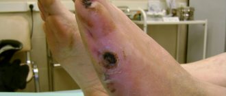
To treat a patient with CLI, the doctor chooses one of two options: amputation of the affected limb or an attempt to restore blood flow through it. Currently, from 70 to 90% of amputations in the world are performed due to CLI; in Russia this figure is even higher [8, 9, 22]. When a leg is amputated, the quality of life sharply decreases and its duration decreases [32, 33, 41]. From a surgical point of view, amputation in patients of this category cannot be the operation of choice. With amputation below the knee joint, up to 60% of purulent-necrotic complications develop [38], repeated amputation in this case is required in another 15% of patients [29], and the mortality rate when performed exceeds 30% [12]. The wound after amputation above the knee heals better, but the procedure worsens the patient’s ability to rehabilitate.
The most important task for limb preservation is maintaining blood flow at the minimum required level. This is achieved in three ways [26, 28, 31, 45]:
— direct revascularization ( bypass surgery or percutaneous angioplasty of the great arteries (PCMA)
;
— indirect revascularization: denervating interventions, stimulation of neoangiogenesis, surgical (perforating osteotrepanation, transplantation of the greater omentum to a limb) and therapeutic (introduction of bone marrow mononuclear cells, gene preparations, platelet suspension);
- drug treatment.
A unified tactic for treating such patients has not yet been determined; each of the authors tries to show the most optimal method from his point of view as a “panacea”.
It is generally accepted that the most effective reconstruction option is direct limb revascularization. However, when performing interventions, the doctor is faced with a number of questions about the cost-effectiveness of the procedure, long-term results and the greatest safety for the patient.
Most patients with CLI have severe concomitant diseases, which increase mortality in the immediate postoperative period [21]. The best treatment option must be: minimally invasive, safe, easy to perform and accessible to use.
CLI is a life-threatening condition in which the main goal is to preserve the limb and life of the patient. From our point of view, treatment of CLI can and should be divided into two stages:
— Stage 1: direct limb salvage
the least invasive method with subsequent stabilization of the clinical condition. This stage should provide compensation for blood circulation in the leg. We consider the optimal condition to be the presence of main blood flow within 3 months after reconstruction. This period is sufficient to conduct a thorough examination and correction of concomitant diseases, for example, performing myocardial revascularization and/or reconstruction of brachycephalic arteries (BCA), as well as rehabilitation after these interventions;
— Stage 2: surgical reconstruction aimed at maintaining limb function “indefinitely” for a long time
.
Based on the above, we will analyze treatment options in 2 clinical situations: damage at the level of the femoropopliteal segment and at the level of the popliteal tibial segment. The aortofemoral segment was excluded from this review, since, according to the general opinion, this issue has been resolved in the international community: restoration of adequate flow to the leg is the key to the quality of care and does not depend on the treatment method (bypass operations have similar long-term patency rates compared to PCI) [24 ].
Restoration of blood flow in the femoral-popliteal segment
When planning reconstruction of the femoropopliteal segment, the surgeon is faced with a choice of 3 technologies: bypass surgery, PCI, or PCI with stenting. Modern surgical science believes that the most optimal situation for performing PCI is a short (up to 10 cm) stenosis or occlusion of the superficial femoral artery (SFA) [47]. Recently, work has been carried out to assess the safety of PCI in patients with extensive lesions, as well as lesions extending to the popliteal artery (class C and D according to the TASC II classification). The authors demonstrate a high percentage of successful PCI (about 94%) and satisfactory long-term results up to 2 years after the intervention [23]. Such results became possible with the advent of hydrophilic guidewires and subintimal angioplasty techniques. A large meta-analysis [18] demonstrated that primary PCI shows similar long-term results compared with PCI + stenting. The same work noted similar indicators of quality of life in patients of both groups. Other studies have shown that the 2-year patency of the stented segment without damage to the popliteal artery reaches 87%, and with its damage, only 75% of reconstructions work after 1 year [20, 46]. G. Rigatelli et al. [45] suggested using long-term balloon angioplasty with a pressure of 18 atmospheres for 2 minutes with repeated sessions until less than 30% of residual stenoses remain on the angiogram. According to their data, the average duration of dilatation was 5.9±1.9 minutes with the need for stent implantation in 11.9% due to the development of dissection. Primary patency of the reconstruction zone was 86.7% within 2 years, which corresponds to the results with implantation of covered stents. It should be noted that more than 60% of patients in this group had TASC C lesions.
Primary surgical revascularization is accompanied by low mortality and almost 100% effectiveness in saving the patient's limb. Based on literature data [35], it can be argued that the degree of preservation of the limb in the early stages after surgery is the same, regardless of the material used for bypass.
Femoropopliteal bypass (FPB) is currently a standard vascular operation that provides satisfactory results regardless of the extent of the FPA lesion [42]. When assessing long-term results (up to 5 years), it was shown that the patency of the autovenous vein is almost twice as high as when using a prosthesis (75% versus 42%, respectively) [49]. Taking these data into account, when performing reconstruction, it is recommended to use an autovenous shunt from the great saphenous vein [2], however, the results of the effective use of cadaveric allografts in this position have been described [26, 39].
The latter are more often used for infection of a previously installed shunt, but their standard use as biological material will reduce the time of surgery with satisfactory long-term results. The preserved native saphenous vein can be subsequently used for myocardial revascularization.
To improve long-term results, hybrid operations can be used: PCI in the proximal part and BPS with a short bypass from the distal part of the SFA to the popliteal artery (RCA) [43]. Another study [51] demonstrated that this approach reduces the number of postoperative complications by 2 times, but in the long term, limb salvage rates remain similar to surgical revascularization.
The difficulty of reconstructing the femoral-popliteal segment is its dependence on the condition of the tibial arteries. The better the distal channel, the better the results. Considering that about 17% of patients have an unsatisfactory course, it is necessary to think through the tactics of simultaneous treatment in this area of the limb [1].
In our opinion, in case of CLI, stage-by-stage correction of the channel is the most justified. This is due to the systemic reactions of the body to the start of blood flow after prolonged ischemia of the limb [1]. The main task of the surgeon is to select a group of patients in whom, immediately after restoration of blood flow through the SFA, reconstruction of the tibial arteries is necessary due to the high risk of early thrombosis of the reconstruction zone. For these patients, in our opinion, PCI of the tibial arteries should be performed. For patients with satisfactory blood flow, isolated reconstruction of the femoral-popliteal segment with further measures aimed at improving blood flow in the leg is indicated.
We believe that in patients with CLI at stage 1, PCI of the SFA should be performed without stenting. Indications for stenting are cases of intimal dissection and significant residual stenosis. Even with unsatisfactory mid-term results, this approach will allow you to save the limb, begin rehabilitation and continue treatment in conditions of chronic ischemia, using all available methods of correction of the distal bed and/or repeated endovascular interventions. It is also worth considering that PCI can be repeated an “unlimited” number of times to improve long-term results [3].
Restoration of blood flow along the popliteal tibial segment
With atherosclerotic lesions of the arteries below the level of the knee joint, surgeons are faced with the problem of low effectiveness of reconstructive operations. Surgical treatment in this area is associated with frequent limb loss as a result of rapid thrombosis of the shunt [5, 11].
The use of endovascular methods makes it possible to improve the outflow through the arteries of the leg, open occlusions and, importantly, restore blood flow along the plantar arch [7]. The latter is most significant for patients with diabetes, as well as in cases of threat of minor amputation. The problem with PCI is the rapid “restenosis” of the tibial arteries. When blood flow in the lower leg is corrected, the condition of the limb returns to the preoperative level in 48% of those operated on within 1 year [15, 33, 34]. Signs of restenosis during a follow-up examination are found in 2/3 of patients after 3 months [48]. For patients with diabetes, these results are of less importance - the main task in diabetic foot is to eliminate trophic changes (as the main infectious agent) during this period. The indicated 3 months are sufficient to resolve this issue in most patients [7]. Limb preservation in patients with CLI was observed in 96.7% after PCI was performed at the level of the leg [17].
In patients with CLI, amputation is performed due to lack of blood supply to the tissues. The surgeon is faced with a choice: to save the limb, it is necessary to perform angioplasty every 3-6 months, which is not economically justified, or during this period it is necessary to provide an alternative path of blood flow through the lower leg and foot.
The first option has high labor intensity and cost. To perform isolated PCI, it is necessary to improve modern technical support and drug approaches to treatment. The most common practice to improve the results of PCI is the implantation of stents in the area of stenosis after angioplasty, but a large meta-analysis [50] demonstrated the lack of effectiveness of this approach in peripheral arteries.
After peripheral angioplasty, all patients are prescribed dual antiplatelet therapy for 3 months, which is believed to improve long-term results [25]. At the same time, such studies have not been conducted directly for stenting the tibial arteries. Cilostazol may also be a drug that improves the results of PCI at the tibial level. In other vascular territories, it can significantly reduce the incidence of restenosis and amputation [37, 40].
The second way involves a hybrid approach. The use of angiogenesis stimulation seems to be a justified method. To date, two methods have been described: therapeutic angiogenesis - the introduction of bone marrow mononuclear cells, platelet-rich plasma or gene drugs [5, 13, 16, 30], and surgical angiogenesis - through transplantation of the greater omentum [10].
Therapeutic angiogenesis makes it possible to increase the distance of pain-free walking, reduce pain in CLI and reduce the frequency of amputations [6]. At the same time, the method is quite new and scientists still do not have an accurate understanding of the mechanism of action and the frequency of side effects, especially the development of malignant neoplasms. Surgical angiogenesis, as we understand it, is the use of the greater omentum to improve blood supply to the tissues of the leg and foot. Implantation of the omentum on the vascular pedicle has two places of application: closing skin defects and “sprouting” the muscles of the limb with vessels from adipose tissue. The main limitation for the successful use of greater omentum transplantation for ischemia of the lower extremities is the functional state of the regional circulation [14]. Freedom from amputation in patients with CLI for up to 5 years using combined treatment, shunt surgery together with implantation of the greater omentum on the lower leg, reaches 87%, while 42% had no restrictions on the duration of walking due to intermittent claudication [36].
Tactics of revascularization of the lower limb in critical ischemia
The effectiveness of the fight to save a limb in CLI in most cases depends on the condition of the distal vascular bed. With minimal changes in the tibial arteries, any method of reconstruction of the femoral-popliteal segment is suitable. A large study [19] demonstrated that in periods of up to 2 years, limb preservation is similar between BPS and PCI of the SFA; in longer periods, a clear advantage of using an autovein was revealed over other methods. Considering the severity of the patient's condition, PCI is probably the operation of choice with further preparation of the patient for distal bypass surgery.
If the distal flow is unsatisfactory, it is necessary to restore blood flow through the maximum number of tibial arteries using PCI. This allows the surgeon to stabilize the clinical situation and gives time to select a balanced tactic and correct concomitant pathology. Restenosis often develops within 3 to 6 months. During this time, it is necessary to compensate the limb and, if possible, improve the volume of the receiving distal channel. The most justified tactic may be the use of angiogenesis methods. Therapeutic angiogenesis has been poorly studied, so we propose the use of greater omentum transplantation. In our opinion, it needs to be transplanted on a vascular pedicle. The tributary artery can be any distal branch of the deep femoral artery. These branches are rarely affected by atherosclerosis and are located away from the access points to the SFA. We consider an important surgical technique to be the distribution of the mass of the greater omentum in the area of accumulation of the main muscle mass and its passage to the distal parts of the foot.
Based on the analyzed literature, it is not advisable to perform this operation in the acute period of ischemia, since for the best engraftment of the transplanted tissue, compensation of blood flow through the limb is necessary. In our opinion, this particular operation will allow the limb to be preserved for the longest time.
As a possible option, hybrid surgery should be considered: BPS + PCI of the tibial arteries. Among the disadvantages of the method, one can note the relatively short period of restoration of blood flow in the lower leg, the need for monthly monitoring of the patency of the tibial arteries, and repeated PCI in case of significant stenosis. These shortcomings can probably be solved through the use of modern antiplatelet agents.
Advantages of treatment at the ISC
The technique of femoral-popliteal bypass in our clinic has been developed to pinpoint precision. We use this operation most often as an element of hybrid surgery for severe lesions of the arteries of the lower limb. To improve the results of treatment of patients with multi-story lesions, we use a femoropopliteal bypass, through which we then perform angioplasty and stenting of the arteries of the leg. There is no need to perform femoral-popliteal bypass as an independent operation, since with isolated blockage of the femoral artery, patients most often do not have serious complaints.
A special feature of the operation in our clinic is mandatory ultrasound monitoring of blood flow during surgery. If we identify problems with the shunt, we perform X-ray contrast angiography and can use angioplasty to improve the results of the intervention.
Preparing for treatment
Preparation for surgery consists of a special examination to determine the nature of vascular damage:
- Ultrasound of the arteries of the lower extremities
- MSCT of the aorta and arteries of the extremities
General examination, determining concomitant pathology and risks of surgery:
- Echocardiography (ultrasound of the heart)
- Gastroscopy (EGD)
- X-ray of the lungs
- A set of tests for hospitalization
Direct preparation for the intervention:
- Shaving of the surgical field from the groin to the middle third of the leg is carried out on the day of surgery
- Do not eat after seven o'clock on the evening before surgery
- Cleansing enema at night
- Installation of a urinary catheter (directly on the operating table)
Why is the procedure necessary?
This operation is performed in the presence of steno-occlusive lesions of the femoral and iliac arteries. Helps prevent the development of atherosclerotic processes and avoid:
- lack of blood supply to the lower extremities;
- intermittent claudication;
- fatigue of the muscles of the lower leg, thigh and buttocks;
- obliterating atherosclerosis;
- ulcers;
- ischemia;
- gangrene;
- amputation.
Restoring the aorta eliminates pain in the buttock area, and the femoral and iliac arteries – in the lower leg and thigh area. The operation is also a prevention of foot ischemia.
Pain relief during treatment
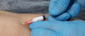
The operation can be performed under local, spinal or epidural anesthesia. In our clinic, epidural anesthesia is used predominantly, since an epidural catheter allows for effective pain relief in the postoperative period.
The catheter is inserted into the lumbar region using a special needle. After inserting the catheter, a small amount of anesthetic is injected into the epidural space, and the patient talks about his sensations. Gradually, the dose increases and sensitivity in the legs is turned off, and then movement.
During the operation, the doctor connects a tracking monitor to the patient, which measures blood pressure using a cuff on the shoulder and takes a three-lead ECG. In addition, a special sensor is placed on the finger of the hand - a pulse oximeter, which measures the pulse wave and blood oxygen saturation (saturation).
How the treatment method works
The patient is placed on the operating table on his back. A bolster is placed under the knee of the affected leg. The entire leg and groin area is treated with a special antiseptic solution. The foot is placed in a special diaper, the groin area is also covered with a diaper.
Revision of the outflow artery
The incision to evaluate the artery can be in the lower third of the thigh (above the knee) or just below the knee joint, depending on the patency of a particular part of the artery according to the preliminary examination. The length of the incision is usually 7-10 centimeters.
After isolating the artery, the surgeon evaluates its density, the presence of atherosclerotic plaques, and using a two-pincer test, the occupancy of the artery from the lower leg is assessed. If the artery is found suitable for bypass, then it is taken on a holder; if not, then it is isolated in another area.
Revision of the tributary artery and proximal (upper) anastomosis
The next incision is made in the groin area. The common femoral artery should be isolated there, along with its branches - deep and superficial. This section of the artery is where the shunt will be powered from.
If the common femoral artery is suitable as a donor vessel, then it is clamped with vascular clamps. Before this, a heparin solution is administered intravenously. After clamping the artery, it is dissected longitudinally, by 2-3 cm. A vascular prosthesis is sewn into the incision of the artery with a blanket suture. Immediately after it is sewn in, the clamps are removed and the prosthesis is filled with saline solution with heparin.
Carrying out a shunt
The next step is to create a tunnel under the skin to accommodate the vascular graft. We try to place the prosthesis as deep as possible, under the muscles, since in case of infection in the wounds, it does not reach the infected area.
Distal anastomosis (lower anastomosis)
The prosthesis is removed into the lower wound, where a longitudinal incision is also made in the artery, and blood flow from the underlying sections is assessed. The prosthesis is sewn with a vascular suture into the artery wound. All vascular clamps are removed and blood flow starts.
Control study after reconstruction
In our clinic, this is always followed by intraoperative ultrasound of the restored arteries. Blood flow through the shunt and popliteal artery, and blood flow closer to the foot is also assessed. Tests with clamping of the shunt are required to assess how dependent the blood flow in the leg is on this shunt.
If a control ultrasound reveals problems with the patency of the artery in the anastomosis or in the arteries below, high resistance along the shunt, then we perform X-ray angiography to identify possible problems. If vascular narrowing is detected, we perform angioplasty or stenting of the problematic arteries.
Completing the operation
The bleeding of anastomoses is assessed. If no bleeding is observed, then special silicone tubes (drains) are installed in the wounds, which are connected to plastic bulbs. This is necessary to remove fluid accumulation near the anastomoses and control possible bleeding.
The patient's wounds are sealed with special bandages and he is transferred to the department for further observation.
Method for treating occlusions of the femoropopliteal arterial segment
The invention relates to medicine, namely to vascular surgery. The prosthesis and stent are selected on a balloon catheter, the end of the prosthesis is turned out in the form of a cuff, a balloon catheter with a stent is passed through the prosthesis to 2/3 of the length of the stent, the connected ends of the vessel and prosthesis are compared by inflating the balloon. In this case, the anastomosis is formed at the border of the initial and middle sections of the popliteal artery. To perform an end-to-end anastomosis, the posterolateral wall of the artery is dissected and the anteromedial semicircle of the anastomosis is formed using the arterial wall. When performing an end-to-side anastomosis, an arteriotomy is made in the lower corner of the wound, the anastomosis is formed with occlusion of the outlet portion of the popliteal artery with an inflated balloon. The method expands the arsenal of means for treating occlusion of the femoral artery during alloplasty of the femoropopliteal arterial segment. 4 ill.
The invention relates to medicine, namely to vascular surgery, and can be used in the formation and reconstruction of the distal anastomosis during alloplasty of the femoropopliteal arterial segment.
Surgical treatment of patients with obliterating atherosclerosis of the vessels of the lower extremities has a long history and is widely used in clinical practice. However, early retrothrombosis is observed in 4-25% of patients, and femoropopliteal reconstructions are complicated 10 times more often than aortoiliac reconstructions. Late thrombotic reocclusion of the femoropopliteal segment occurs in 22-60% of cases. Currently, many authors consider synthetic prostheses made of polytetrafluoroethylene with a predominant subcutaneous method of graft placement as an option for choosing a shunt in the femoropopliteal position above the knee joint gap. At the same time, the maximum patency of GORE-TEX prostheses for up to 5 years does not exceed 60%. Most often, the causes of early thrombosis are technical (anastomotic stenosis, angulation of the prosthesis) and tactical errors; the etiology of late reocclusions is dominated by the progression of atherosclerosis, neointimal hyperplasia with stenosis of anastomoses, slowdown and distortion of the process of endothelialization of shunts, and bends of the prosthesis with scars.
From clinical practice, the authors of the present invention know that a polytetrafluoroethylene prosthesis does not have corrugations and does not stretch well; when forming an end-to-side anastomosis, it is necessary to achieve an acute angle between the prosthesis and the vessel. Otherwise, the prosthesis may fold at the heel of the anastomosis, leading to stenosis or occlusion in this area. To ensure an acute angle, it is necessary to make a wide arteriotomy with a long anastomosis. When forming an anastomosis with the popliteal artery: the prosthesis, when exiting the tunnel in the subcutaneous fatty tissue, goes to the artery at an angle of 60°, there is no need to make a continued arteriotomy, you can limit yourself to a 10-12 mm incision. On the inner surface of rigid porous synthetic prostheses made of polytetrafluoroethylene, which are not capable of tangential stretching, intimal thickening occurs, which is largely determined by longitudinal stretching. Due to the bidirectional blood flow in the end-to-side anastomosis, intimal hyperplasia, according to Robichek's theory, is more pronounced due to the local hydrodynamic impact of the blood flow into the vessel wall, which is why wide anastomoses up to two diameters of the prosthesis have advantages. When the graft is carried out subcutaneously and a wide end-to-end anastomosis is formed in the gill fossa, the folding of the prosthesis occurs not at the heel, but on the opposite side, and the fold is subsequently fixed with scars, which is often revealed during thrombectomy from the prosthesis.
The study of patent and scientific-medical literature made it possible to identify the following methods for the formation and reconstruction of anastomoses during alloplasty of the femoropopliteal arterial segment.
There is a known method for treating occlusion of the femoral artery [A 93031511], including taking a vein, forming a new bed with a combined shunt from a synthetic prosthesis in the distal part and an autovenous vein at the edges, pulling the shunt into the subcutaneous layer and suturing it into the artery above and below the site of occlusion.
The disadvantage of this method is the impossibility of its use for reconstruction of femoral-popliteal polytetrafluoroethylene prosthesis anastomoses. When using this method, it is possible to fold the prosthesis at the heel of the anastomosis until there is stenosis or occlusion in this area; the method does not allow for an acute angle when forming an anastomosis with the popliteal artery.
There is a known method of surgical treatment of ischemia of the lower extremities with occlusive lesions of the femoral-popliteal segment [A 2000132996], which includes isolating a section of the trunk of the great saphenous vein in the area of the knee joint, maximally corresponding in length and diameter of the popliteal artery, placing it in the lumen of the trunk of the great saphenous vein, which remains at this, in its bed of a thin-walled polytetrafluoroethylene prosthesis, made conical with a decrease in diameter in the proximal direction, the formation of a distal anastomosis of the trunk of the great saphenous vein and the lower edge of the prosthesis with the popliteal artery, the performance of a proximal anastomosis of the mouth of the great saphenous vein and the upper edge of the prosthesis with the common femoral artery.
The method cannot be used for reconstruction of femoral-popliteal polytetrafluoroethylene prosthesis anastomoses; the method does not prevent folding of the prosthesis at the heel of the anastomosis, the development of stenosis or occlusion in the distal anastomosis area.
There is a known method for surgical correction of regional hemodynamic disturbances in obliterating diseases of the arteries of the extremities [A 2001113636], in which, after direct revascularization of the extremity, a distal arteriovenous fistula is formed. The proximal portion of the hemodynamically less significant artery and the distal accompanying vein are ligated. An anastomosis is created between the distal end of the artery and the proximal portion of the vein.
The method cannot be used for the formation and reconstruction of anastomoses during femoral-popliteal bypass surgery with a polytetrafluoroethylene prosthesis.
There is a known method of treating patients with occlusive diseases of the arteries of the lower extremities [A 2004120892] by bypassing arteries and surgical intervention aimed at improving microcirculation, characterized in that in case of occlusion of the popliteal and tibial arteries, lumbar sympathectomy and (or) revascularizing osteotrephination are performed, and in case of superficial occlusion Femoral-popliteal bypass is performed in the femoral artery.
The disadvantage of this method is the lack of technical techniques aimed at preventing reocclusions during the formation and reconstruction of distal anastomoses during alloplasty of the femoropopliteal arterial segment.
There is a known method for the treatment of chronic ischemia of the lower extremities due to occlusive lesions of the arteries of the leg [C 2257162]. A femoral-tibial bypass is performed using an autologous vein, which is left in its bed without isolation. Proximal and distal anastomoses of the great saphenous vein with the common femoral and posterior tibial arteries are formed. A distal anastomosis is performed in the lower third of the leg between the posterior tibial artery and the great saphenous vein side-to-side. In this case, the segment of the great saphenous vein distal to the anastomosis is left unligated, and the valves in it and in the medial marginal vein are destroyed up to the venous arch of the foot. Leave the origins of the great saphenous vein patent. Perforating veins on the foot are not ligated.
The disadvantages of this method are that it is not intended for the treatment of occlusion of the femoral artery and distal anastomosis during alloplasty of the femoropopliteal arterial segment; in addition, the implementation of the method is associated with a high probability of developing thromboembolic complications in the postoperative period.
There is also a known method of surgical treatment of ischemia of the lower extremities with occlusive lesions of the distal arteries [A 97118252], including autovenous shunting by forming distal and proximal anastomoses of the great saphenous vein with the tibial and common femoral arteries, characterized in that the great saphenous vein is left in its bed, highlighting its mouth for the formation of a proximal anastomosis and the distal part of its trunk with a tributary of sufficient length and diameter, maximally corresponding to the tibial artery, which, after destruction of the valves in them, are used to form distal anastomoses, respectively, with the posterior and anterior tibial arteries.
The disadvantages of this method are that it is not intended for the treatment of occlusion of the femoral artery and distal anastomosis during alloplasty of the femoral-popliteal arterial segment; the implementation of the method is not always possible due to the anatomical characteristics of the patient (lack of corresponding inflow of the great saphenous vein) and is associated with a high likelihood of developing thrombotic complications in the postoperative period.
There is a known method for restoring the main arterial blood flow of the lower extremities [A 2004115305], characterized in that the popliteal artery is isolated from the patient in the prone position through the popliteal fossa, a semi-open endarterectomy is performed from it, and then after turning the patient onto his back, a femoral-popliteal bypass is performed above knee joint gaps.
The disadvantage of this method is that it is not intended for the treatment of occlusion of the distal anastomosis during alloplasty of the femoropopliteal arterial segment; in addition, the implementation of the method does not provide for technical techniques that improve hemodynamic conditions in the area of the femoropopliteal shunt, in particular “datalization” of the outlet anastomosis.
The prototype of the present invention, as the closest in terms of the set of characteristics to the claimed method, selected the method of connecting a blood vessel with a prosthesis according to G.L. Ratner. [A 97121426], in which a prosthesis and a stent are selected on a balloon catheter. The end of the prosthesis is turned out to form a cuff. A balloon catheter with a stent is passed through the prosthesis to 2/3 of the length of the stent. The end of the prosthesis is moved close to the end of the vessel. The balloon is moderately inflated, matching the connected ends of the vessel and the prosthesis. By maximally inflating the balloon, the stent is completely inflated, tightly pressing the vessel wall to the prosthesis.
In the modification declared by the authors, the method has the following disadvantages. The method is not directly intended for the treatment of occlusion of the femoral artery and distal anastomosis during alloplasty of the femoropopliteal arterial segment; the technical methods of the method do not help prevent occlusion and reocclusion during the formation and reconstruction of distal anastomoses during alloplasty of the femoropopliteal arterial segment.
The objective of the claimed invention is to prevent occlusion and reocclusion during the formation and reconstruction of distal anastomoses during alloplasty of the femoropopliteal arterial segment.
The goal is achieved by the fact that the anastomosis is formed at the border of the initial and middle sections of the popliteal artery to apply an end-to-end anastomosis, the posterolateral wall of the artery is dissected and the anteromedial semicircle of the anastomosis is formed due to the arterial wall, when performing an end-to-end anastomosis side arteriotomy is performed in the lower corner of the wound, an anastomosis is formed with occlusion of the efferent section of the popliteal artery with an inflated balloon, the anastomosis is “distalized” and an acute angle is formed between the graft and the artery, during repeated surgery of the stenosing fold of the anteromedial or posterolateral wall of the prosthesis near the existing “ In a standard end-to-side anastomosis, the narrowed section of the prosthesis is excised, the incision is extended onto the artery, after applying “safety” Z-shaped sutures at the intersection with the anastomosis line, thrombinthymectomy is performed, and an autovenous patch is sewn into the formed defect.
The method is carried out as follows. Access to the femoral-popliteal segment is provided. The anastomosis is formed at the border of the initial and middle sections of the popliteal artery. To facilitate blocking of the distal section when forming a distal anastomosis, a Fogarty probe is inserted into the distal artery through the prosthesis through the lumen of the prosthesis (Fig. 1, 2, item 1), the balloon is inflated, due to which blood flow is maintained and the application of a distal clamp is not required, which allows perform anastomosis more distally. An end-to-end anastomosis is applied (Fig. 1), while the posterolateral wall of the artery is cut and the anteromedial semicircle of the anastomosis is formed due to the more elastic arterial wall. When performing an end-to-side anastomosis (Fig. 2), an arteriotomy is performed in the lower corner of the wound, the anastomosis is formed with occlusion of the efferent section of the popliteal artery with an inflated balloon of the Fogarty probe (Fig. 2, item 1) without using a vascular clamp, due to which “distalization” of the anastomosis with the formation of an acute angle between the graft and the artery. If, during a repeat operation, a stenosing fold of the anteromedial wall (Fig. 3, item 1) of the prosthesis is identified near the existing “standard” end-to-side anastomosis, the narrowed section of the prosthesis is excised, the incision is extended to the artery, after applying “safety” Z- thrombinthymectomy is performed using shaped sutures at the intersection with the anastomosis line, and an autovenous patch is sewn into the formed defect (Fig. 3, item 2). If, during a repeat operation, a stenosing fold of the posterolateral wall (Fig. 4, item 1) of the prosthesis is identified near the existing “standard” end-to-side anastomosis, the narrowed section of the prosthesis is excised, the incision is extended to the artery, after applying “safety” Z- thrombinthymectomy is performed using shaped sutures at the intersection with the anastomosis line, and an autovenous patch is sewn into the formed defect (Fig. 4, item 2).
The method was tested in 16 patients in the department of emergency thoracic-vascular surgery of the City Emergency Hospital No. 2, Rostov-on-Don.
Example No. 1
Patient K, 56 years old, medical history No. 20358, was admitted on 03/12/05. Due to obliterating atherosclerosis of the vessels of the lower extremities, occlusion of the left femoral artery was established. Access was made to the femoral-popliteal segment. The anastomosis was formed at the border of the initial and middle sections of the popliteal artery. To facilitate blocking of the distal section during the formation of the distal anastomosis, a Fogarty probe was inserted into the distal artery through the prosthesis through the lumen of the prosthesis, the balloon was inflated, due to which the blood flow was maintained and a distal clamp was not applied. An end-to-end anastomosis was performed, in which the posterolateral wall of the artery was cut and the anteromedial semicircle of the anastomosis was formed due to a more elastic arterial wall. The course of the postoperative period was smooth; no thrombosis of the anastomosis was noted over the course of 1 year.
Example No. 2
Patient D, 67 years old, medical history No. 190014, admitted 09/01/04. Due to obliterating atherosclerosis of the vessels of the lower extremities, occlusion of the right femoral artery was established. Access was made to the femoral-popliteal segment. When performing an end-to-side anastomosis, an arteriotomy was made in the lower corner of the wound, the anastomosis was formed with occlusion of the efferent section of the popliteal artery with an inflated balloon of the Fogarty probe without the use of a vascular clamp, due to which the anastomosis was “distalized” with the formation of an acute angle between the graft and the artery. The course of the postoperative period was smooth; no thrombosis of the anastomosis was noted over the course of 1 year.
Example No. 3
Patient F, 62 years old, medical history No. 232452, was admitted on 08/13/05. Against the background of obliterating atherosclerosis of the vessels of the lower extremities, a condition after femoropopliteal bypass surgery on the right, reocclusion of the distal bypass anastomosis was established. Access was made to the femoral-popliteal segment. A stenosing fold of the anteromedial wall of the prosthesis was identified near the existing “standard” end-to-side anastomosis, the narrowed section of the prosthesis was excised, the incision was extended onto the artery, after applying “safety” Z-shaped sutures at the intersection with the anastomosis line, thrombinthymectomy was performed, into the formed The defect was sewn with an autovenous patch. The course of the postoperative period was smooth; no rethmbosis of the anastomosis was observed for 1 year.
Example No. 4
Patient P, 63 years old, medical history No. 218743, admitted on 04/18/05. Against the background of obliterating atherosclerosis of the vessels of the lower extremities, a condition after femoropopliteal bypass surgery on the left, reocclusion of the distal bypass anastomosis was established. Access was made to the femoral-popliteal segment. A stenosing fold of the posterolateral wall of the prosthesis was identified near the existing “standard” end-to-side anastomosis, the narrowed section of the prosthesis was excised, the incision was extended onto the artery, after applying “safety” Z-shaped sutures at the intersection with the anastomosis line, thrombinthymectomy was performed, and the formed The defect was sewn with an autovenous patch. The course of the postoperative period was smooth; no rethmbosis of the anastomosis was observed for 1 year.
Compared with the prototype, the proposed method allows us to eliminate additional bending of a thin-walled polytetrafluoroethylene prosthesis placed in the subcutaneous tunnel along an arc of small radius when forming a distal anastomosis in the gill fossa and eliminate it in previously formed anastomoses. The method allows during the operation of femoral-proximal-popliteal prosthetics (bypass) with subcutaneous insertion of a graft made of thin-walled polytetrafluoroethylene when performing a distal anastomosis, end-to-end, to dissect the posterolateral wall of the artery, and to form the anteromedial semicircle of the anastomosis due to a more elastic arterial wall , which will allow the formation of an anastomosis in more favorable hemodynamic conditions. The proposed method allows one to avoid the formation of a stenotic fold along the anteromedial wall of a subcutaneously placed graft near the anastomosis with subsequent intimal hyperplastic reaction and deepening stenosis of the anastomosis zone, that is, to eliminate the immediate cause of thrombosis and reocclusion during reconstruction of the femoropopliteal segment.
A method for treating occlusion of the femoral artery during alloplasty of the femoropopliteal arterial segment, including selecting a prosthesis and a stent on a balloon catheter, everting the end of the prosthesis in the form of a cuff, passing a balloon catheter with a stent through the prosthesis for 2/3 of the length of the stent, matching the connected ends of the vessel and the prosthesis by inflating balloon, characterized in that the anastomosis is formed at the border of the initial and middle sections of the popliteal artery; to apply an end-to-end anastomosis, the posterolateral wall of the artery is dissected and the anteromedial semicircle of the anastomosis is formed due to the arterial wall; when performing an end-to-side anastomosis, the arteriotomy is performed in in the lower corner of the wound, the anastomosis is formed with occlusion of the outlet portion of the popliteal artery with an inflated balloon, the anastomosis is “distalized” and an acute angle is formed between the graft and the artery.
Possible complications during treatment
Femoropopliteal bypass surgery is a well-established operation and complications are rare. However, sometimes they still happen. We divided them into several groups.
Complications during surgery:
- Bleeding is very rare, more often with severe damage to the arterial wall by calcification.
- Damage to the deep veins is an extremely rare complication associated with holding a holder under the artery with a pronounced adhesive process. Leads to massive venous bleeding.
- Damage to nearby nerves - with a good knowledge of anatomy, practically does not occur. As a result, there may be a decrease in the sensitivity of the skin on the leg.
- Damage to the lymphatic vessels - can be observed with an incorrect approach to the vascular bundle in the groin area, or with repeated interventions through scar tissue.
General complications:
- Hemorrhagic shock - can occur when there is bleeding during surgery.
- Myocardial infarction is a rare complication observed in patients with initial severe coronary artery disease.
Postoperative complications:
- Bleeding in the postoperative period occurs most often on the first day after surgery, which is why drainage is installed for control.
- Lymphorrhea or lymphocele is the leakage of lymphatic fluid from a postoperative wound or its accumulation in the subcutaneous tissue. Correction requires puncture of fluid accumulations or careful dressings. Gradually goes away on its own.
- Lymphatic edema - there may be an increase in the volume of the leg by several centimeters. It goes away within 2-3 months.
- Suppuration of the prosthesis in the late postoperative period can occur due to poor wound healing and when an infection occurs on the vascular prosthesis. The complication is very dangerous and is fraught with additional complications. If detected, it requires removal of the vascular prosthesis and repeated vascular surgery to restore blood flow.
Publications in the media
Acute arterial occlusion is an acute circulatory disorder distal to the site of arterial occlusion by an embolus or thrombus. The condition is considered urgent. Proximal and distal to the occlusion site, normal blood flow is disrupted, which leads to additional thrombus formation. The process can involve collaterals, and the thrombus may spread up to the venous system. The condition is considered reversible within 4–6 hours from its onset (in the English literature this time period is called the “golden period”). After this time, deep ischemia leads to irreversible necrotic changes.
Statistical data . The frequency of hospitalizations is 5–10:10,000 population. Leading cause of death and limb loss in old age. The predominant age is over 60 years. The predominant gender is male.
Etiology • Arterial embolism - obstruction of a vessel by an embolus that has migrated through the bloodstream. Emboli are classified depending on the primary source of damage •• Source - the left half of the heart ••• Parietal thrombus as a result of arrhythmia, infarction, surgical trauma, mitral valve stenosis, endocarditis and cardiac weakness of any etiology ••• Vegetations on the valves ••• Foreign bodies ••• Tumors •• Source - aorta ••• Sclerotic plaques ••• Trauma followed by thrombosis ••• Aneurysm ••• Foreign bodies •• Source - pulmonary veins ••• Thrombosis ••• Trauma followed by thrombosis ••• Tumors •• Source - right heart: with defects of the interventricular and interatrial septa •• Source - veins of the systemic circulation: with defects of the interventricular and interatrial septa. • Arterial thrombosis. Virchow's pathogenetic triad: damage to the vascular wall, changes in blood composition, disturbances of blood flow (its laminar flow) •• Damage to the vascular wall ••• Obliterating atherosclerosis ••• Arteritis: systemic allergic vasculitis (thromboangiitis obliterans, nonspecific aortoarteritis, periarteritis nodosa), infectious arteritis ••• Trauma ••• Iatrogenic vascular damage ••• Other (due to frostbite, exposure to electric current, etc.) •• Blood diseases: polycythemia vera, leukemia •• Diseases of internal organs (atherosclerosis, hypertension, malignant tumors and etc.) •• Blood flow disturbances ••• Extravasal compression ••• Aneurysm ••• Spasm ••• Acute circulatory failure, collapse ••• Previous surgery on the arteries.
Classification of acute limb ischemia • Stress ischemia: absence of signs of ischemia at rest and their appearance during exercise. • Stage I ischemia. Sensitivity and movement in the affected limb are preserved •• Grade I ischemia is characterized by a feeling of numbness, coldness, paresthesia •• With grade IB ischemia, pain appears in the distal parts of the limb. • Ischemia degree II. Characteristic disorders of sensitivity, as well as active movements of the limb: from paresis (grade IIA) to plegia (IIB). • Ischemia of the III degree is characterized by the beginning of necrobiotic phenomena, which is clinically expressed in the appearance of subfascial edema (IIIA), and later - muscle contracture: partial (IIIB) or total (IIIB).
Genetic aspects . A combination with hereditarily caused increased blood clotting and hyperlipidemia syndromes is possible. Clinical manifestations
• Five main symptoms - in the English literature the symptom complex of the “five Ps”. (If any of these signs are present, routine testing to identify occlusion is indicated. Occlusion of vessels located more proximally leads to more rapid progression of symptoms. Occlusion at the level of the aortic bifurcation can cause symptoms on both sides.) •• Pain ( Pain) - localized distal to the site of occlusion, diffuse, gradually intensifies (sometimes disappears with spontaneous resolution of occlusion). Most often, this is the first sign of embolism. Does not improve when changing the position of the limb •• Pulselessness is mandatory for the diagnosis of embolism or thrombosis. Doctors often lack the skill to determine the pulse on a. dorsalis pedis, which leads to diagnostic errors. When determining the pulse, it is necessary to compare it on both limbs •• Pale (Pallor) - the skin color is pale at first, then cyanosis occurs. The temperature of the limb should be checked sequentially from top to bottom. There may be signs of chronic ischemia (skin atrophy [dryness, wrinkling, peeling], lack of hair, thickening and brittleness of nails) •• Paresthesia (Paresthesia) - numbness, tingling sensation, crawling “goosebumps” appear in the early stages of thrombosis. First, tactile sensitivity (the sensation of touch) disappears. In diabetes, tactile sensitivity may be initially reduced. The disappearance of pain and deep sensitivity indicates severe ischemia •• Paralysis - motor function is impaired in the later stages and indicates deep ischemia.
• The most common localization of embolism (along all main branches of the aorta) •• Femoral artery - 30% •• Iliac - 15% •• Popliteal - 10% •• Brachial - 10% •• Aortic bifurcation - 10% •• Mesenteric - 5% •• Renal - 5%. • Determination of the level of occlusion in the arteries •• Absence of pulse below and its intensification above the level of occlusion •• Symptoms usually appear one joint below the level of occlusion. • Differences in the clinical picture of embolism and thrombosis •• Embolism ••• Often preceded by heart disease: rheumatic mitral stenosis, MI, left atrial myxoma ••• Embolism is often provoked by cardiac arrhythmias ••• In other cases there are aneurysms of large arteries ••• The onset of the disease is sudden with severe pain ••• Above the level of embolism – increased pulsation of the artery •• Thrombosis ••• History of chronic vascular diseases (atherosclerosis of the arteries or endarteritis) ••• Preceded by trophic disorders: hypotrichosis of the limbs with atherosclerosis, hyperkeratosis of the feet , deformation of nail plates, etc. ••• Symptoms appear gradually. More often, paresthesia occurs first, and then pain ••• Systolic murmur on auscultation above the site of blockage and above the arteries of the unaffected side ••• May be preceded by injury or intervention on the vessels.
Laboratory data • PTI increases • Bleeding time decreases • Fibrinogen content increases • In the postoperative period it is necessary to monitor: •• Daily diuresis •• TAM •• ACR •• Serum myoglobin •• Serum urea •• Serum electrolytes, first of all potassium.
Special studies • Doppler sonography: presence or absence of blood flow • Preoperative angiography. If the onset is acute, there is a source of emboli in the heart, and the patient has no previous history of intermittent claudication, then there is no need for preoperative angiography. Differential diagnosis • Dissecting aneurysm of the abdominal aorta • Acute thrombophlebitis of the deep veins of the limb (white painful phlegmasia).
TREATMENT Stationary mode. Treatment tactics depend on the degree of ischemia • Tension ischemia and grade IA - you can limit yourself to conservative treatment. If there is no effect within 24 hours in a patient with vascular embolism or within 7 days in a patient with thrombosis, organ-preserving surgery is necessary • Ischemia of degrees IB–IIB is an emergency operation aimed at restoring blood flow (thrombectomy or embolectomy, reconstructive bypass surgery) • Ischemia of IIIA–IIIB degrees - emergency thrombus or embolectomy, bypass surgery, necessarily supplemented by fasciotomy. In some cases, the operation is accompanied by regional perfusion of the limb • Ischemia IIIB degree - primary amputation of the affected limb, because restoration of blood flow can lead to autointoxication and death of the patient.
Conservative therapy. If drug therapy is ineffective, one should not hesitate with surgical intervention, since passive tactics can lead to the death of the patient from increasing intoxication. • Antithrombotic therapy •• Heparin - first 100 IU/kg (5000–10,000 IU) IV or 20,000–30,000 IU (1000–1500 IU/h) IV drip, then 60,000–80,000 IU /day (under the control of blood clotting time, PTT or INR). Fractional heparinization is continued for up to 10 days. 1–3 days before discontinuation of heparin, indirect anticoagulants are started. Contraindications: allergic reactions, hemorrhagic diathesis, trauma (for example, head injury), hematuria, hemoptysis, acutely developed aortic aneurysm •• Indirect anticoagulants: ethyl biscoumacetate, phenindione. The effect of indirect anticoagulants is monitored by determining the PTI (kept at 50–40% as long as there is a risk of thrombosis). In case of bleeding, discontinue the drug, begin the administration of menadione sodium bisulfite, vitamin P, ascorbic acid, calcium chloride, transfusion of platelet mass, fresh frozen blood plasma •• Fibrinolysis activators, for example xanthinol nicotinate •• Antiplatelet agents ••• Pentoxifylline ••• Acetylsalicylic acid (cannot be prescribed together with indirect anticoagulants) ••• Dipyridamole.
• Fibrinolytic agents (fibrinolysin, streptokinase, streptodecase, alteplase [tissue plasminogen activator]). Contraindicated in patients with intracardiac thrombi due to the risk of developing repeated embolisms, as well as in recent MI, aneurysms, dissecting aortic aneurysm, stroke, trauma, severe arterial hypertension, and after recent surgery. • To improve blood circulation in an ischemic limb •• alprostadil is quite effective - it has a vasodilating, aniagregant effect, improves microcirculation •• antispasmodics (papaverine hydrochloride, drotaverine) are much less effective •• physiotherapeutic procedures (diadynamic currents, magnetic therapy, regional barotherapy) in satisfactory condition patient. • To improve tissue metabolism in the ischemic zone - protease inhibitors (aprotinin), antioxidants. • To ensure high diuresis (preferably 100 ml/h) - infusion therapy. Surgical treatment • Indirect embolism and thrombectomy. The Fogarty balloon catheter is most often used • Endarterectomy and bypass surgery - if the Fogarty method is not applicable.
Postoperative management is anticoagulant therapy to prevent recurrent embolism and rethrombosis. Complications • Acidosis, myoglobinuria, hyperkalemia • Relapse of occlusion • Persistent occlusion due to the inability to eliminate a thrombus or embolus • Reperfusion syndrome - a syndrome that occurs when blood flow is restored in an ischemic limb; its manifestations are similar to positional trauma and partly long-term compression syndrome •• Predisposing factors: combined arterial damage, deep and prolonged ischemia, arterial hypotension •• Observed in both the upper and lower extremities •• Clinical signs ••• Severe pain in rest ••• Hypoesthesia of the areas of innervation of the affected nerves ••• Paralysis of the muscles of the affected limb distal to the former occlusion ••• Painful tense swelling ••• Intoxication (vomiting, severe headache, impaired consciousness) ••• Oliguria •• Early consequences of unrecognized reperfusion syndrome : sepsis, myoglobinuria and renal failure, shock, multiple organ failure syndrome •• Late consequences of unrecognized reperfusion syndrome: ischemic contractures, infections, causalgia, gangrene.
Course and prognosis • 90% of favorable outcomes with timely treatment • Late initiation of treatment or its absence causes death or loss of a limb • Hospital mortality - 20–30% depending on causative factors.
ICD-10 • I74 Embolism and thrombosis of the arteries • I74.2 Embolism and thrombosis of the arteries of the upper extremities • I74.3 Embolism and thrombosis of the arteries of the lower extremities • I74.9 Embolism and thrombosis of unspecified arteries.
Prognosis after treatment method
If the operation is performed according to strict indications, then its results are very good. More than 80% of femoral-popliteal shunts function within 5 years. If the closure of this shunt gradually develops, then the body compensates for blood flow bypassing this segment and the patient may not feel that the shunt has stopped working.
Secondary complications after femoral-popliteal bypass surgery develop in 10% of patients. Most often this is early thrombosis of the shunt. This outcome occurs when the operating surgeon underestimates the condition of the vascular bed, or when antithrombotic drugs are not taken. To prevent this complication, the condition of the shunt is regularly monitored using ultrasound.
Early thrombosis of the shunt can lead to the development of critical ischemia or gangrene. Timely re-intervention allows you to save the leg and prevent amputation.
Observation program after treatment method
Effective and long-term operation of the femoral-popliteal shunt is impossible without a detailed assessment of its performance. Most often, ultrasound angioscanning of the arteries of the operated limb is performed.
The first examination after discharge should be carried out one month after the operation. The condition of the wounds on the thigh, the presence of swelling and sensitivity disorders, the patient’s range of motion, and the distance of pain-free walking are assessed. Ultrasound of the arteries assesses the condition of the anastomoses and the speed of blood flow through the arteries in the foot.
Follow-up examinations are recommended every 6 months after surgery. A year later, it is advisable to perform MSCT of the arteries of the lower extremities with contrast. Or this study may be ordered by your vascular surgeon based on the results of a periodic examination.