**
**
There is little blood -> danger of infection -> wash and bandage
Example: skinned knee
- Any colorless liquid that can be drunk is suitable for rinsing
- for the bandage, clean (relatively) cloth
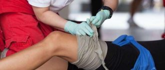
There is a lot of blood -> danger of blood loss -> pressure bandage
Example: struck a knife on a finger
- if blood continues to ooze, then apply another bandage and press harder
- do not remove the already soaked bandage
Fountain -> very rapid blood loss -> pinch the artery, tourniquet
Places of arterial compression:
- Lower third of the shoulder
- Upper third of thigh
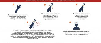
Rules for applying a tourniquet
A tourniquet is applied only in extreme cases (fountain), as it very often causes irreversible damage.
- applied above the wound
- placed on clothing (if there is no clothing, put it on).
- 1 round of the tourniquet - fasten it, then stretch it and apply 3-4 rounds
- Apply the tourniquet quickly, remove it slowly, gradually.
- write the date and time of application of the tourniquet on the forehead (with anything)
- time: in winter - 1 hour, in summer - 2 hours,
- then loosen for 5-10 minutes and apply a tourniquet just above the previous application site
- the tourniquet must be visible!
- check that the tourniquet is applied correctly - there is no pulse in the limb
- see a doctor immediately
What not to do
- Let's not put our hands into the wound!
- We’re not getting anything out of the wound!
- do not remove the already soaked bandage
What to do next
- take the victim to a doctor
- If you are unable to move independently, call an ambulance.
First aid
The most important role in emergency care for bleeding of arterial origin is played by the time factor: for maximum effectiveness it should be provided no later than 2-3 minutes from the moment of injury. If it concerns the main arterial trunks, then bleeding from them must be stopped no later than 1-2 minutes after the injury. Otherwise, the chances of a successful outcome will decrease every second with every milliliter of blood lost.
Important! No matter how critical the conditions, before helping others, protect yourself first - put on rubber gloves from your travel first aid kit, and if you don’t have them, minimize contact with blood using available items (for example, cellophane).
The algorithm for stopping any arterial bleeding is as follows:
- Assessing the type of bleeding.
- Finger pressure on an artery that is damaged.
- Applying a tourniquet, applying maximum limb flexion or a pressure bandage.
- Applying an aseptic dressing to the wound.
This sequence of actions may vary slightly depending on the characteristics of the damaged anatomical area.
Methods to stop bleeding can be temporary or permanent. Temporary arrest of arterial bleeding is used at the stage of first premedical and medical care. The final stage is carried out in a hospital and is part of the hospital stage of care. It is worth noting that in some cases, temporary stopping measures are enough to completely stop the bleeding.
Internal bleeding
- call an ambulance
- cold on the abdominal area
- anti-shock measures
- transport in a sitting position
Signs and symptoms of internal bleeding
- the person becomes weaker, there may be no pain
- paleness, cold sweat, chills
- "floaters before the eyes", dizziness
- breathing is weak, shallow
- swollen, hard, painful belly when pressed, “fetal position”
- there may be a bruise on the stomach
What not to do
- do not anesthetize!
- don't feed
- don't give anything to drink
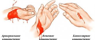
Distinctive signs of arterial bleeding
The classification of bleeding implies its division into three main types:
- arterial,
- venous,
- capillary.
With extensive traumatic injuries, mixed bleeding, for example, venous and arterial, can be observed. In addition, any bleeding, relative to where the blood flows, is divided into internal (in the body cavity) and external (into the external environment). First aid for internal bleeding, as well as its diagnosis itself, is carried out exclusively by medical staff. External bleeding is easier to diagnose and can be treated by anyone.
Arterial bleeding occurs due to damage to the arterial trunks - the vessels that carry oxygenated blood from the cavities of the heart to all tissues of the body. Venous-type bleeding develops when the integrity of the veins that collect blood saturated with carbon dioxide and carry it to the heart is disrupted. Capillary bleeding occurs due to traumatization of capillaries - small vessels that are directly involved in tissue gas exchange.
External differences in types of bleeding
With arterial bleeding, the color of the flowing blood is bright red or scarlet, in contrast to venous bleeding, in which the blood is dark red and comes out slowly. In the case of arterial damage, blood is released rapidly, in a gushing stream. At the same time, the blood stream pulsates, each portion of it comes out synchronously with the pulse and heartbeat. This is due to high pressure in the arterial vessels that come directly from the heart.
In case of arterial bleeding, if help is not provided in time, the phenomena of hemorrhagic shock quickly increase - a pathological condition due to significant blood loss. It has the following symptoms:
- drop in blood pressure;
- increased heart rate;
- pallor and marbling of the skin;
- cyanosis of the extremities;
- respiratory disorders;
- decreased diuresis;
- severe weakness;
- dizziness;
- cold extremities;
- loss of consciousness.
Attention! The faster a person loses blood, the more pronounced the symptoms of shock are, since the body does not have time to compensate for the blood loss.
Penetrating chest injury
- close the hole tightly
- apply cold
- not allowed to speak
- breathe deeply
Signs and symptoms of penetrating chest injury
- white or bloody foam at the mouth
- the same one at the wound site
Glue the bag, a piece of plastic with tape or adhesive tape, check: if there is an entrance hole, then there may be an exit hole (in the case of a gunshot wound, it can be 10 times larger than the entrance hole), if it is small, you can seal it.
Using improvised means
If you don’t have a medical tourniquet at hand, you can use any available material. First aid for arterial bleeding of a limb can be provided with a tie or belt, a bandage or belt from clothes, a rubber tube or a scarf, but this should not be done with wire or thin twine.
If you use fabric, then you need to adjust the twist for tightening, as in the figure above in the article. Be sure to ensure that when twisting the fabric, no skin gets between the layers of fabric or bandage.
- Applying a tourniquet for arterial bleeding in summer and winter - rules and possible mistakes
Long-term crush syndrome (Crush syndrome)
When a person gets caught in a rubble
- apply a tourniquet above the point of pressure and only then release
- released - tightly bandage the limb and remove the tourniquet
- plenty of warm drinks
- anti-shock measures and urgent hospitalization
This material is part of the School of First Aid course “Space of Safety”. We recommend signing up for classes at the school by calling +7 495 760—9092 or on the website https://www.allsafety.ru
Source: https://www.allsafety.ru/
Maximum fixed limb flexion
In some cases, you can use maximum flexion of the limbs as a method of temporarily stopping bleeding from the artery. It should be performed after making sure that the victim does not have a fracture of the injured limb.
A thick pad should be placed at the bend of the limb (popliteal, elbow and groin areas) to compress the damaged artery at maximum flexion
After inserting the roller, the bent arm or leg is fixed to the patient’s body. Such actions are aimed at temporarily stopping bleeding, and if they are ineffective, one should prepare to apply an arterial tourniquet. This same technique, even when performed correctly, has questionable effectiveness.
Sources
- Zideman DA., Singletary EM., Borra V., Cassan P., Cimpoesu CD., De Buck E., Djärv T., Handley AJ., Klaassen B., Meyran D., Oliver E., Poole K. European Resuscitation Council Guidelines 2021: First aid. // Resuscitation - 2021 - Vol161 - NNULL - p.270-290; PMID:33773828
- Gyedu A., Stewart B., Otupiri E., Donkor P., Mock C. First Aid Practices for Injured Children in Rural Ghana: A Cluster-Random Population-Based Survey. // Prehosp Disaster Med - 2021 - Vol36 - N1 - p.79-85; PMID:33491619
- Singletary EM., Zideman DA., Bendall JC., Berry DA., Borra V., Carlson JN., Cassan P., Chang WT., Charlton NP., Djärv T., Douma MJ., Epstein JL., Hood NA ., Markenson DS., Meyran D., Orkin A., Sakamoto T., Swain JM., Woodin JA., De Buck E., De Brier N., O D., Picard C., Goolsby C., Oliver E ., Klaassen B., Poole K., Aves T., Lin S., Handley AJ., Jensen J., Allan KS., Lee CC., De Buck E., De Brier N., O D., Picard C ., Goolsby C., Oliver E., Klaassen B., Poole K., Aves T., Lin S., Handley AJ., Jensen J., Allan KS., Lee CC. 2021 International Consensus on First Aid Science With Treatment Recommendations. // Resuscitation - 2021 - Vol156 - NNULL - p.A240-A282; PMID:33098920
- Singletary EM., Zideman DA., Bendall JC., Berry DC., Borra V., Carlson JN., Cassan P., Chang WT., Charlton NP., Djärv T., Douma MJ., Epstein JL., Hood NA ., Markenson DS., Meyran D., Orkin AM., Sakamoto T., Swain JM., Woodin JA., De Buck E., De Brier N., O D., Picard C., Goolsby C., Oliver E ., Klaassen B., Poole K., Aves T., Lin S., Handley AJ., Jensen J., Allan KS., Lee CC., Schmölzer GM., Morley PT., Nieuwlaat R., Lang E. 2021 International Consensus on First Aid Science With Treatment Recommendations. // Circulation - 2020 - Vol142 - N16_suppl_1 - p.S284-S334; PMID:33084394
- Chang KC., Huang YK., Chen YW., Chen MH., Tu AT., Chen YC. Venom Ophthalmia and Ocular Complications Caused by Snake Venom. // Toxins (Basel) - 2021 - Vol12 - N9 - p.; PMID:32911777
- Coughlin DJ., Boulter JH., Miller CA., Curry BP., Glaser J., Fernandez N., Bell RS., Schuette AJ. An Endovascular Surgery Experience in Far-Forward Military Healthcare-A Case Series. // Mil Med - 2021 - Vol185 - N11-12 - p.2183-2188; PMID:32812042
- Sumardino S., Widodo., Poddar. Analysis of pre hospital emergency management in case of head injury. // Enferm Clin - 2020 - Vol30 Suppl 5 - NNULL - p.228-233; PMID:32713577
- Goolsby C., Rojas LE., Rodzik RH., Gausche-Hill M., Neal MD., Levy MJ. High-School Students Can Stop the Bleed: A Randomized, Controlled Educational Trial. // Acad Pediatr - 2021 - Vol21 - N2 - p.321-328; PMID:32473216
- Menger R., Lin J., Cerpa M., Lenke LG. Epidural hematoma due to Gardner-Wells Tongs placement during pediatric spinal deformity surgery. // Spine Deform - 2021 - Vol8 - N5 - p.1139-1142; PMID:32314179
- Zhao YF., Zhao JY., Hu WZ., Ma K., Chao Y., Sun PJ., Fu XB., Zhang H. Synthetic poly(vinyl alcohol)-chitosan as a new type of highly efficient hemostatic sponge with blood -triggered swelling and high biocompatibility. // J Mater Chem B - 2021 - Vol7 - N11 - p.1855-1866; PMID:32255048
First aid for venous bleeding
With venous bleeding, a stream of blood comes to the surface at high speed so that it does not allow a blood clot to form and stop the bleeding.
As a result, a person may lose a large amount of blood.
Algorithm for providing assistance:
- Raise the injured limb upward;
- Cover the wound with a bandage or clean cloth folded several times;
- Cover the wound tightly with a bandage.
In case of severe venous bleeding, a tourniquet is applied and then cold is applied to the wound. Next, go to the hospital for help.
Technique for performing a hypertensive enema
Indication: swelling of various origins.
Contraindication: acute inflammatory and ulcerative processes in the lower parts of the colon, fissures in the anal area.
Prepare sterile:
- pear-shaped canister or Janet syringe,
- gas outlet pipe,
- medicine,
- 10% sodium chloride solution in an amount of 100-150 ml (dilute 1 tablespoon of table salt in a glass of water),
- latex gloves,
- Vaseline oil,
- tray,
- oilcloth,
- large diaper,
- container with disinfectant solution.
1. Warm the bottle with the medicine in a water bath to 38°C.
2. Fill a pear-shaped balloon with 100 (200) ml of heated solution.
3. Help the patient lie on his left side, the right leg should be bent at the knee and pressed to the stomach (if it is impossible to lay the patient on the left side, the enema is given in a supine position).
4. Place an oilcloth and a large napkin under the patient.
5. Spread the patient’s buttocks and insert the gas tube into the rectum to a depth of 20-30 cm.
6. Attach a pear-shaped balloon to the tube, releasing the air from it, and slowly introduce the heated solution.
7. Disconnect, without unclenching, the pear-shaped cylinder from the gas outlet pipe.
8. Remove the gas outlet tube.
9. Remind the client to try to retain the solution in the intestines for 15-20 minutes.
26 TECHNIQUES FOR APPLYING VENOUS TURNIFIES
Purpose: carried out to reduce venous blood flow to the heart, the volume of circulating blood, especially in situations where it is not possible to carry out drug therapy.
Indications: acute left ventricular failure.
Contraindications: thrombophlebitis, in the acute phase.
Workplace equipment : 3 venous tourniquets.
Execution sequence:
Preparatory stage of performing the manipulation.
- Sit the patient with his legs down for 10-15 minutes (a blood depot is created in the lower extremities, blood flow to the heart decreases).
The main stage of the manipulation.
- Apply two tourniquets simultaneously on both lower extremities, 15 cm below the inguinal fold and on the arm in the upper third of the shoulder. You need to place a napkin under the tourniquet or straighten your underwear.
- Check for the presence of a pulse below the application of tourniquets (arterial pulse should be detected).
- The position of the harnesses changes clockwise every 20 minutes.
- Monitor the general condition of the patient. Monitor the color of the skin below the application of the tourniquets (the color of the skin below the application of the tourniquets should not be pale).
The final stage of the manipulation.
- Remove tourniquets after pulmonary edema has resolved.
27 Assisting a patient with vomiting