Diagnosing various pathologies of cerebral vessels is a pressing issue, since cases of strokes, which are accompanied by loss of ability to work, disability and death, have become more frequent. Statistics show that only 40% of people after a stroke can return to a full life.
Doppler ultrasound is the leading examination method that can be used to identify various lesions of the brachiocephalic arteries.
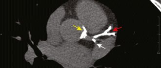
To understand what is meant by the words “brachiocephalic arteries,” you need to trace their path to the brain from the aorta. The blood flow that comes from the aortic arch is distributed into 3 large arteries: the common carotid, the left subclavian artery, and the brasiocephalic trunk. The latter is divided into 3 more arteries, which are located on the right side: subclavian, carotid, vertebral.
All of these vessels, in one way or another, take part in the blood supply to the shoulder girdle and, directly, to the brain. At the same time, they form a complex system in the form of a closed circle, which allows evenly distributing the entire volume of blood in order to ensure adequate blood supply. Impaired blood flow can lead to blood redistribution.
Ultrasound diagnostics
Content:
- Ultrasound diagnostics
- Reasons for the study
- Causes of vascular system disorders
- Indications
- Decoding the results
Examination of arteries using ultrasound is the most informative diagnostic method that allows you to identify physiological changes. In addition, you can determine the qualitative and quantitative parameters of blood flow.
Ultrasound scanning, which is used to diagnose pathologies of the above arteries, is divided into 2 types: Doppler ultrasound and duplex scanning.
The basis of the two methods is the Doppler effect. Its essence is to capture ultrasonic waves from moving objects. Moving objects that move and reflect ultrasound are red blood cells.
The ultrasound monitor displays the change in frequency as a color image. Red is a positive shift, blue is a negative shift.
The positive side of the BCA ultrasound method is the ability to examine the arteries that are located inside the cranium. However, classical ultrasound does not have such capabilities.
The negative side is the inability to accurately determine the position of the vessel. Therefore, diagnosis is made based on the probable location and changes in scanning depth.
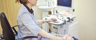
Ultrasound of the brachiocephalic arteries is performed by specialists using an ultrasound scanner, which combines classical research in β-mode and Dopplerography.
The condition of the arteries and the quality of blood flow can be assessed based on two-dimensional and three-dimensional images. The vessel can be seen both in the transverse plane and in length.
It is also possible to use extracranial ultrasound. This method allows you to examine vessels that are located outside the cranium and obtain the following information about them:
- condition of the walls;
- thickness, structure and number of atherosclerotic plaques;
- the size of the lumen;
- blood flow speed;
- the presence of aneurysms and other pathologies.
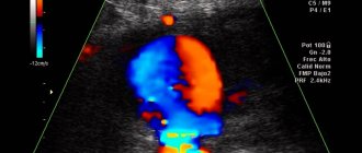
Tandem lesions on duplex scanning of the BCA
Despite its rarity, tandem lesions of the CCA and proximal ICA can occur. Poststenotic disruption of blood flow from a more proximal lesion can extend to a distal lesion, which distorts the results of their study using only peak blood flow velocity and makes this method less meaningful. In this case, the ICA/CCA velocity ratio may be a more useful criterion for defining stenoses less than or greater than 70%. For tandem lesions, additional imaging techniques are advisable. Cardiac rhythm disturbances Arrhythmias, such as atrial fibrillation, frequent extrasystole, accompanied by changes in the duration of cardiac cycles, can lead to the formation of systolic peaks of varying heights. This makes it difficult to accurately measure blood flow velocity on duplex scanning and requires the average of at least 4 or 5 consecutive cardiac cycles to be calculated when studying blood flow velocities.
Causes of vascular system disorders
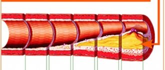
The main cause of pathological changes in the brachiocephalic vascular system is atherosclerotic lesions of the arteries. A study of people in relatively good health showed the presence of atherosclerosis in the initial stage in 3 percent of those examined. Their age ranged from 40 to 50 years. This result may indicate a fairly high probability of stroke in people after 55-60 years of age.
Atherosclerotic changes are considered an indirect cause of structural changes in blood vessels. For example, the appearance of tortuosity.
Paragangliomas
Paragangliomas (chemodectomas) are rare (less than 1% of all neck tumors). These are slow-growing tumors, in 95% of cases they are detected on one side on ultrasound of the brachiocephalic vessels. Typically, the pathology occurs in the carotid bodies and in approximately 10% of cases it becomes malignant. Often they are first identified by palpation in the form of painless formations in the neck. Ultrasound of the brachiocephalic vessels is visualized as heterogeneous and hypervascular formations with clear contours, leading to bevelling of the contour of the CCA bifurcation with displacement of the ICA and ECA. Paragangliomas are distinguished from other neck formations by their localization in the bifurcation of the carotid arteries, as well as their solid structure. In most cases, the tumor is supplied by the ascending pharyngeal artery, but duplex studies show an increase in diastolic blood flow velocity in the ECA.
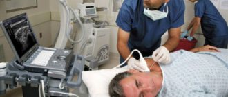
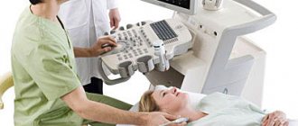
Carrying out an ultrasound
The procedure is performed with the patient in a horizontal position. A special cushion is placed under the neck, the head is turned to the side. The neck area is freed from clothing and jewelry. A medical gel that conducts ultrasound is applied to the skin and transducer. The doctor moves the sensor along the neck according to the location of the vessels. Such movements are carried out in the longitudinal anterolateral and posterolateral planes and the transverse one.
During the study, the patient is prohibited from turning his head or talking. First of all, the common carotid arteries are examined, since it is there that the accumulation of atherosclerotic plaques is most likely to occur. For indications such as genetic or anatomical abnormalities, additional functional tests may be prescribed:
- conducting the examination in a vertical position;
- holding your breath;
- use of antianginal medications (Anaprilin, Nitroglycerin).
The time interval for the procedure is from half an hour to 45 minutes. The examination protocol is given to the patient for further consultation with the doctor who referred for the ultrasound.
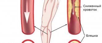
Atherosclerotic plaques are located mainly in the carotid arteries
results
Ultrasound interpretation is performed by comparing the indicators of a particular patient with standards according to gender and age. Initially, the intimate-media complex of the carotid artery is assessed as an indicator of the presence of atherosclerosis and the establishment of its stage. This complex consists of three layers: the inner shell, which is in contact with the bloodstream, the middle (media) - the thickest, the outer (adventitia), containing the smallest blood vessels.
Ultrasound of the BCS clearly visualizes the inner and middle membranes. The presence of atherosclerosis is evidenced by the following characteristics: there are areas with increased conductivity (echogenicity), the layers of the artery merge with each other, the artery wall has thickening zones. The average permissible thickness of the intimate-media complex of the carotid artery is 1.1 mm. When this figure increases by more than 1.3 mm, the presence of an atherosclerotic growth (plaque) is diagnosed.
Paired vertebral vessels should normally be identical in diameter. The permissible diameter of the arteries is:
- general carotid – 4.2–6.9 mm;
- internal carotid – 3.0–6.3 mm;
- external carotid – 3.0–6.0 mm;
- vertebrates – 3–4 mm.
Ideally, the vessels should not be deformed. The speed of blood flow in the arteries is measured: systolic (during periods of tension) and diastolic (during periods of relaxation) of cardiac activity. The obtained indicators are compared with the standards. In the common carotid artery, systolic velocity should remain within the range of 50–104 cm/sec., diastolic – 9–36 cm/sec. Limits of systolic blood flow velocity:
- in the internal carotid artery – 32–100 cm/sec;
- in the outside – 37–105 cm/sec.
- in the vertebral arteries – 20–61 cm/sec.
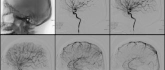
If a patient has serious pathologies of arterial blockage, blood supply to the brain structures can be restored through surgery
Diastolic velocity has the following limits: internal carotid artery – 9–35 cm/sec, external – 6–27 cm/sec, vertebral arteries – 6–27 cm/sec. The final protocol should display all study parameters and indicate the degree of vasoconstriction.
Indications and contraindications for sonography
In newborns, duplex ultrasound (synonymous with sonography) of the brachiocephalic vessels is performed if defects of the BCA and brain, or birth trauma are suspected. Regardless of age, ultrasonography is prescribed for all pathological conditions with a likelihood of deterioration of the blood supply to intracranial tissues. In surgery it is also used in the preparatory period for surgery and during rehabilitation.
Direct indications of duplex sonography of the BCA:
- hyperlipidemia, increased cholesterol;
- blood diseases;
- atherosclerosis;
- vegetative-vascular dystonia (VSD);
- arterial or intracranial hypertension;
- heart disease, aorta;
- head and neck injuries;
- pathologies with the likelihood of compression/damage to the brachiocephalic vessels;
- inflammation of the walls of the BCA (vasculitis, arteritis, other types);
- neoplasms in the area of the BCA;
- history of stroke, cardiac ischemia, heart attack.
Examination of the great arteries of the head (MAG) and neck is also indicated for people at risk. These are people suffering from diabetes, osteochondrosis, autoimmune vascular pathologies, metabolic disorders, cardiomyopathies, and obesity.
Ultrasound scanning of the brachiocephalic arteries is indicated when:
- frequent attacks of cephalalgia (headache);
- migraines;
- fainting;
- dizziness;
- darkening of the eyes, lethargy, persistent desire to sleep;
- deterioration of vision, hearing, mental activity;
- violations of motor abilities, coordination;
- areas of the body with numbness, loss of sensitivity;
- persistent deviation of blood pressure (BP) from the norm;
- unequal blood pressure readings on the right and left arms.
Duplex BCA is also indicated for elderly smokers with nicotine addiction for more than 10 years. It is advisable to undergo ultrasound scanning for people whose relatives have had cases of cerebral circulatory disorders (stroke, ischemia, etc.).
There are no contraindications to brachiocephalic vessel duplex. A temporary limitation of sonography in the area of BCA scanning is acute inflammation or deep damage to the skin due to injury or disease.
Pay attention to the video review from a specialist about diagnosing BCA:
Preliminary preparation
No special preparation is provided, but fulfilling certain conditions will help make the procedure easier and get more accurate results. Firstly, on the day of the examination you need to stop smoking, drinking coffee and strong tea. It is advisable to limit salty foods in your diet. You should not drink alcohol for three days. Because such eating behavior can have a negative impact on vascular tone and the filling of blood vessels.
Secondly, you must notify your doctor if you are taking any medications. A day before the procedure, it would be better to stop using medications that affect concentration, nootropic and psychostimulants. Such medications affect the state of the vascular system and can distort the actual results. Doctors also advise taking a walk in the fresh air immediately before the test.
Possible diagnoses
Using the results of the study, the doctor can make a diagnosis with extreme accuracy. The most common pathologies diagnosed by ultrasound of the BCA are:
- systemic damage to the arteries - atherosclerosis. The primary manifestation is recorded according to IMT indicators;
- narrowing of the vessel (stenosis) or blockage (occlusion);
- pathological tortuosity, otherwise deformation of the arterial bed;
- protrusion of the vascular wall (aneurysm);
- fistula (fistula) in the intervascular space;
- dissection or longitudinal tear and dissection of the arterial wall, accompanied by hemorrhage and hematoma;
- an inflammatory autoimmune disease characterized by thickening of the vascular wall, unevenly distributed (nonspecific aortoarteritis);
- steel syndrome (an anomaly in which the blood supply to the arm is carried out through the vertebral arteries);
- functional failure of the vertebral artery canals due to their compression (extravasal compression).
During the examination, the doctor has the opportunity to assess the dynamics of rehabilitation after surgical operations on blood vessels.
If the doctor has prescribed an ultrasound of the BCA, you cannot ignore the recommendations, because this is an examination with which you can predict the patient’s vascular health. The risk of stroke directly depends on the degree of narrowing of the BCA. Today, diagnostics using this method is available to most patients. Where to have an ultrasound done, in a large diagnostic center or a regional hospital, is decided by the patient himself. The cost of diagnostics does not exceed 3,000 rubles.