Triplex ultrasound of head and neck vessels
(triplex scanning of head and neck vessels) is a diagnostic ultrasound method for studying the condition and function of the vessels that supply blood to the head and brain area.
Triplex scanning of vessels has a number of advantages compared to ultrasound and duplex scanning , which will be discussed below.
The method includes the study of the carotid, subclavian and vertebral arteries, veins of the neck, as well as the main arteries of the brain. Very often, a triplex scan of the neck vessels (brachiocephalic vessels) and a triplex scan of the cerebral vessels are performed together, which creates a holistic view of the vessels that supply blood to the brain.
Ultrasound of cerebral vessels
Dopplerography of cerebral vessels is prescribed for suspected neurological diseases. The following symptoms may indicate this:
- Panic attacks.
- Difficulty speaking.
- Difficulties in perceiving information.
- Impaired coordination of movements.
- Decreased visual, auditory, kinesthetic sensations.
Based on the results of the ultrasound, the doctor can determine what changes in the structure of the blood vessels could cause these symptoms and prescribe treatment, if necessary.
The procedure is simple and safe and has no age restrictions. It can be carried out at home using a portable scanner.
When and why is duplex scanning of the vessels of the neck and head performed?
Indications for the examination are varied, primarily dizziness, headaches, lightheadedness, episodes of loss of consciousness, swaying when walking, osteochondrosis of the cervical spine, numbness or weakness in the arm or leg, asymmetry of blood pressure and pulse in the arms, decreased memory and concentration attention, speech impairment, previous stroke or ischemic attack, condition after surgery on the carotid arteries, increased blood pressure (arterial hypertension), coronary heart disease, diabetes mellitus. In case of cervical osteochondrosis, duplex scanning can reveal signs of compression of the vertebral arteries, including when performing tests with head turns.
Duplex scanning is recommended for people over 40 years of age
, even if they feel healthy, but have risk factors (high cholesterol levels in the blood; family history of cardiovascular diseases; smoking; sedentary lifestyle, etc.). The purpose of the examination is to determine the patency of blood vessels, assess the presence, size and structure of intraluminal formations (atherosclerotic plaques), study the condition of the vascular wall, the geometry of the vessels (the presence of deformations, tortuosities), and assess the quantitative and qualitative characteristics of blood flow in the vessels under study. Detection of the pathological process is possible in almost any area of the vascular system. That is why it is important to conduct a detailed study of extracranial and intracranial vessels along the entire length available for study.
Up
How is the procedure done?
Ultrasound duplex scanning of the vessels of the neck and head does not require special preparation. Before the procedure, it is advisable to refrain from smoking, drinking strong tea, coffee, alcohol, and energy drinks. If you are taking medications that improve cerebral circulation, they should not be discontinued without the consent of your doctor. The examination is carried out lying on your back, with your head slightly turned to the side opposite to the side being examined. The doctor conducting the duplex scan moves a sensor on the surface of the neck, on which a special gel is applied to improve contact between the sensor and the skin. When performing the examination, slight pressure from the sensor may be applied; most people tolerate this easily. The monitor screen of an ultrasound scanner displays the information received by the sensor using ultrasound and converted by the device into the form of a digital signal. During the test, you may also hear special noises that help assess the flow of blood in your body.
Thus, the doctor assesses the condition of all the main vessels of the neck - the carotid, vertebral and subclavian arteries, internal jugular and vertebral veins on the left and right. Then transcranial duplex scanning (TCDS) is performed,
that is, the study of intracranial vessels. The doctor alternately installs a special sensor on different areas of the head: temporal, occipital, and sometimes supraorbital. Additionally, rotational and other functional tests are carried out. A comprehensive study of the vessels of the neck and brain is optimal for problems solved by neurologists, chiropractors, cardiologists, and cardiovascular surgeons. This scope of research corresponds to modern diagnostic standards. The whole procedure lasts about 30-40 minutes. The protocol, which is handed to the patient, records all blood flow indicators with a full description of the detected pathology, and gives a general conclusion on the study. If you have had a similar study done before, it is advisable to take the previous protocol with you for comparison.
Up
Are there any contraindications to duplex scanning?
Unlike other instrumental research methods, ultrasound duplex scanning of blood vessels has no contraindications. Ultrasound examination is completely harmless for the patient. The safety of the ultrasound diagnostic method is one of its advantages, which allows repeated examinations over time and evaluation of the effectiveness of the treatment.
What pathology can duplex scanning detect?
Up
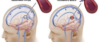
The most common pathology detected by duplex scanning is atherosclerosis. This is the deposition of fatty substances (lipids) and the proliferation of connective tissue in the walls of the arteries. Currently, atherosclerosis is considered as a complex, multicomponent pathological process affecting the arterial wall, lipid metabolism, blood coagulation system, and cellular elements. Although the prevalence of atherosclerosis increases with increasing age, it is a disease and not an age-related change. Atherosclerosis also occurs in young people, and may be absent in old people. Atherosclerosis of the main arteries of the head and neck most often develops in the third or fourth decade of life. An early symptom of atherosclerotic lesions of the carotid arteries is an increase in the thickness of the vessel wall (intima-media). The ultrasound method of duplex scanning will reveal signs of atherosclerotic lesions of the brachiocephalic arteries (thickening of the intima-media, changes in its structure, the presence of atherosclerotic plaques, their nature, extent, degree of narrowing of the vessel and the nature of blood flow at the site of narrowing). Detection of atherosclerotic vascular changes in a patient with arterial hypertension is associated with a high risk of adverse cardiovascular events (acute coronary syndrome, myocardial infarction, stroke, etc.). Early detection of stenosis (narrowing) of the carotid arteries helps prevent such a serious and dangerous disease as stroke. In a number of foreign countries that have achieved success in preventing stroke, an annual examination of the entire population over 40 years of age is used, using ultrasound techniques (duplex scanning of the vessels of the head and neck with color mapping of blood flow). This allows you to determine the risk group.
In addition to atherosclerotic changes, duplex scanning reveals various congenital vascular anomalies: deformations of the carotid and vertebral arteries, pathological tortuosity; hypoplasia (congenital reduction in the diameter of the vessel), anomalies of entry and exit of the vertebral arteries. In pathology of the cervical spine, disturbances in the course of the vertebral arteries, asymmetry of blood flow and signs of compression of the vertebral arteries in the canals of the transverse processes of the cervical vertebrae are most often detected.
Thus, ultrasound duplex scanning with color flow is a highly informative and completely safe diagnostic method; it helps to identify vascular pathology at the initial stage. The results of the examination will help your doctor create an individual plan of treatment and preventive measures in order to maintain your health.
Calcified atherosclerotic plaque at the mouth of the internal carotid artery (two-dimensional mode).
Characteristics of blood flow in the common carotid artery (two-dimensional echography, spectral Doppler and color flow mode).
Characteristics of blood flow in the middle cerebral artery (two-dimensional echography, spectral Doppler and color flow mode).
The essence of Doppler ultrasound
Based on the name of the procedure, it is performed using ultrasonic waves. Their work is based on the Doppler effect. The procedure differs from a standard ultrasound: a vessel with blood passing through it is displayed on the monitor screen. Looking at the resulting image, the specialist assesses the state of blood flow and vascular patency, which affects the quality of blood supply to the brain.
Dopplerography of cerebral vessels makes it possible to study the work of the veins, which “supply” blood to the brain, and the arteries, which are responsible for the outflow of waste products from it. The vessels being examined are located in the cervical and cranial regions. The type of ultrasound will depend on their location:
- Dopplerography of extracranial vessels located on the surface of the neck (jugular veins, carotid, vertebral and subclavian arteries). Any disturbances in their structure affect the state of the brain, which is why these vessels are called “maternal” vessels.
- Dopplerography of transcranial, or “daughter” vessels. The doctor installs an ultrasound sensor in the thinnest parts of the skull and evaluates the functioning of the veins and arteries that supply the brain.
Ultrasound of the BCA - what is the procedure?
Ultrasound of the brachiocephalic arteries is a type of ultrasound examination that gives an accurate picture of the condition of the vessels directly responsible for feeding the brain:
- main (extracranial) sections of the cervical arteries (subclavian, carotid and vertebral);
- intracranial sections of the brachiocephalic arteries (located inside the cranium).
The procedure can be performed in three ways:
- Ultrasound Dopplerography of the extracranial sections of the brachiocephalic arteries - study of the general condition of the great vessels, as well as their patency. It is used to identify the causes of blood flow disorders: cholesterol plaques, blood clots, etc.
- Duplex (triplex) scanning of the extracranial sections of the brachiocephalic arteries helps, among other things, to determine the speed and quality of blood flow, identifying factors that prevent a decrease in vascular tone.
- Transcranial Dopplerography (TCDG) is a scan of the intracranial sections of the brachiocephalic arteries.
In modern medicine, all of the above methods are equally in demand.
In what cases is ultrasound ultrasound prescribed?
The main symptoms signaling a cerebrovascular accident are:
- Impaired coordination of movements.
- Deterioration of hearing and memory.
- Frequent attacks of headache.
- Impairment or partial loss of taste.
- Insomnia, difficulty falling asleep.
- Noise, whistling and ringing in the ears.
- Deterioration of visual function, manifested in loss of visual fields.
- Difficulty concentrating.
- Decreased sensitivity and motor activity of the arms and legs.
A doctor may prescribe a procedure in the following cases:
- The patient has diabetes mellitus.
- For arrhythmia, as this increases the risk of cardiac clot rupture and artery blockage.
- The patient has suffered a heart attack or stroke.
- Osteochondrosis of the cervical spine.
- Before planned heart surgery.
- If the patient is a smoker with many years of experience.
- An area with strong pulsation is visible on the neck (in this case, an ultrasound of the neck is prescribed).
Based on the above symptoms, the doctor can determine the location and name of the vessel in which the patency is impaired. Several pronounced symptoms indicate poor blood flow in a large vessel. In this case, extracranial Doppler ultrasound is prescribed. Violation of only one function will be the reason for conducting transcranial Doppler ultrasound.
Are there any contraindications?
Ultrasound waves are absolutely safe for humans, so there are no special contraindications to this type of ultrasound. The only exception is a violation of the integrity of the skin. The procedure involves contact of sensors with the head or neck, so the examination is postponed until the wound heals.
However, there are a number of special cases that make duplex examination of blood vessels difficult:
- cardiac arrhythmia, which changes the speed of blood circulation through healthy arteries and veins;
- the area under study is hidden under bone tissue;
- slow blood flow in the patient.
These are not contraindications, but in this case changes in the location of the sensors are required. It will be necessary to navigate non-standard conditions, which requires professionalism on the part of the doctor.
Rules for preparing for research
On the day of the procedure, you must refrain from:
- Taking antispasmodics such as “Riabal”, “No-shpa”, “Drotaverine”, “Papaverine”, “Baralgin”, “Cinnarizine”.
- Smoking.
- Drinking black tea and drinks containing caffeine.
- Staying in unventilated, stuffy rooms with large crowds of people, which can have a detrimental effect on vascular tone.
If the patient is undergoing treatment with cardiovascular medications, then the advisability of discontinuing them before the procedure must be agreed with a neurologist. In any case, the sonologist must be informed about the treatment being performed before the ultrasound.
Stages of extracranial Dopplerography:
1. In the office, remove all jewelry from your neck and head.
2. Remove clothing from those parts of the body through which the sensor will be passed: neck, collarbones, shoulder blades.
3. Lie on the couch with your head facing the monitor. The doctor will apply acoustic gel to open areas of the body and begin to move the device over them, placing it in areas where medium and large arteries and veins are located. To obtain more accurate data on the state of vascular tone, the patient will need to hold his breath at the doctor’s request, take special medications and change body position.
Doppler ultrasound of vessels located in the cranial area has a different specificity. Acoustic gel is applied to the temples, the back of the head and the area above the eyes. The device is installed there because in these places the bones of the skull are the thinnest and are able to transmit ultrasonic waves.
Literature
- Ahuja Anil T., Dai Younis Y. L. Ultrasound diagnostics. Head and neck. – M.: Panfilov Publishing House, 2021. – 540 p.
- Velkoborski H. Yu. Ultrasound diagnosis of diseases of the head and neck. – M.: MEDpress-inform, 2021 – 176 p.
- Kulikov V.P. Fundamentals of ultrasound examination of blood vessels. – M.: Vidar-M, 2015. – 392 p.
- Stuart J. Hutchison, Catherine K. Holmes. Ultrasound diagnostics in angiology and vascular surgery. – M.: GEOTAR-Media, 2018 – 400 p.
- Zwibel V., Pellerito G. Ultrasound examination of blood vessels. – M.: Vidar-M, 2008 – 656 p.
Author: Korolev E. S.
Reviewer: reflexologist Kurus A. N.
By what principle is the received data decrypted?
Each vessel has its own parameters, on the basis of which calculations are made and the degree of their discrepancy with the norm is assessed.
Transcranial and extracranial Dopplerography of arteries compares digital data in each vessel segment:
- Artery wall thickness.
- Vessel diameter.
- Phases of blood flow, its features.
- The degree of symmetry of blood flow through identical arteries located on different sides.
- Diastolic blood velocity.
- Maximum systolic blood flow velocity.
- Pulsation and resistive indices, systole and diastole ratio.
- The degree of pathological narrowing of blood vessels (stenosis), the condition of the artery beyond its limits.
Dopplerography of veins evaluates the following parameters:
- Condition of the venous wall of the vessel.
- The nature of blood movement through the studied vessels.
- Vein diameter
Data obtained at rest and after functional tests are compared with normal values. Any deviations are marked with a special abbreviation of letters and numbers. Based on them, the neurologist prescribes further studies or forms a course of treatment.
Ultrasound of the BCA: norm and deviations
The functionality of the brachiocephalic arteries is assessed by the thickness of their walls, lumen diameter and blood flow speed. The obtained data are compared with the normative ones. Typical signs of abnormalities in the circulatory system are: cholesterol plaques, blood clots or the presence of extraneous vascular branches. The sonologist will provide all the necessary recommendations to eliminate these and other disorders.
Where can I get Doppler ultrasound of cerebral vessels?
The study is carried out in municipal clinics and public hospitals, specialized clinics, and general medical centers. Depending on the location of the ultrasound, the cost of each type of Doppler ultrasound will be in the range of 500-6000 rubles. The procedure has many advantages, including painlessness and the absence of side effects. Within 20 minutes, a sonologist will be able to determine the presence of anomalies in venous or arterial inflow and assess the degree of development of danger to blood vessels.
However, the procedure will not help determine the cause of compression of the thin arteries, since the capabilities of the device do not allow them to be seen from the outside.