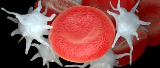
The non-straightness of the vertebral arteries is often determined by scanning or duplex examination of the cerebral blood vessels. This pathology is very dangerous in terms of the development of transient or permanent cerebrovascular accident. Patients experience serious difficulties with mental performance, mood swings occur, and secondary hypertensive syndrome may develop.
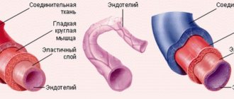
Non-straightness of the vertebral arteries is always a consequence of pathology of the spinal column in the cervical region. This may be curvature, rotation of the vertebral body, destruction of intervertebral discs against the background of long-term osteochondrosis, deposition of calcium salts and many other diseases. The normal course of the vertebral artery is ensured by the anatomical structure of the cervical vertebrae, in which there are special openings for these blood vessels. They are reliably protected from external traumatic influences. However, when the vertebral bodies or the cartilage tissue separating them, the fibrous ring of the intervertebral disc, is deformed, serious deformation and curvature occurs.
Any deformation of the course of the vertebral arteries leads to serious consequences, manifested by various cerebral symptoms. We will talk about how to recognize the disease, diagnose it and treat it without surgery using manual therapy techniques.
Symptoms and treatment of tortuosity of the vertebral arteries
Very often, people susceptible to high blood pressure and neurocirculatory dystonia do not know the main cause of the disease.
Behind the pathology is often tortuosity of the vertebral arteries, which increases the risk of stroke several times due to deterioration of blood flow in vital vessels. Such consequences can disrupt the activity of the brain and the entire central nervous system. Typically, tortuosity of the vertebral arteries is a hereditary disease and develops when the arterial tissue contains predominantly elastic fibers. As a result, the vascular walls quickly wear out, become thinner and become deformed.
The situation is aggravated if a person suffers from atherosclerosis. In this situation, plaques form on the walls, reducing the overall patency of the vessels. This in turn causes improper blood flow in the brain and other organs of the body. Usually, the bend does not manifest itself in any way, and only over time does the patient experience a circulatory disorder.
As a result, if the pathology is not diagnosed in time, the risk of strokes increases. It happens that the disease is detected during a standard medical examination. In this case, it is necessary to immediately begin competent treatment.
Prevention
To avoid a problem such as a deformed artery, it is recommended to take the following measures:
- control the level of cholesterol in the blood, exclude unhealthy foods (spicy, salty, fatty, fried, etc.) from the daily diet;
- get rid of bad habits (nicotine addiction). Smoking has a negative effect on the condition of the vascular walls and is the main cause of the formation of plaques and narrowing of the arteries;
- control body weight. For these purposes, physical exercises are used for all parts of the spine.
In addition, try not to lift heavy objects and limit your participation in professional sports. Also, you should not make sudden rotations, turns or tilts of your head.
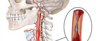
Anatomical features of the vertebral arteries
The vertebral artery branches from the subclavian towards the central part of the inner edge of the scalene muscle in the neck.
It is important that no more than 1–1.5 cm remains to the adjacent mouth of the thyrocervical trunk, which is also a branch of the subclavian artery. This creates an additional mechanism of “stealing” (redistribution of blood) in case of hypoplasia or stenosis of the vertebral artery
Heading upward, the artery at the level of the sixth cervical vertebra (less often the fifth) enters a protected bone canal formed by the spinous processes of the vertebrae.
It is customary to distinguish sections or segments of the vertebral artery:
- I - the entire area from VI to II cervical vertebrae, where the vessel exits the foramen;
- II - outside the canal, at an angle of 450, it deflects posteriorly and goes to the transverse process of the first cervical vertebra (atlas);
- III - passing through the opening of the atlas on its back side, the artery forms loops, their role is to prevent disruption of blood flow when turning the head;
- IV - heading into the foramen magnum, the artery is located inside a dense ligament, with ossification of the receptacle or bone outgrowths on the occipital bone, conditions are created for traumatizing the walls of the vessel during movements in the cervical region;
- V - inside the foramen magnum (intracranial segment), the vertebral artery passes through the dura mater and lies on the surface of the medulla oblongata.
The fusion of the left and right arteries into a single trunk (basilar artery) ensures participation in the formation of the circle of Willis at the base of the brain
20% of cases of pathology of vertebral arteries are due to developmental anomalies:
- originating directly from the aorta;
- entry into the bony spinal canal higher than usual (at the level of the third to fifth cervical vertebrae);
- displacement of the mouth outward.
More often, the lesions are combined and are divided into the following options:
- up to 34% is due to the combined effect of developmental anomalies and extravasal muscle compression;
- 39% are stenoses of atherosclerotic and thrombotic nature;
- the maximum part - 57% - represents compression by various displacements of the vertebrae in combination with atherosclerosis.
Based on the presence of symptoms and the determination of its connection with neck movements, a suspicion of pathology of the vertebral arteries arises from a general practitioner or therapist. Timely referral to a neurologist and examination is a matter of experience.
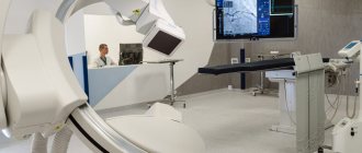
Duplex scanning allows you to see the structure of the vessel, determine the nature of the stenosis, the degree of damage to the artery walls
- Ultrasound Dopplerography - an assessment of all anatomical characteristics of the vertebral arteries on both sides, diameter along the length, speed of the blood flow wave is carried out; it is important as a way to determine cerebral circulatory reserve;
- Magnetic resonance imaging of the brain and neck vessels will indicate emerging lesions with impaired blood supply, the formation of cysts, aneurysms;
- An x-ray of the cervical spine can be used to judge the participation of pathological growths of bone tissue in pinching of the vertebral arteries;
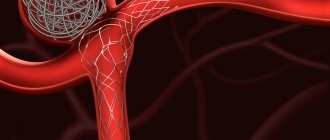
- Angiography of the neck vessels is performed by injecting a contrast agent into the subclavian artery. The technique is informative, but is carried out only in specialized departments.
The vertebral artery, which is a paired vessel and located on both sides of the human body, originates from the subclavian artery. The sections (or segments) of the vertebral artery pass in the canal of the transverse processes of the cervical vertebrae, which is why the vessel got its name.
The following sections of the vertebral artery are distinguished:
- Prevertebral department. This segment occupies the area from the beginning of the artery at the subclavian vessel to the entrance to the canal of the vertebral processes;
- The cervical region occupies a section of the artery passing in the canal of the processes of the cervical vertebrae;
- Cervico-occipital segment. This section is located in the interval from the exit from the canal of the transverse processes of the vertebrae to the entrance to the cranium;
- The intracranial section extends from the skull to the junction of the two vertebral arteries into the basilar vessel.
Thus, more than half of the cases of death from cerebral vascular insufficiency are associated with pathology of the vertebral arteries. One of these disorders is the non-straightness of the vertebral arteries between the transverse processes of the cervical vertebrae.
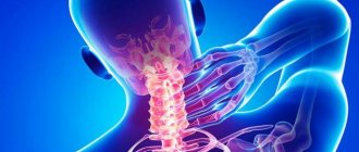
Symptoms of pathology
Timely identification of the problem can significantly speed up the diagnostic and treatment processes. For this reason, it is worth remembering a number of symptoms, if they occur, you should contact a neurologist and orthopedist.
- Frequent headaches in the back of the head;
- Neck pain;
- The appearance of a pre-fainting state with sudden movements;
- Increased intracranial pressure;
- Constant or intermittent weakness in the arms, legs, or fingers;
- Spontaneous attacks of nausea;
- Loss of coordination and problems with the vestibular system;
- Systematic visual impairment.
These symptoms do not directly indicate that the problem lies specifically in the vertebral arteries, they only indicate a violation of the blood circulation in the brain. However, it is often associated with the vertebral arteries.
The non-linearity of the course means that instead of the direct and shortest path to the brain, the artery is located along a tortuous path. To correct the pathology, it is necessary to act not on the arteries, but on the primary problem - the cervical spine. Let's look at the causes of the disease in more detail.
Symptoms
The non-straightness of the vertebral arteries, which supply the vessels of the head, can affect many functions. Main symptoms:
- Non-straightness of the vertebral arteries
- Visual impairment: double vision, spots in the eyes, darkening, photophobia.
- Noise, ringing in the ears.
- Headaches, migraines, even nausea.
- Problems with the vestibular system, vertigo, fainting.
- Increased intracranial pressure, sometimes even to the point of seizures.
- Orthostatic hypotension is a decrease in blood pressure when standing up.
Impaired blood flow can cause both plethora (hot flashes) and transient attacks with pale skin. There is a risk of ischemic and hemorrhagic stroke. Insufficient blood flow to the brain is a signal to increase blood pressure in order to increase the supply of nutrition to the main organ of the body. The sympathoadrenal system is activated.
Read about a lump in the throat with cervical osteochondrosis: symptoms, causes, diagnosis and treatment.
Find out whether a headache can occur with cervical osteochondrosis and how to treat the disease.
Vegetative-vascular dystonia often develops against the background of non-linearity of the course, since the work of the vasomotor and respiratory centers located in the medulla oblongata is disrupted.
Migraine develops due to blood redistribution. This vascular disorder is characterized by a disorder in the circulation of cerebrospinal fluid in the ventricles of the brain. Light sensitivity in one eye is associated with paralysis of the muscles that constrict the pupil.
How is the non-linearity of the course of the vertebral arteries between the transverse processes manifested?
It is important to know how the non-linearity of the course of the vertebral arteries between the transverse processes manifests itself, what clinical signs may indicate the presence of such a pathological change. Most often, non-straightness of both vertebral arteries develops simultaneously, which is due to anatomical features. Very rarely, unilateral pathology develops after a serious injury to the cervical spine with the growth of callus.
In most cases, pathological deformation is diagnosed simultaneously on both sides. This causes a total decrease in blood flow on both sides at once, which is difficult not to notice based on clinical signs. If deformation of the course of the vertebral artery develops only on one side, then a compensatory reaction occurs to increase blood flow in the second blood vessel. It will be very difficult to see the pathology clinically in this situation, since there will be no symptoms of cerebral circulatory insufficiency. Only random functional diagnostics are possible when performing duplex scanning of blood vessels for a completely different reason.
Typical symptoms of non-straightness of the vertebral arteries include the following:
- aching and bursting pain in the back of the head;
- a feeling of strong pressure from the inside on the bone structures of the skull in the occipital area;
- regularly occurring severe pain in the collar area and neck associated with prolonged static muscle tension;
- orthostatic dizziness and development of a pre-fainting state upon sudden rise from a sitting or lying position;
- unmotivated rises in blood pressure that are not amenable to standard pharmacological correction with drugs;
- increased intracranial pressure for no apparent reason;
- decreased mental performance and a feeling of constant drowsiness, lack of freshness in the morning after waking up;
- nosebleeds and attacks of nausea not associated with food intake and diseases of the digestive system;
- a feeling of weakness in the upper and lower extremities, not associated with physical activity and its intensity;
- decreased hearing and visual acuity;
- the presence of extraneous noise in the ears;
- flickering of spots or circles before the eyes.
With serious deformation of the course of the vertebral arteries, the development of a spinal type of stroke (acute cerebrovascular accident) is possible. This pathology can develop even in young people (30–45 years old). Typical signs are unsteadiness of gait, changes in facial expressions, deviation of the tongue to one side, paralysis of the limbs on the right or left side, impaired speech function, etc. Immediate medical attention and placement of the patient in a special vascular or neurological hospital is required. This condition is life-threatening.
Crisis prevention
Once the diagnosis is established, the patient is able to prevent vascular crises. To do this you need:
- do gymnastic exercises;
- wean yourself from sleeping on your stomach;
- undergo physiotherapy and massage courses at least twice a year;
- purchase an orthopedic pillow to ensure a level position of the cervical spine during sleep;
- wear a Shants collar;
- get rid of factors that narrow arteries (smoking, drinking alcohol).
The clinical picture of a stroke is not necessarily caused by intracerebral vessels. Extracranial disorders should always be kept in mind when making a diagnosis and prescribing treatment. This tactic helps prevent life-threatening complications.
LSC on the left – 19 cm/s; LSC on the right – 30 cm/s.
3. In the left supraclavicular region, the internal jugular vein, dilated to 34 mm, is located. The right internal jugular vein is not dilated. The veins are not dilated, are passable, and phasic blood flow is detected.
1. Atherosclerosis of the extracranial parts of the brachiocephalic arteries with stenosis of the area of the carotid bifurcation on the left by 20%, the mouth of the right internal carotid artery by 30%.
2. Non-straightness of the course of the vertebral arteries between the transverse processes of the cervical vertebrae, which is obviously due to the presence of osteochondrosis of the cervical spine, C-shaped tortuosity of the extravertebral section of the right vertebral artery.
3. Ectasia of the internal jugular vein on the left.
Extravasal compression of the vertebral arteries is one of the causes of a whole complex of manifestations of serious disturbances in the blood supply to the brain. The fact is that blood enters the brain through two main channels: the carotid (two carotid arteries) and the vertebrobasilar (two vertebral arteries).
In cases where the functions of one or more of these blood vessels are disrupted, normal nutrition of the brain stops: it receives an insufficient amount of useful substances, including oxygen, and the patient in this case suffers from a whole complex of neurocirculatory disorders known as “Vertebral Artery Syndrome.” "
Arteries originating from the subclavian arteries are responsible for supplying blood to the brain, cerebellum and inner ear. They are located in a special canal, in the cervical vertebrae, close to the elements of the spinal column. Therefore, if the structure of the spine is disturbed, there is a danger of reducing the lumen of the channel through which this blood vessel passes - there is a possibility of extravasal influence of nearby tissues on it, its compression and disruption of blood flow.
Fundamentals of Anatomy and Physiology
Blood enters the brain through four large arteries: the left and right common carotid and the left and right vertebral. It is worth noting that 70-85% of the blood passes through the carotid arteries, so disruption of blood flow in them often leads to acute cerebrovascular accidents, that is, ischemic strokes.
cerebellum, hypothalamus, corpus callosum, midbrain, partly the temporal, parietal, occipital lobes, as well as the dura mater of the posterior cranial fossa. Before entering the cranial cavity, branches depart from the vertebral artery, carrying blood to the spinal cord and its membranes. Consequently, when blood flow in the vertebral artery is disrupted, symptoms arise that indicate hypoxia (oxygen starvation) of the areas of the brain that it supplies.
- Treatment of arthrosis and osteoarthrosis of the costovertebral joints
Diagnostics
When making a diagnosis, the specialist interviews the patient about the presence of any chronic diseases or symptoms.
In addition, to exclude errors, the doctor prescribes a number of examinations:
- Doppler ultrasound, with the help of which a specialist obtains data on the speed and direction of blood flow, as well as the patency of the vertebral arteries;
- Compression-functional tests help to find a way to avoid brain hypoxia during the period of vessel compression;
- Duplex scanning allows you to view the condition of the vessel walls, as well as the structure and nature of the narrowing of the lumen;
- MRI and Angiography allow you to study the condition of the great vessels of the head;
- Radiography.