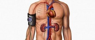
Blood circulation is a continuous flow of blood that moves through the vessels and cavities of the heart. This system is responsible for metabolic processes in the organs and tissues of the human body. Circulating blood transports oxygen and nutrients to the cells, taking carbon dioxide and metabolites from there. That is why any circulatory disorders threaten with dangerous consequences.
The blood circulation consists of a large (systemic) and a small (pulmonary) circle. Each turn has a complex structure and functions. The systemic circle originates from the left ventricle and ends in the right atrium, and the pulmonary circle originates from the right ventricle and ends in the left atrium.
Types of Blood Vessels
Blood circulation is a complex system that consists of the heart and blood vessels. The heart constantly contracts, pushing blood through the vessels to all organs and tissues. The circulatory system consists of arteries, veins, and capillaries.
The circulatory system is formed by arteries, veins and capillaries
The arteries of the systemic circulation are the largest vessels; they have a cylindrical shape and transport blood from the heart to the organs.
Structure of the walls of arterial vessels:
- outer connective tissue membrane;
- middle layer of smooth muscle fibers with elastic veins;
- strong elastic inner endothelial membrane.
Arteries have elastic walls that constantly contract, allowing blood to move evenly.
With the help of the veins of the systemic circulation, blood moves from the capillaries to the heart. Veins have the same structure as arteries, but they are less strong, since their middle layer contains less smooth muscle and elastic fibers. That is why the speed of blood movement in the venous vessels is largely influenced by nearby tissues, especially skeletal muscles. All veins, except the vena cava, are equipped with valves that prevent the backflow of blood.
Capillaries are small vessels that consist of endothelium (a single layer of flat cells). They are quite thin (about 1 micron) and short (from 0.2 to 0.7 mm). Due to their structure, microvessels saturate tissues with oxygen and useful substances, carrying away carbon dioxide, as well as metabolic products. Blood moves slowly through them; in the arterial part of the capillaries, water is removed into the intercellular space. In the venous part, blood pressure decreases and water flows back into the capillaries.
Heart and blood vessels
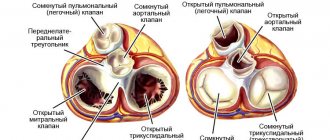
The basis of the circulatory system is the heart, which, like a pump, pumps blood through the arteries, ensuring the delivery of oxygen and nutrients to all organs and tissues. The heart is located in the chest along the projection of the sternum, slightly to the left, and is a hollow muscular organ the size of a fist. The heart has 4 chambers separated by septa. Between the left atrium and the left ventricle, as well as the right atrium and the right ventricle, there are openings with valves that regulate the direction of blood flow from the atria to the ventricles. Blood circulation occurs through the systemic and pulmonary circulation. Blood collects from all organs and tissues and enters the right atrium through the veins. From the right atrium, through the corresponding opening, venous blood flows into the right ventricle. Both of these right reservoirs (atrium and ventricle) are also called the right heart (“venous”). The pulmonary circulation begins in the right ventricle. From the right ventricle, due to the contraction of its muscular wall, dark, oxygen-poor and carbon dioxide-rich blood is pushed into the pulmonary artery and flows through it into the lungs. There it enters small arteries and capillaries, is cleared of carbon dioxide by diffusion and enriched with oxygen, acquires a bright red color and is already called arterial blood. Through four pulmonary veins, arterial blood enters the left atrium, and this completes the pulmonary circulation. Considering that the left cavities of the heart (atrium and ventricle) or the left heart are already filled with arterial blood, the left heart is called “arterial”. The systemic circulation begins in the left atrium. From the left atrium, arterial blood flows into the left ventricle, which is an even more powerful pump than the right ventricle. Contracting, the left ventricle pushes blood into the aorta and its branches, through which it enters all organs and tissues, down to the smallest capillaries. Having given oxygen to the tissues and taken away carbon dioxide from them, the blood again becomes venous. Venous capillaries gradually connect with each other into larger veins, which, in turn, form two wide ones: the superior and inferior vena cava. The superior vena cava collects blood from the head, neck, upper limbs and torso walls, and the inferior vena cava collects blood from the lower limbs, abdominal organs and pelvic region. Both vena cavae carry blood to the right atrium, where the systemic circulation ends.
In this way, 2 closed circles of blood circulation are obtained, which are connected by the motor of the human body - the heart. So, the systemic circulation begins in the left ventricle of the heart and ends in the right atrium. Its function is to supply all organs and tissues with nutrients and oxygen. The pulmonary circulation begins from the right ventricle and ends in the left atrium. Its function is to enrich the blood with oxygen in the lungs. The heart muscle, constantly performing enormous work, itself needs nutrition and oxygen. Blood flows to the heart through vessels that extend directly from the aorta and surround the heart like a crown or crown, which is why they are called coronary or coronary arteries.
Movement of blood through vessels.
The heart does a lot of work. So, in one minute it pumps 4.5–5 liters of blood in only one direction. Blood movement is ensured by valves located between the atria and ventricles, between the left ventricle and the aorta, pulmonary vessels and the right atrium. The speed of blood movement through the vessels depends on their diameter: if in the aorta the blood moves at high speed, then in the capillaries this speed is minimal. When the cardiovascular system is damaged by an atherosclerotic, inflammatory or degenerative process, both general and local circulatory disorders can be observed. An example of general circulatory disorders is heart failure with shortness of breath, palpitations, cough, bluish skin and swelling. An example of local circulatory disorders, when the blood supply to any organ is affected, is a heart attack (of the heart, lung or kidneys), or gangrene of a limb. But since the circulatory system functions as a single whole, even local circulatory disorders in any organ eventually affect the entire system. The activity of the heart is regulated by the central nervous system. In addition, the heart also has its own intracardiac regulatory mechanisms that promote rhythmic contraction (systole phase) and relaxation (diastole phase) of the heart. In an adult, the number of heartbeats per minute normally ranges from 60 to 80 beats; in athletes, the heart works more economically. Their heart rate is 40–50 beats per minute.
Arteries of our body
Before talking about atherosclerosis, let us recall the anatomical features of the arteries. Arteries are cylindrical elastic tubes of various diameters. The wall of arteries is much thicker than that of veins, the vessels that carry blood back to the heart. This difference in the thickness of the vessels is not accidental and is due to the fact that the blood pressure in the arteries is much greater than in the veins. The arterial wall consists of three layers: outer, middle and inner. The outer layer or serosa is a framework of connective tissue; the middle (muscle) layer consists of smooth muscle fibers; the inner layer (intima) is lined with a single layer of cells and is called endothelium. It is the endothelium, or rather its damage or dysfunction, that plays a major role in the development of atherosclerosis. However, we will talk about this later. The lumen of the arteries can change as a result of contraction or relaxation of the smooth muscle fibers of the middle layer. The dilation of blood vessels (for example, in hot conditions) helps to increase blood flow and more intense metabolism, and conversely, their narrowing (in low temperatures) slows down these processes in the body. If the vessels are constantly narrowed for some reason, then the organs and tissues receive little blood, and therefore oxygen. Over time, this leads to disruption of the functioning of those organs and tissues that are nourished by narrowed arteries.
Vascular changes in atherosclerosis
Atherosclerosis (from the Greek words “ather” - gruel and “sclerosis” - hardening), exactly as the name suggests, is the process of accumulation of soft deposits of lipids (fats, fat-like substances, primarily cholesterol) on the walls of arteries.
It has been established that atherosclerosis develops in response to damage to the endothelium (the inner lining of blood vessels).
Damage or dysfunction of the endothelium can be caused by a number of reasons, in particular, smoking, significant increases in blood lipid levels, high blood pressure, acute or chronic psycho-emotional stress, viral or bacterial infection. Following damage to the endothelium, fats, fat-like substances, and cholesterol begin to penetrate into the arterial wall. Leukocytes, or rather their special varieties, monocytes and macrophages, rush here from the blood. This is the beginning of the formation of an atherosclerotic plaque. The “mush” formed on the wall of the artery is covered with a thin connective tissue capsule consisting of fibrin threads. Such an atherosclerotic plaque is called young. Over time, as the atherosclerotic process progresses, calcium begins to accumulate in the plaques, and fibrous and connective tissue grows. The plaque becomes covered with a thick capsule (this is a formed plaque), increases in size and significantly narrows the lumen of the artery. Often several plaques form and merge with each other, further narrowing the lumen of the vessel. Due to the narrowing of the lumen of the vessel, the organ supplied with blood does not receive the required amount of oxygen and chronic ischemia occurs (from the Greek words “ishe” - I retain and “hemo” - blood). Thus, when one or more coronary arteries narrows, chronic myocardial ischemia occurs (chronic coronary heart disease).
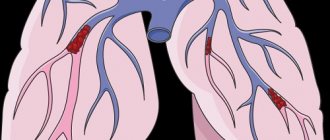
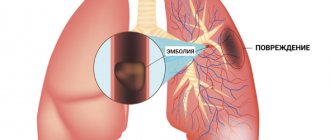
Acute ischemia (acute vascular insufficiency) develops differently. The fact is that the body produces special enzymes that “eat away” the connective tissue membrane of the atherosclerotic plaque from the edges, reaching its soft, mushy core. When the plaque capsule is opened, this mass enters the blood. An open wound on the capsule of an ulcerated plaque becomes covered with sticky platelets. Gradually, a blood clot forms - a thrombus, which bulges into the lumen of the vessel, sharply narrowing it. A thrombus can break away from the vessel wall and, moving with the blood flow, clog a smaller vessel, creating acute local vascular insufficiency and stopping tissue nutrition, which leads to tissue necrosis (death). For example, when one of the coronary arteries of the heart is blocked, myocardial infarction occurs; when thrombosis of the femoral artery or leg artery occurs, foot necrosis (gangrene) occurs. Atherosclerosis can completely affect the aorta and its branches, but more often arteries of various sizes are affected, and not throughout, but in certain areas. Atherosclerotic plaques “love” to form in places where arteries bend and branch, which are especially numerous in vital organs. Thus, the coronary arteries of the heart, the vessels of the brain, the abdominal aorta and its branches that supply blood to the abdominal organs, kidneys, pelvic organs, and lower extremities are most susceptible to atherosclerosis. Often, atherosclerosis initially favors one organ. For example, with damage to the coronary arteries, coronary heart disease develops; with atherosclerosis of the cerebral arteries, coronary artery disease develops, including stroke. When the iliac or femoral vessels are narrowed by atherosclerotic plaques, obliterating atherosclerosis of the vessels of the lower extremities occurs. If the renal vessels are affected by atherosclerosis, then hypertension may develop with a predominant increase in the “lower” numbers of blood pressure. Depending on the location of the affected vessels, clinical manifestations of atherosclerosis of a particular vital organ are formed. Atherosclerosis in its development is not similar to any other disease, since its first signs appear in early childhood. Thus, at six months of age, some babies begin to develop fatty spots and stripes on the inner wall of blood vessels. During the growth process, a significant part of these spots resolves and only a few remain. If you do not engage in prevention, then by the age of 35-50 in men and somewhat later in women, clinical signs of atherosclerosis appear in the form of coronary heart disease, coronary disease of the brain and other organs. The resulting disease requires many years, and in fact lifelong treatment. Meanwhile, in medicine it has long been proven that serious diseases, including atherosclerosis, are better and easier to prevent than to treat later for many, many years. In order to prevent atherosclerosis, you need to know everything about the substrate of atherosclerotic plaques - cholesterol and its assistants - lipoproteins.
Structure of the systemic circulation
The aorta is the largest vessel of the great circle, with a diameter of 2.5 cm. This is a kind of source from which all other arteries emerge. The vessels branch, their size decreases, they go to the periphery, where they give oxygen to organs and tissues.
The largest vessel in the systemic circulation is the aorta
The aorta is divided into the following sections:
- ascending;
- descending;
- the arc that connects them.
The ascending section is the shortest, its length is no more than 6 cm. The coronary arteries emanate from it, which supply oxygen-rich blood to the myocardial tissues. Sometimes the term “cardiac circulation” is used to name the ascending section. From the most convex surface of the aortic arch, arterial branches depart that supply blood to the arms, neck, and head: on the right side there is the brachiocephalic trunk, divided in two, and on the left there is the common carotid, subclavian artery.
The descending aorta is divided into 2 groups of branches:
- Parietal arteries that supply blood to the chest, spinal column, and spinal cord.
- Visceral (splanchnic) arteries that transport blood and nutrients to the bronchi, lungs, esophagus, etc.
Under the diaphragm is the abdominal aorta, the parietal branches of which supply the abdominal cavity, the lower surface of the diaphragm, and the spine.
The internal branches of the abdominal aorta are divided into paired and unpaired. The vessels that extend from the unpaired trunks transport oxygen to the liver, spleen, stomach, intestines, and pancreas. The unpaired branches include the celiac trunk, as well as the superior and inferior mesenteric arteries.
There are only two paired trunks: renal, ovarian or testicular. These arterial vessels are adjacent to the organs of the same name.
The aorta ends with the left and right iliac arteries. Their branches extend to the pelvic organs and legs.
Many people are interested in the question of how the systemic circulatory system works. In the lungs, the blood is saturated with oxygen, after which it is transported to the left atrium, and then to the left ventricle. The iliac arteries supply blood to the legs, and the remaining branches supply blood to the chest, arms, and organs of the upper half of the body.
The veins of the systemic circulation carry oxygen-poor blood. The systemic circle ends with the superior and inferior vena cava.
The diagram of the veins of the systemic circle is quite clear. The femoral veins in the legs unite to form the iliac vein, which becomes the inferior vena cava. In the head, venous blood collects in the jugular veins, and in the arms - in the subclavian veins. The jugular as well as the subclavian vessels unite to form the innominate vein, which gives rise to the superior vena cava.
The heart is the basis of the circulatory system
The heart is a muscular organ about the size of a human fist that is located on the left side of the chest, just in front of the lungs. This organ is actually a powerful double pump with four chambers that pumps blood and keeps it moving throughout the body.
The right side of the heart consists of an upper (atrium) and lower (ventricle) chamber. The atrium receives processed venous blood, saturated with carbon dioxide, and then sends it to the ventricle. From there it enters the pulmonary arteries, where it is again saturated with oxygen. “Fresh” blood circulates to the left upper chamber (atrium), from where it enters the aorta and begins renewed transportation throughout the body.
The heart muscle beats more than 3 billion times during a lifetime.
Blood supply to the head
The circulatory system of the head is the most complex structure of the body. The carotid artery, which is divided into 2 branches, is responsible for the blood supply to the parts of the head. The external carotid arterial vessel saturates the face, temporal region, oral cavity, nose, thyroid gland, etc. with oxygen and useful substances.
The main vessel supplying blood to the head is the carotid artery
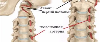
The internal branch of the carotid artery goes deeper, forming the Circle of Wallisian, which transports blood to the brain. In the cranium, the internal carotid artery branches into the ophthalmic, anterior, middle cerebral, and communicating arteries.
This is how only ⅔ of the systemic circle is formed, which ends with the posterior cerebral arterial vessel. It has a different origin, the scheme of its formation is as follows: subclavian artery - vertebral - basilar - posterior cerebral. In this case, the brain is supplied with blood by the carotid and subclavian arteries, which are connected to each other. Thanks to anastomoses (vascular anastomoses), the brain survives minor disturbances in blood flow.
Conduction system of the heart
The heart, like any organ, has its own nervous system. The nervous system of the heart has several levels. The first and main pacemaker of the heart is the sinus node, located in the right atrium. It is subject to the atrioventricular node, which is located on the border between the atria and ventricles and quite often slows down the heart rate set by the sinus node. Then the nerve impulse goes to the ventricles of the heart along the branches of the Hiss bundle, which are divided into the smallest nerve endings - Purkinje fibers.
Principle of placement of arteries
The circulatory system of each body structure is approximately similar to that described above. Arterial vessels always approach organs along the shortest path. The vessels in the limbs pass precisely along the flexion side, since the extensor part is longer. Each artery originates at the embryonic site of the organ, and not at its actual location. For example, the arterial vessel of the testicle emerges from the abdominal aorta. Thus, all vessels are connected to their organs from the inside.
The arrangement of vessels resembles the structure of the skeleton
The placement of arteries is also related to the structure of the skeleton. For example, the brachial branch runs along the upper limb, which corresponds to the humerus; the ulnar and radial arteries also pass next to the bones of the same name. And in the skull there are openings through which arterial vessels transport blood to the brain.
Arterial vessels of the systemic circulation form networks in the joint area using anastomoses. Thanks to this scheme, the joints are continuously supplied with blood during movement. The size of the vessels and their number depend not on the size of the organ, but on its functional activity. Organs that work more intensively are saturated with a large number of arteries. Their placement around the organ depends on its structure. For example, the diagram of the vessels of parenchymal organs (liver, kidneys, lungs, spleen) corresponds to their shape.
Cellular structure of blood
Blood consists of two components: plasma (50-60%) and suspended formed elements (40-50%).
The second category includes:
· Erythrocytes (red blood cells) are the most numerous of the formed elements. According to official studies, one drop of blood contains about 5 million red blood cells. Red blood cells are responsible for transporting gases - oxygen and carbon dioxide. They contain the protein hemoglobin, which binds oxygen molecules in the lungs. Red blood cells deliver oxygen to all tissues and organs, after which they absorb carbon dioxide and carry it to the lungs. It is removed from the body during respiration.
· Leukocytes (white blood cells) - elements that protect our body from foreign bodies and compounds, are part of the immune system. White blood cells recognize and attack pathogens through the production of antibodies and macrophages. When an infection enters the body, the production of leukocytes increases significantly. Normally, their quantity is inferior to the concentration in the blood of other formed elements.
· Platelets (blood platelets) are cells that provide coagulation (clotting) of blood flowing from a damaged vessel and protect the body from heavy blood loss. They stick to the hole in the damaged vessel, forming a “sealing” plug to stop bleeding. It is the platelets that can stick together and form pathological blood clots inside the vessels, called thrombi.
All formed elements are synthesized by the bone marrow and distributed through plasma, the liquid part of the blood.