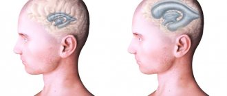
1.What is ventriculo-peritoneal shunting?
Ventriculo-peritoneal shunt
– a surgical operation that allows you to successfully combat
hydrocephalus
.
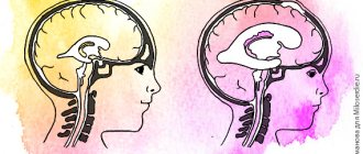
Hydrocephalus, popularly called dropsy of the brain, is a serious disease, the essence of which is a violation of the systematic outflow of cerebrospinal fluid (CSF), which, in turn, leads to an increase in the volume of the ventricles of the brain.
A must read! Help with treatment and hospitalization!
Hydrocephalus
The essence of hydrocephalus is an increase in the volume of cerebrospinal fluid, and, as a rule, an increase in intracranial pressure, due to an imbalance between the production and absorption of cerebrospinal fluid.
Hydrocephalus can be either congenital or acquired. Acquired hydrocephalus occurs due to tumor obstruction of the cerebrospinal fluid pathways, trauma, intracranial hemorrhage, or infectious lesions of the meninges.
Pathophysiology – hydrocephalus develops for the following reasons:
- Excessive production of cerebrospinal fluid (CSF) (due to papilloma, choroid plexus carcinoma)
- Impaired fluid circulation - it occurs due to obstruction (blockage) of the circulation pathways of fluid produced in the ventricles of the brain. Typically, this situation occurs when tumor occlusion of the natural openings connecting the ventricles of the brain with each other and with the subarachnoid space.
- Impaired absorption (absorption) of cerebrospinal fluid. Often, impaired absorption occurs due to blockage of the pachyonic granulations located along the superior sagittal sinus (parasagittal).
In different age groups, hydrocephalus clinically manifests itself differently.
Acute hydrocephalus is clinically manifested by severe headache, gait disturbance, nausea, and visual disturbances. In children, early signs of hydrocephalus may include irritability and an inability to hold the head up. When the third ventricle expands, the patient develops Parinaud's syndrome (upward gaze paresis - inability to raise the eyes upward) or the setting sun symptom (Parinaud's syndrome with retraction of the eyelids and forced downward rotation of the gaze). Rarely, there may be a focal symptom in the form of abducens nerve paresis. In children, the fontanel bulges, the veins of the scalp expand, and the head circumference progressively increases. As the pathology progresses, symptoms of brain stem damage, coma, and hemodynamic disturbances occur.
With normal pressure hydrocephalus (hydrocephalus with normal intracranial pressure), the symptoms are quite clear. It usually develops in elderly patients.
Indications
For symptomatic hydrocephalus, the underlying disease must be treated.
Contraindications
Hydrocephalus does not always need to be treated.
- Ventriculomegaly in the elderly who do not have a triad of clinical symptoms
- Hydrocephalus en vacuo, when brain tissue is replaced by cerebrospinal fluid. Since there is no imbalance between the production and absorption of cerebrospinal fluid, the condition is not technically hydrocephalus.
- Arrested hydrocephalus – which is characterized by the fact that the neurological condition of the patient is stable with stable ventriculomegaly. The diagnosis must be made very carefully, as neurological deterioration in children occurs very slowly and is difficult to document.
- Benign hydrocephalus of children occurs in newborns and infants. The children have no neurological symptoms, and their head sizes are normal. A CT scan of the brain reveals moderately dilated cerebral ventricles and subarachnoid fissures.
Research
- CT scan shows the extent of ventriculomegaly and in many cases determines the etiology. With intravenous administration of a contrast agent, tumors and infectious processes leading to obstruction can be detected. This fact helps to plan surgical treatment. The ventricles are dilated proximal to the point of obstruction. With pseudotumor of the brain, no pathological foci are detected on a CT scan of the brain.
- MRI will show congenital causes of hydrocephalus in many, but not all, cases. It shows pathologies such as agenasia of the corpus callosum, Chiari malformation, neuronal migration disorders, and vascular malformations. Some tumors, for example, gliomas of the midbrain plate, can only be identified with this study. T2-weighted studies may show transependymal fluid flow.
- Fetal and neonatal ultrasound is a good method for monitoring ventricular size and intraventricular hemorrhage.
Diagnostic procedures
Lumbar puncture is used to measure intracranial pressure. But it must be performed after an MRI or CT scan of the brain.
Drug treatment
Has a temporary effect. In transient cases such as sinus occlusion, meningitis, neonatal ventricular hemorrhage, drug treatment may be effective. The following drugs are used.
- Acetazolamide (25 mg/kg/d x 3). Respiratory status and electrolyte balance must be monitored. Treatment is not recommended to continue for more than 6 months.
- Furosemide (1 mg/kg/d x 3) – monitoring of electrolyte and fluid balance is necessary.
- Lumbar puncture - in newborns it is used for intraventricular hemorrhage, in some cases it can lead to cure.
- In many cases, the solution to the problem is to remove the cause of hydrocephalus.
Preoperative details
- Before performing invasive procedures, it is necessary to clarify the cause of hydrocephalus. If possible, hydrocephalus should be treated conservatively (with medication).
- Patients with suspected infection or with elevated protein levels in the cerebrospinal fluid (more than 150 mg/dL) should not undergo surgery.
- It is necessary to preoperatively assess the nature of hydrocephalus (level of intracranial pressure) to select the appropriate valve pressure for shunt operations. The most optimal is the use of programmable shunts.
- Antibiotic prophylaxis is mandatory before surgery.
Surgery
- Endoscopic ventriculostomy of the third ventricle (ventriculostomy) - this procedure is used in patients with normal or almost normal ability to absorb cerebrospinal fluid. Surgery is performed using endoscopic technology. High precision of the operation is ensured by the use of frameless steriotaxic equipment.
- Ventriculoperitoneal shunt is the most commonly performed operation for hydrocephalus. After preoperative assessment of intracranial pressure, an appropriate pressure valve is selected. The most optimal is the use of programmable shunts, which allows, if necessary, to change the valve pressure without repeated surgical intervention .
- If there are certain problems in the absorption of fluid in the abdominal cavity (previous peritonitis, adhesive disease), ventriculoatrial (the distal end of the catheter is installed into the appendage of the heart through the jugular vein) and ventriculopleural (the distal end of the shunt is installed into the pleural cavity) shunting can be performed.
- Torkildsen shunt - using a catheter without a valve, the ventricular cavity is connected to the occipital cistern of the brain.
- Lumboperitoneal shunting is used for connecting hydrocephalus (non-occlusive), especially if the ventricles of the brain are narrow. The classical indication for this type of shunting is cerebral pseudotumor.
Postoperative management of patients
- After shunt surgery, it is advisable to activate patients and bring them to an upright position slowly, to avoid the occurrence of a subdural hematoma.
- An X-ray of the skull is performed to assess the correct location of the ventricular catheter.
- It is necessary to monitor the healing of the wound; sutures are removed 10–14 days after surgery
- A CT scan of the brain is performed 2-3 weeks after surgery.
Complications
- Infectious complications are most likely during the first 6 months after surgery.
- Postoperative subdural hematomas are rare. This complication can be avoided by slowly mobilizing the patient.
- Impaired absorption in the abdominal cavity.
- Excessive drainage - most often occurs with lumboperioneal shunting, manifested by headache in an upright position. More often than not, this problem resolves on its own. Otherwise, the operation is repeated and the valve is changed to a higher pressure (or, with a programmable shunt, the valve is reprogrammed non-surgically)
results
The results depend on many factors - the nature of the underlying pathology, the duration of hydrocephalus, and the initial condition of the patient.
With timely and correctly performed surgery, the results are generally good.
2.Basic methods of conducting operations
Ventriculoperitoneal shunt surgery has been in practice for more than fifty years, being the main standard method for getting rid of almost any form of hydrocephalus. The essence of this operation is to use a special system of tubes equipped with a valve to remove excess cerebrospinal fluid into the natural cavities of the body. Depending on which cavity is chosen for this, the following types of bypass can be distinguished:
- ventriculo-peritoneal (cerebrospinal fluid enters the abdominal cavity);
- ventriculoatrial (cerebrospinal fluid is drained into the right atrium);
- ventriculo-pleural (cerebrospinal fluid enters the pleural cavity).
Today, the most common method of treating hydrocephalus is the ventriculoperitoneal shunt
. To carry out such an operation, the surgeon has the opportunity to choose from two hundred types of shunt systems that currently exist.
As already mentioned, the main goal of ventriculoperitoneal shunting is to create an artificial pathway for the outflow of excess cerebrospinal fluid from the ventricular system of the brain into the abdominal cavity. In some cases, the doctor may decide to perform surgery using a minimally invasive laparoscopic method.
, i.e. without resorting to wide incisions of the anterior abdominal wall - this reduces the risk of trauma to the internal organs located in the abdominal cavity and shortens the patient’s rehabilitation period.
Visit our Neurosurgery page
Hydrocephalus of the brain: diagnosis
A neurologist, even during a visual examination, diagnoses hydrocephalus, based on its characteristic external signs. However, to organize effective treatment, it is important for neurosurgeons to establish the type and form of hydrocephalus.
For a more detailed study of the pathology, children and adults are offered to undergo examinations:
- MRI
(allows us to establish the causes of development and the form of the pathology; children sometimes have to be given a light anesthesia to ensure immobility during the procedure); - (allows you to determine the contours of the brain, skull, and detect tumors);
- Ultrasound
(indicated only for infants with an open fontanel; for adults this method is not very informative); - Angiography
(carried out using a contrast agent to detect problems in blood circulation); - Lumbar puncture
(allows you to determine the cause of the pathology).
3.Types of shunt systems
We have already mentioned that modern medicine has a whole arsenal of various shunt systems - there are more than 200 types. All shunt models equipped with valves can be divided into three types depending on their throughput, or, in other words, on the level of cerebrospinal fluid pressure.
Shunt systems with preset pressure valves
represent a certain difficulty for the surgeon, since an error in selecting the parameters of the shunt system can lead to both excessive and insufficient levels of cerebrospinal fluid drainage.
by shunt systems that appeared not so long ago
. The complexity of the installation of such a system is practically no different from a similar operation during which a system with a valve of a given pressure is installed. The “highlight” of the programmable valve is that it is equipped with an anti-siphon device designed to allow remote control of the level of drainage and intracranial pressure of the patient.
About our clinic Chistye Prudy metro station Medintercom page!
Symptoms
The external symptoms of congenital hydrocephalus in a newborn are so obvious that the pathology is determined not only by doctors, but also by parents. The baby has:
- excessive and disproportionate enlargement of the head (diameter can reach 80 cm);
- enlargement and bulging of the fontanel (it closes much later than in healthy babies);
- persistent drowsiness or irritability;
- thinning of the skin (veins are easily visible);
- poor appetite;
- inhibition of psychomotor development;
- low location of the pupils;
- vomiting and frequent regurgitation;
- poor weight gain;
- convulsions;
- decreased activity.
In adults and older children, almost the same symptoms are observed, only several additional signs are added to it:
- intense pain;
- vision problems;
- double vision;
- uncontrolled urine output;
- labored breathing;
- problems with remembering.
Postoperative period
In the early postoperative period, medical supervision is necessary until the next morning. The main attention is paid to the level of blood pressure, the absence of neurological disorders, swallowing and voice disorders. It is imperative to monitor the discharge from the drainage and the condition of the dressing (dry or wet with blood). Pain after surgery is mild and easily relieved with conventional analgesics. The day after surgery, the drainage is removed. The day after surgery, if the course is smooth, the patient can be discharged. Sutures are removed on the 7th day after surgery.
Our advantages
Surgeons at the Innovative Vascular Center have been dealing with the problem of treating pathological tortuosity of the carotid arteries since 2001. During this time, a lot of scientific work was carried out to determine indications for treatment and a diagnostic algorithm. The technique of surgical interventions was developed. Since that time, more than 200 surgical interventions for pathological tortuosity have been performed without complications with good immediate and long-term results. The results of operations in all patients were analyzed. Constant postoperative monitoring of the patency of the operated arteries and neurological functions is carried out.
Possible complications
According to the literature, the following complications are possible after surgery on the carotid artery:
- Bleeding from the surgical site is a very rare complication, occurring in no more than 0.2% of all operations. If detected in a timely manner, the wound is opened, the source of bleeding is identified and it is finally stopped.
- Postoperative ischemic stroke - after surgery for pathological tortuosity is quite rare, not more than 1% of all surgical cases. Most often associated with thrombosis of the reconstructed carotid artery. If a cerebral circulatory disorder is detected in a timely manner, repeated surgery to remove the blood clot can restore cerebral blood flow.
- Damage to the recurrent or hypoglossal nerve. When the recurrent nerve is injured, hoarseness occurs. The recurrent nerve may be damaged by stretching the wound during access or accidentally cut. In the first case, the voice disorder will be reversible, in the other case it will be permanent. If the hypoglossal nerve is damaged, the tongue deviates to the side and choking occurs when eating liquid food. The only way to prevent these complications is an impeccable technique for operating the carotid arteries. In our clinical practice, such complications were not encountered.
- Suppuration of a postoperative wound is an extremely rare complication that can develop in patients with chronic purulent processes in the lungs and mediastinum and weakened immunity. We have not encountered this in our practice.
Operation options
Redressing the internal carotid artery - the operation involves isolating the artery from the adhesions, straightening all bends and fixing the artery to the surrounding tissues in a straightened position.
Resection of a tortuous carotid artery with end-to-end anastomosis - this option involves isolating a loop, excision of the altered area and suturing the artery end-to-end. This option is used when the loop is located high from the mouth of the internal carotid artery and with the same diameters of the efferent and adductor ends of the artery.
Resection with redressing of the internal carotid artery is performed when the bends are located near the mouth of the carotid artery. The artery is cut off from the common carotid artery, after which the excess is resected and the artery is sutured to its old place.
Resection of pathological tortuosity with prosthetics of the carotid artery - the tortuous area is excised, and an artificial vessel or the own saphenous vein is sewn in place of this area. This type of operation is used when there is a sharp change in the arterial wall, or when tortuosity is combined with severe atherosclerosis of the artery.
Progress of the operation
The incision is made along the inner edge of the sternocleidomastoid muscle on the neck, the skin, subcutaneous tissue, and subcutaneous muscle are sequentially dissected. The edge of the sternocleidomastoid muscle is retracted outward. The bifurcation (bifurcation) of the common carotid artery is highlighted. The latter is divided into internal and external. The external carotid artery is distinguished by the presence of branches, the internal one does not have them. After this, the internal carotid artery is carefully isolated up to and above the tortuosity. Care must be taken when isolating the superior section, as this is where the hypoglossal nerve and pharyngeal plexus are located. After identifying the tortuosity, it is straightened out and a decision is made on the correction method. After excision of the excess artery, the course of the artery is restored. After starting the blood flow, the restored vessel is examined using an ultrasound scanner, and the nature of the blood flow (laminar, turbulent) and its speed are studied. After making sure that blood flow has been restored, they begin to suture the wound. The operation ends with the installation of drainage.
Anesthesia
Surgery for pathological tortuosity is performed under local anesthesia or general anesthesia. The choice of anesthesia method in our clinic depends on the volume of the upcoming operation. We prefer to perform this operation under local anesthesia, as it makes it possible to determine the brain's response to temporary compression of the carotid artery. In case of signs of cerebral circulatory insufficiency (impaired consciousness, implementation of commands), methods of protecting the brain (temporary shunt) can be used during test clamping. For operations under general anesthesia, a clamp test of the internal carotid artery is performed during an ultrasound examination before the operation.