December 12, 2013
The extensive network of the human circulatory system consists not only of large veins and arteries, but also of the smallest capillaries
The extensive network of the human circulatory system consists not only of large veins and arteries, but also of the smallest capillaries, thanks to which all the substances necessary for optimal life are delivered to each of our cells along with blood. It is not surprising that a person’s health largely depends on the state of his cardiovascular system.
Vessels
Arteries are vessels that deliver blood from the heart to the organs. The arterial system, which permeates the entire body, includes the arteries of the heart, pulmonary, renal, splenic arteries, aorta, brachiocephalic arteries, arteries of the extremities, etc. Since the arteries need to withstand considerable blood pressure, their walls are initially elastic and elastic. However, as a result of atherosclerosis, the walls of the arteries are weakened. Impaired elasticity of the walls of blood vessels and a decrease in the lumen inside the arteries lead to heart failure and coronary heart disease. As a result of the changes, the arterial walls become stratified and aneurysms form on them. Such ailments are dangerous, because disruption of just one link in the chain of the cardiovascular system will lead to pathological processes inside the body.>
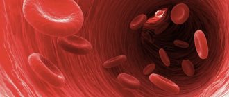
Capillaries are vessels that connect the arterial and venous systems. Thanks to these thinnest vessels, blood and tissues exchange substances with each other. Capillaries permeate tissues that need oxygen.
Veins are vessels that carry blood obtained from capillaries to the heart.
What is blood pressure?
Blood pressure is the force with which blood presses against the walls of the arteries. It increases when the heart contracts and pumps out blood, and decreases when the heart muscle relaxes. Blood pressure is stronger in the arteries and weaker in the veins. Blood pressure is measured with a special device - tonometer
.
Pressure readings are usually recorded in two numbers. Thus, normal blood pressure for an adult is considered to be 120/80
.
The first number, systolic pressure
, is a measure of the pressure during a heartbeat.
The second is diastolic pressure
- the pressure during relaxation of the heart.
Such dangerous excess weight
Professor Sergei Boytsov, chief specialist in preventive medicine at the Russian Ministry of Health and Social Development, talks about how extra pounds affect the heart and blood vessels.
Pressure is measured in the arteries and expressed in millimeters of mercury. In the capillaries, the pulsation of the heart becomes invisible and the pressure in them drops to approximately 30 mm Hg. Art. A blood pressure reading can tell your doctor how your heart is working. If one or both numbers are higher than normal, this indicates high blood pressure. If it’s lower, it means it’s reduced. High blood pressure indicates that the heart is working too hard: it requires more effort to push blood through the vessels. It also indicates that a person has an increased risk of heart disease.
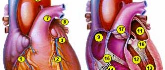
Heart
The heart is the organ that pumps blood from the veins to the arteries. Thanks to the work of the heart, blood is supplied to the organs through arteries and capillaries. It is like a powerful pump and consists of muscle tissue - the myocardium. The heart pumps about 5 liters of blood per minute. An abrupt stop in its work is fraught with fatal consequences - myocardial infarction or heart attack often ends in the death of a person.
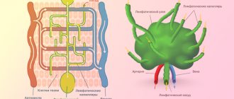
Foundation of life
The circulatory system consists of more than just the heart, blood, and blood vessels. This is only one of two complementary systems - cardiovascular and lymphatic. The latter serves to transport lymph - a colorless liquid with many lymphocytes. The lymphatic system is also extremely important, because human immunity largely depends on it. It is these two systems - cardiovascular and lymphatic - that make up the largest human circulatory system with a total length of more than 100,000 km. This complex mechanism is driven by the heart. This living motor, consisting of muscles, works with amazing performance, pumping more than 9,500 liters of blood per day. In this way, blood is supplied to every cell.
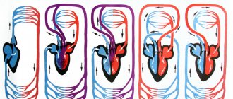
Circulation circles
The structure of the human cardiovascular system is characterized by closedness. It forms blood circulation circles. The pulmonary circulation carries blood from the heart to the lungs, and also in the opposite direction. The small circle begins in the right ventricle and ends in the left atrium. In this circle, the blood is saturated with oxygen and removes carbon dioxide. The systemic circulation ensures the movement of blood from the left ventricle to the right atrium, and all organs and tissues are saturated with blood.
Arteries
Not a single cell can survive without nutrients and oxygen. They are delivered by arteries. They carry oxygen-rich blood throughout the body. When you breathe, oxygen enters your lungs. where the delivery of oxygen throughout the body begins. First to the heart, then through the systemic circulation to all parts of the body. There, the blood exchanges oxygen for carbon dioxide and then returns to the heart. The heart pumps it back to the lungs, which take in carbon dioxide and give out oxygen, and so on endlessly. There are also pulmonary arteries of the pulmonary circulation, they are located in the lungs and through them blood, poor in oxygen and rich in carbon dioxide, enters the lungs, where gas exchange occurs. This blood then returns to the heart through the pulmonary veins.
Anatomy
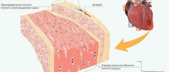
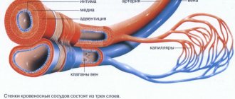
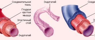
Veins and arteries consist of three layers:
- Tunica adventitia : The outer layer of a blood vessel, composed of collagen and elastin. This layer allows the vessel to expand or contract, depending on the type of vein or artery. This function is important for controlling blood pressure.
- Tunica media : This is the middle of the blood vessel. Elastin and muscle fibers form the shell of the carriers. The amount of elastin or muscle varies depending on the type of blood vessel. For example, elastic arteries contain few muscle fibers in their membranes.
- Tunica Intima: This name refers to the inner layer of a blood vessel. It mainly contains elastic membranes and tissues and may include valves that help blood move in the right direction.
Popular message topics
- Pushkin in the village of Mikhailovskoye
The exile of the poet to his mother’s family estate in the village of Mikhailovskoye followed immediately upon his return from southern exile. While in exile in Odessa, Pushkin wrote the ode “Liberty,” which the emperor did not like. - Organ of hearing
A person has several organs that help him navigate in space: eyes, ears, nose and others. The ear is the organ with which we hear. The organ of hearing has a complex structure. It consists of three parts: the outer ear, the middle - Glaciers
Glaciers are huge masses of ice. They are formed at low temperatures from large amounts of fallen snow. Such conditions are created on the peaks and slopes of mountains, and this is how mountain glaciers are formed. The largest of them are in the Himalayas
Types of Veins
Arteries and veins are designed roughly the same, but veins are thinner and have less muscle, which allows them to hold more blood. Veins typically contain about 70% of the body's blood at one time.
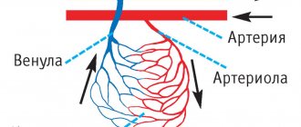
Venules are the smallest type of vein. They have very thin walls in order to hold a lot of blood. They pump low-oxygen blood through capillaries from the arteries directly into the veins. The blood then returns to the heart through a series of veins of increasing size and muscle.
There are two main types of veins: pulmonary and systemic.
Systemic veins are further classified into:
- Deep veins : These veins usually have a corresponding artery nearby and are found in muscle tissue. These veins may have a one-way valve to prevent blood from flowing backwards.
- Superficial veins : These veins do not have an artery of the same name nearby and are found close to the surface of the skin. They may also have a one-way valve.
- Connecting veins : These small veins allow blood to flow from the superficial veins to the deep veins.
Why does hypotension occur?
Low blood pressure (hypotension) is not dangerous for humans. However, people experiencing hypotension feel weak in the body. Dizziness often occurs, concentration decreases and fatigue increases. Therefore, hypotension can cause great discomfort. The most unpleasant consequences of this phenomenon are considered to be a decrease in intellectual activity. Girls face hypotension, and the World Health Organization states that a low blood pressure level is 100/60 mmHg. Art. for women and 110/70 mmHg. for men. It is worth noting that some feel quite comfortable even with constant hypotension.
WORK OF THE CIRCULATORY SYSTEM
Pulmonary circulation.
It is convenient to begin describing the normal movement of blood throughout the body from the moment when it returns to the right half of the heart through two large veins. One of them, the superior vena cava, brings blood from the upper half of the body, and the second, the inferior vena cava, brings blood from the lower half. Blood from both veins enters the collecting compartment of the right side of the heart, the right atrium, where it mixes with blood brought by the coronary veins, which open into the right atrium through the coronary sinus. The coronary arteries and veins circulate the blood necessary for the functioning of the heart itself. The atrium fills, contracts, and pushes blood into the right ventricle, which contracts to force blood through the pulmonary arteries into the lungs. The constant flow of blood in this direction is maintained by the operation of two important valves. One of them, the tricuspid valve, located between the ventricle and the atrium, prevents the return of blood to the atrium, and the second, the pulmonary valve, closes when the ventricle relaxes and thereby prevents the return of blood from the pulmonary arteries. In the lungs, blood passes through the branches of the vessels, entering a network of thin capillaries that are in direct contact with the smallest air sacs - the alveoli. An exchange of gases occurs between the capillary blood and the alveoli, which completes the pulmonary phase of blood circulation, i.e. the phase of blood entering the lungs (see also RESPIRATORY ORGANS).
Systemic circulation.
From this moment the systemic phase of blood circulation begins, i.e. phase of blood transfer to all tissues of the body. Cleared of carbon dioxide and enriched with oxygen (oxygenated), blood returns to the heart through four pulmonary veins (two from each lung) and enters the left atrium at low pressure. The path of blood flow from the right ventricle of the heart to the lungs and return from them to the left atrium is the so-called. pulmonary circulation. The left atrium, filled with blood, contracts simultaneously with the right and pushes it into the massive left ventricle. The latter, when filled, contracts, sending blood under high pressure into the artery of the largest diameter - the aorta. All arterial branches supplying the tissues of the body depart from the aorta. Just like on the right side of the heart, there are two valves on the left. The bicuspid (mitral) valve directs blood flow into the aorta and prevents blood from returning to the ventricle. The entire path of blood from the left ventricle until it returns (through the superior and inferior vena cava) to the right atrium is designated as the systemic circulation.
Arteries.
In a healthy person, the diameter of the aorta is approximately 2.5 cm. This large vessel extends upward from the heart, forms an arch, and then descends through the chest into the abdominal cavity. Along the course of the aorta, all the large arteries that enter the systemic circulation branch off from it. The first two branches, extending from the aorta almost at the very heart, are the coronary arteries, which supply blood to the heart tissue. Apart from them, the ascending aorta (the first part of the arch) does not give off branches. However, at the top of the arch, three important vessels branch off from it. The first, the innominate artery, immediately divides into the right carotid artery, which supplies blood to the right side of the head and brain, and the right subclavian artery, which passes under the collarbone into the right arm. The second branch from the aortic arch is the left carotid artery, the third is the left subclavian artery; These branches carry blood to the head, neck and left arm.
From the aortic arch begins the descending aorta, which supplies blood to the organs of the chest, and then enters the abdominal cavity through an opening in the diaphragm. Separated from the abdominal aorta are two renal arteries that supply the kidneys, as well as the abdominal trunk with the superior and inferior mesenteric arteries, which extend to the intestines, spleen and liver. The aorta then divides into two iliac arteries, which supply blood to the pelvic organs. In the groin area, the iliac arteries become femoral; the latter, going down the thighs, at the level of the knee joint pass into the popliteal arteries. Each of them, in turn, is divided into three arteries - the anterior tibial, posterior tibial and peroneal arteries, which nourish the tissues of the legs and feet.
Along the entire length of the bloodstream, the arteries become smaller and smaller as they branch, and finally acquire a caliber that is only several times larger than the size of the blood cells they contain. These vessels are called arterioles; as they continue to divide, they form a diffuse network of vessels (capillaries), the diameter of which is approximately equal to the diameter of a red blood cell (7 μm).
Structure of arteries.
Although large and small arteries differ somewhat in their structure, the walls of both consist of three layers. The outer layer (adventitia) is a relatively loose layer of fibrous, elastic connective tissue; the smallest blood vessels (the so-called vascular vessels) pass through it, feeding the vascular wall, as well as branches of the autonomic nervous system that regulate the lumen of the vessel. The middle layer (media) consists of elastic tissue and smooth muscles, which provide elasticity and contractility of the vascular wall. These properties are essential for regulating blood flow and maintaining normal blood pressure under changing physiological conditions. Typically, the walls of large vessels, such as the aorta, contain more elastic tissue than the walls of smaller arteries, which are predominantly muscle tissue. Based on this tissue feature, arteries are divided into elastic and muscular. The thickness of the inner layer (intima) rarely exceeds the diameter of several cells; It is this layer, lined with endothelium, that gives the inner surface of the vessel a smoothness that facilitates blood flow. Through it, nutrients flow to the deep layers of the media.
As the diameter of the arteries decreases, the walls become thinner and the three layers become less distinguishable until—at the arteriolar level—they contain mostly spiral muscle fibers, some elastic tissue, and an inner lining of endothelial cells.
Capillaries.
Finally, the arterioles imperceptibly turn into capillaries, the walls of which are lined only with endothelium. Although these tiny tubes contain less than 5% of the volume of circulating blood, they are extremely important. The capillaries form an intermediate system between the arterioles and venules, and their networks are so dense and wide that no part of the body can be pierced without piercing a huge number of them. It is in these networks that, under the influence of osmotic forces, oxygen and nutrients are transferred to individual cells of the body, and in return, products of cellular metabolism enter the blood.
In addition, this network (the so-called capillary bed) plays a critical role in regulating and maintaining body temperature. The constancy of the internal environment (homeostasis) of the human body depends on maintaining body temperature within narrow limits of normal (36.8–37°). Normally, blood from the arterioles enters the venules through the capillary bed, but in cold conditions the capillaries close and blood flow decreases, primarily in the skin; in this case, blood from the arterioles enters the venules, bypassing many branches of the capillary bed (bypass). On the contrary, when there is a need for heat transfer, for example in the tropics, all capillaries open and skin blood flow increases, which promotes heat loss and maintains normal body temperature. This mechanism exists in all warm-blooded animals.
Vienna.
On the opposite side of the capillary bed, the vessels merge into numerous small channels, venules, which are comparable in size to arterioles. They continue to connect to form larger veins that carry blood from all parts of the body back to the heart. Constant blood flow in this direction is facilitated by a system of valves found in most veins. Venous pressure, unlike pressure in the arteries, does not directly depend on the tension of the muscles of the vascular wall, so blood flow in the desired direction is determined mainly by other factors: the pushing force created by the arterial pressure of the systemic circulation; the “suction” effect of negative pressure that occurs in the chest during inhalation; the pumping action of the muscles of the limbs, which, during normal contractions, push venous blood to the heart.
The walls of veins are similar in structure to arterial ones in that they also consist of three layers, however, much less pronounced. For the movement of blood through the veins, which occurs practically without pulsation and at relatively low pressure, it does not require such thick and elastic walls as those of the arteries. Another important difference between veins and arteries is the presence of valves in them, which maintain blood flow in one direction at low pressure. The valves are found in greatest numbers in the veins of the extremities, where muscle contractions play a particularly important role in moving blood back to the heart; large veins, such as the cava, portal and iliac veins, lack valves.
On their way to the heart, the veins collect blood flowing from the gastrointestinal tract through the portal vein, from the liver through the hepatic veins, from the kidneys through the renal veins, and from the upper extremities through the subclavian veins. Two vena cavae form near the heart, through which blood enters the right atrium.
The vessels of the pulmonary circulation (pulmonary) resemble the vessels of the systemic circulation, with the only exception that they lack valves, and the walls of both arteries and veins are much thinner. In contrast to the systemic circulation, venous, non-oxygenated blood flows through the pulmonary arteries into the lungs, and arterial, i.e., flows through the pulmonary veins. saturated with oxygen. The terms “arteries” and “veins” refer to the direction of blood movement in the vessels - from the heart or to the heart, and not to the type of blood they contain.
Auxiliary organs.
A number of organs perform functions that complement the work of the circulatory system. The spleen, liver and kidneys are most closely associated with it.
Spleen.
As red blood cells (erythrocytes) pass through the circulatory system repeatedly, they are damaged. Such “waste” cells are removed from the blood in many ways, but the main role here belongs to the spleen. The spleen not only destroys damaged red blood cells, but also produces lymphocytes (which are white blood cells). In lower vertebrates, the spleen also plays the role of a reservoir of red blood cells, but in humans this function is weakly expressed. See also SPLEEN.
Liver.
To carry out its more than 500 functions, the liver needs a good blood supply. Therefore, it occupies an important place in the circulatory system and is provided by its own vascular system, which is called the portal system. A number of liver functions are directly related to the blood, such as removing waste red blood cells from the blood, producing clotting factors, and regulating blood sugar levels by storing excess sugar in the form of glycogen. See also LIVER.
Kidneys.
The kidneys receive approximately 25% of the total blood volume ejected by the heart every minute. Their special role is to cleanse the blood of nitrogen-containing waste. When this function is disrupted, a dangerous condition develops - uremia. Loss of blood supply or kidney damage causes a sharp rise in blood pressure, which, if left untreated, can lead to premature death from heart failure or stroke. See also KIDNEYS; UREMIA.