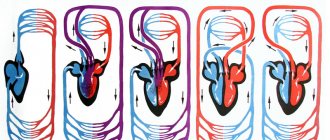
Circulation circles
The blood flowing through the cardiovascular system can be compared to an athlete running different distances. When it passes through the pulmonary circulation, it is a sprint. And a big circle is already a marathon. The Englishman William Harvey described these circles back in 1628. During a large circle, blood is distributed throughout the body, not forgetting to provide it with oxygen and take away carbon dioxide. During this “race”, arterial blood becomes venous.
The pulmonary circulation is responsible for the flow of blood into the lungs, where the blood releases carbon dioxide and is enriched with oxygen. Blood from the pulmonary circulation returns to the left atrium. The systemic circulation, starting in the left ventricle, transports blood throughout the body. Oxygenated blood is pumped by the left ventricle into the aorta and its many branches - the various arteries. Then it enters the capillary vessels of organs and tissues, where oxygen from the blood is exchanged for carbon dioxide. The systemic circulation ends in small veins that merge into two large veins (vena cava) and return blood to the right atrium. The superior vena cava drains blood from the head, neck and upper extremities, and the inferior vena cava drains blood from the torso and lower extremities.
Topic 9.3 Veins of the systemic circulation Lymphatic system
Topic 9.3 Veins of the systemic circulation
Lymphatic system
Venous vessels of the systemic circulation are represented by: the superior vena cava system, the inferior vena cava system and the portal vein system
To the superior vena cava system
These include all venous vessels that collect blood from the head, brain, neck, upper limbs, from the walls of the chest cavity, organs of the chest cavity, and also partially from the abdominal cavity. They are represented by the brachiocephalic veins, internal jugular, subclavian, veins of the upper extremities, azygos and semi-gypsy veins.
Inferior vena cava
collects blood from the lower extremities, walls and organs of the pelvis and abdominal cavity.
Portal vein
collects blood from unpaired abdominal organs; spleen, stomach, gall bladder, pancreas, small and large intestines. This is a short thick trunk that runs deep into the hepatoduodenal ligament.
| Vein | Main branches | Area, organ from which blood is collected |
| I. Superior vena cava system | Head, neck, upper limbs, upper body | |
| 1. Veins head and neck | Superficial veins: external jugular vein | Temporal, parietal and occipital areas of the head, ears, anterior and lateral areas of the neck |
| Deep veins: internal jugular vein | The brain and its membranes; anterior and lateral areas of the face, tongue, pharynx, larynx, thyroid gland | |
| 2. Veins of the upper limb | Superficial veins | |
| Lateral saphenous vein | Skin, subcutaneous tissue of the lateral parts of the upper limb | |
| Medial saphenous vein | Skin, subcutaneous tissue of the medial parts of the upper limb | |
| Deep veins | ||
| Radial vein - steam room | Muscles, ligaments, bones of the lateral sides of the hand and forearm | |
| Ulnar vein - steam room | Muscles, ligaments, bones of the medial sides of the hand and forearm | |
| The brachial vein is initially a steam vein, then the two veins merge into one trunk | Free part of the upper limb (skin, ligaments, muscles, hand bones, forearm, shoulder) | |
| Axillary vein | Free part of the upper limb, skin, subcutaneous tissue of the lateral sections of the chest wall | |
| Subclavian vein | Upper limb, upper anterior and lateral chest wall | |
| 3. Veins of the chest | Azygos vein | Posterior wall of the abdomen and chest cavity, mediastinal organs |
| Hemizygos vein | Posterior wall of the abdomen and left half of the chest cavity, mediastinal organs | |
| Brachiocephalic vein | The anterior wall of the abdomen and chest cavity, mediastinal organs, thyroid gland, thymus, larynx, cervical spinal cord and its membranes, deep muscles of the neck, head, neck, upper limbs | |
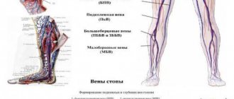
| II. Inferior vena cava system | Lower limbs, walls and organs of the pelvis, diaphragm (partially), posterior, lateral and part of the anterior wall of the abdominal cavity, paired abdominal organs, unpaired abdominal organs (through the venous vascular system of the liver) | |
| Vein | Main branches | Area, organ from which blood is collected |
| 1. Veins of the lower limb | Superficial veins | |
| Great saphenous vein legs | Skin and subcutaneous tissue of the anteromedial parts of the foot, lower leg and thigh, external genitalia, anterior abdominal wall | |
| Small subcutaneous leg vein | Skin and subcutaneous tissue posterolateral sections of the bone and lower leg | |
| Deep veins | ||
| Anterior tibial vein—paired | Muscles, ligaments, bones of the dorsum of the foot and the front of the leg | |
| Posterior tibial vein - steamed | Muscles, ligaments, bones of the sole of the foot and the back of the leg | |
| Popliteal vein | Skin, ligaments, muscles, bones of the foot, leg and knee | |
| Femoral vein | Skin, ligaments, muscles, bones of the foot, lower leg, thigh, skin and subcutaneous tissue of the external genitalia, anterior abdominal wall | |
| 2. Veins of the pelvis | External iliac vein | Free part of the lower limb, anterior abdominal wall, external genitalia |
| Internal iliac vein | Walls and organs of the pelvis, external and internal genitalia | |
| Common iliac vein | Walls and organs of the pelvis, external and internal genitalia, lower limb | |
| III. Portal vein system | Unpaired organs of the abdominal cavity (stomach, small and large intestine, pancreas, spleen) | |
| Splenic vein | Spleen, area of the bottom and posterior wall of the body of the stomach, body and tail of the pancreas, left half of the greater omentum | |
| Superior mesenteric vein | Small intestine and its mesentery, cecum, ascending and right half of the transverse colon, appendix, head and part of the body of the pancreas, right half of the body of the stomach and greater omentum | |
| Inferior mesenteric vein | Upper rectum, sigmoid colon, descending colon and left transverse colon | |
| Portal vein | Receives venous blood from the splenic, superior mesenteric and inferior mesenteric veins | |
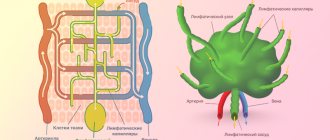
The lymphatic system is a closed part of the vascular system; it complements the venous bed, with which it jointly drains organs through the formation of lymph.
Along with this, the lymphatic system performs specific functions: transport - it transfers metabolic products from tissues into the blood, and into tissues - nutrients and hormones; hematopoietic and lymph-forming - forms lymphocytes; barrier - retains some particles (foreign) and microbial bodies, tumor cells; immune - produces immune bodies responsible for immunity.
The lymphatic system includes: lymph, lymphatic bed (capillaries, intra- and extraorgan vessels, lymphatic trunks and ducts), as well as lymphoid organs (lymph nodes).
Lymphatic system (general diagram)
1 - parotid lymph nodes; 2 - submandibular lymph nodes; 3 - cervical lymph nodes; 4 - superior vena cava; 5 - axillary lymph nodes; 6 - thoracic duct; 7 - cistern of the thoracic duct;
8 - inferior vena cava; 9 - iliac lymph nodes; 10 - superficial lymphatic vessels of the upper limb; 11 - inguinal lymph nodes;
12 - superficial lymphatic vessels of the lower limb; 13 - right lymphatic duct
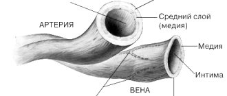
Arteries
Not a single cell can survive without nutrients and oxygen. They are delivered by arteries. They carry oxygen-rich blood throughout the body. When you breathe, oxygen enters your lungs. where the delivery of oxygen throughout the body begins. First to the heart, then through the systemic circulation to all parts of the body. There, the blood exchanges oxygen for carbon dioxide and then returns to the heart. The heart pumps it back to the lungs, which take in carbon dioxide and give out oxygen, and so on endlessly. There are also pulmonary arteries of the pulmonary circulation, they are located in the lungs and through them blood, poor in oxygen and rich in carbon dioxide, enters the lungs, where gas exchange occurs. This blood then returns to the heart through the pulmonary veins.
content .. 150 151 152 153 154 155 156 157 158 159 ..Veins (human anatomy)
Veins of the systemic circulation (human anatomy)
Veins of the systemic circulation
connect to the final large collectors - the superior vena cava, the inferior vena cava and the coronary sinus of the heart, which flow into the right atrium.
The veins of the heart, venae cordis, are mainly collected in the coronary sinus of the heart, which is formed from the veins of the heart wall (see section Veins of the heart, this edition).
Superior vena cava system (human anatomy)
Superior vena cava
, v. cava superior, formed by the brachiocephalic veins (right and left), vv. brachiocephalicae dextra et sinistra, which are formed from the fusion of the right and left internal jugular veins, vv. jugulares internae, and right and left subclavian veins, vv. subclaviae dextra et sinistra (Fig. 169). The azygos vein, v., also flows into the superior vena cava. azygos.
Rice. 169. Superior vena cava, brachiocephalic veins and their tributaries. 1 - a. facialis; 2, 3 - v. facialis; 4 - v. jugularis interna; 5 - v. jugularis externa; 6 - v. jugularis anterior; 7 - arcus venosus juguli; 8 - v. brachiocephalica sinistra; 9 - a. subclavia; 10 - v. subclavia; 11 - v. thoracica interna; 12 - arcus aortae; 13 - v. cava superior; 14 - v. thyreoidea ima; 15 - v. cephalica; 16 - v. transversa colli
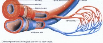
For better assimilation, the structure of the veins is considered according to the blood flow, starting from small veins and ending with large vessels.
Internal jugular vein
, v. jugularis interna, steam room, is formed from the veins of the brain, its membranes and the veins of the face.
Veins of the cerebrum, vv. cerebri, are divided into superficial, formed in the cerebral cortex, and deep, located in the central parts of the hemispheres.
The superficial veins of the brain include the following.
1. Superior cerebral veins, vv. cerebri superiores, collect blood from the cortex of the dorsolateral surface of the cerebral hemispheres, forming a network of veins in the pia mater. Large venous vessels are located mainly in the cortical grooves, vv. cerebri superiores, pierce the arachnoid membrane and flow into the sinus sagittalis superior.
2. Superficial middle cerebral vein, v. cerebri media superficialis, a paired, large vein, passes in the central groove and connects the sinus sagittalis superior and sinus cavernosus.
3. Anterior cerebral vein, v. cerebri anterior, originates on the medial surface of the cerebral hemispheres, extends to the base and connects the great cerebral vein with the inferior sagittal sinus.
4. Inferior cerebral veins, vv. cerebri inferiores, originate in the basal cortex of the brain, flow into the sinus cavernosus, inter cavernosus.
5. Main vein, v. basalis, forms in the area of the substantia perforata anterior, then accompanies the optic nerve tract. Having gone around the cerebral peduncles, it flows over the pineal gland into v. cerebri magna.
6. Superior veins of the cerebellum, vv. cerebelli superiores, begin on the superior surface of the cerebellar hemispheres, flow into the sinus rectus and v. cerebri magna.
7. Inferior veins of the cerebellum, vv. cerebelli inferiores, located on the lower surface of the cerebellum, anastomose with the previous ones. They flow into sinus transversus and sinus petrosus inferior.
The deep veins of the cerebral hemispheres begin in the basal ganglia and white matter. They are represented by the following trunks.
1. Internal veins of the cerebrum, vv. cerebri internae, collect blood from the white matter of the cerebral hemispheres, the walls of the ventricles, the optic thalamus and the basal ganglia. In the transverse fissure of the brain near the quadrigeminal, all branches of the veins merge into the large vein of the brain, v. cerebri magna.
2. Vein of the choroid plexus, v. chorioidea, is formed from the veins of the choroid plexus of the lateral ventricle, penetrating through the foramen interventricularis into the central part of the lateral ventricle and in the transverse sulcus of the brain flows into v. cerebri magna.
3. Veins of the transparent septum, vv. septi pellucidi, begin in the substance of the brain, forming the anterior horn of the lateral ventricle. They flow into v. chorioidea.
4. Great vein of the brain, v. cerebri magna, single, represents a short trunk, 0.5-1 cm long. It is formed from the fusion of the listed branches of the deep veins. In the transverse groove of the brain above the quadrigeminal, it flows into the sinus rectus.
content .. 150 151 152 153 154 155 156 157 158 159 ..
Anatomy of the cardiovascular system
In order to talk about diseases of the cardiovascular system, it is necessary to understand its structure. The circulatory system is divided into arterial and venous. Through the arterial system, blood flows from the heart, through the venous system it flows to the heart. There are large and small circles of blood circulation.The great circle includes the aorta (ascending and descending, aortic arch, thoracic and abdominal sections), through which blood flows from the left side of the heart. From the aorta, blood enters the carotid arteries that supply blood to the brain, subclavian arteries, blood supply to the arms, renal arteries, arteries of the stomach, intestines, liver, spleen, pancreas, pelvic organs, iliac and femoral arteries, and blood supply to the legs. Blood flows from the internal organs through veins that drain into the superior vena cava (collects blood from the upper half of the body) and the inferior vena cava (collects blood from the lower half of the body). The vena cava drains into the right heart.
The pulmonary circulation includes the pulmonary artery (through which, however, venous blood flows). Through the pulmonary artery, blood enters the lungs, where it is enriched with oxygen and becomes arterial. Through the pulmonary veins (four), arterial blood enters the left heart.
The heart pumps blood - a hollow muscular organ consisting of four sections. These are the right atrium and right ventricle, which make up the right heart, and the left atrium and left ventricle, which make up the left heart. Oxygen-rich blood coming from the lungs through the pulmonary veins enters the left atrium, from there into the left ventricle and then into the aorta. Venous blood enters the right atrium through the superior and inferior vena cava, from there into the right ventricle and further through the pulmonary artery into the lungs, where it is enriched with oxygen and again enters the left atrium.
There are pericardium, myocardium and endocardium. The heart is located in the cardiac sac - the pericardium. Cardiac muscle - the myocardium consists of several layers of muscle fibers; there are more of them in the ventricles than in the atria. These fibers, contracting, push blood from the atria into the ventricles and from the ventricles into the vessels. The internal cavities of the heart and valves are lined by the endocardium.
- Right coronary artery
- Anterior descending artery
- Ear
- Superior vena cava
- Inferior vena cava
- Aorta
- Pulmonary artery
- Branches of the aorta
- Right atrium
- Right ventricle
- Left atrium
- Left ventricle
- Trabeculae
- Chords
- Tricuspid valve
- Mitral valve
- Pulmonary valve
Valvular apparatus of the heart.
Between the left atrium and the left ventricle there is a mitral (bicuspid) valve, and between the right atrium and the right ventricle there is a tricuspid (three-leaf) valve. The aortic valve is located between the left ventricle and the aorta, the pulmonary valve is between the pulmonary artery and the right ventricle.
Work of the heart.
From the left and right atria, blood flows into the left and right ventricles, while the mitral and tricuspid valves are open, the aortic and pulmonary valves are closed. This phase in the work of the heart is called diastole. Then the mitral and tricuspid valves close, the ventricles contract and through the opened aortic and pulmonary valves, blood rushes into the aorta and pulmonary artery, respectively. This phase is called systole, systole is shorter than diastole.
Conduction system of the heart.
We can say that the heart works autonomously - it itself generates an electrical impulse that spreads through the heart muscle, causing it to contract. The pulse must be generated at a certain frequency - normally about 50-80 pulses per minute. In the conduction system of the heart, the sinus node is distinguished (located in the right atrium), from which nerve fibers go to the atrioventricular (atrioventricular) node (located in the interventricular septum - the wall between the right and left ventricles). From the atrioventricular node, nerve fibers run in large bundles (right and left bundle branches), dividing into smaller bundles in the walls of the ventricles (Purkinje fibers). An electrical impulse is generated in the sinus node and spreads through the conduction system throughout the myocardium (heart muscle).
Blood supply to the heart.
Like all organs, the heart must receive oxygen. Oxygen is delivered through arteries called coronary arteries. The coronary arteries (right and left) arise from the very beginning of the ascending aorta (at the origin of the aorta from the left ventricle). The trunk of the left coronary artery is divided into the descending artery (also known as the anterior interventricular) and the circumflex artery. These arteries give off branches - the artery of the obtuse edge, diagonal, etc. Sometimes the so-called median artery branches off from the trunk. The branches of the left coronary artery supply blood to the anterior wall of the left ventricle, most of the interventricular septum, the lateral wall of the left ventricle, and the left atrium. The right coronary artery supplies part of the right ventricle and the posterior wall of the left ventricle.
Now that you have become an expert in the anatomy of the cardiovascular system, let's move on to its diseases.