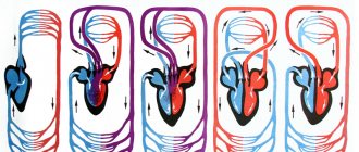
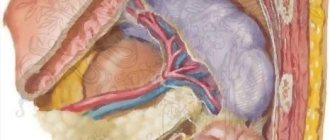
In a normal state, a person's heart is located more on the left side, and on the right. But in a relatively small number of people, about 0.01% of the population, dextrocardia is determined, when the heart is located more on the right side than on the left. This congenital anomaly often does not interfere with a person's normal life, although in some cases special treatment is required.
Dextrocardia (DC) is a condition in which the heart is located on the right side of the chest. Usually the heart is located on the left. The condition is determined immediately after the birth of the child, and is therefore considered a congenital malformation. As a rule, the pathology is not life-threatening, although it often occurs along with more serious complications, such as heart defects and abnormal placement of organs in the abdominal cavity.
The term “dextrocardia” comes from the Latin dexter, which means “right”, that is, right, and the Greek kardia, which means “heart”. It is considered a rare congenital disease.
Dextrocardia, uncomplicated by other developmental defects, may not have any effect on the patient’s quality of life. It is often determined during a random medical examination. If the heart is displaced to the right due to the development of some pathological process in the chest (tumor growth, hydrothorax formation), then this is not true dextrocardia, but a pathological dextroposition of the heart.
Video Mirror Organs
History and statistics
Dextrocardia was first described by the Italian Hieronymus Fabricius, who became famous as an anatomist and surgeon. An inventory of the disease was carried out in 1606. 37 years later it was described again, and this time the anomaly attracted interest all over the world. This was achieved by Marco Aurelio Severino.
- Today, dextrocardia is quite rare, with an incidence of approximately one case per 8-25 thousand newborns.
- Complete dextrocardia occurs in 1 in every 10,000 cases.
- When comparing all abnormalities of the cardiovascular system, the heart is located on the right in about 3% of people.
Consequences of COVID-19 in patients with digestive diseases
A severe infection can lead to exacerbation and decompensation of chronic liver disease
, especially at the stage of liver cirrhosis.
It is rare to experience a severe reaction to drugs used to treat COVID-19 in the form of drug-induced hepatitis. This disease can also develop in patients with a previously healthy liver. Even more rarely, a virus and drugs can trigger the development of autoimmune liver disease
.
Therefore, all patients who have had COVID-19 should monitor a biochemical blood test after the infection resolves and, if abnormalities in biochemical parameters persist, contact a hepatologist
.
Previous COVID-19 and drug exposure can lead to exacerbation and complications (for example, bleeding, diarrhea) of other digestive diseases. Taking antibacterial drugs can lead to disruption of intestinal microflora
with the development of loose stools (so-called antibiotic-induced diarrhea).
This diarrhea goes away on its own and with the use of medications - probiotics, adsorbents. However, in some cases, if a special bacterium (clostridium) is present in the intestines, a severe and dangerous form of diarrhea can develop, which is called pseudomembranous colitis
.
Therefore, in all cases of abdominal pain, heartburn, nausea, diarrhea and bloating after COVID-19, you should consult a gastroenterologist and conduct a full examination to clarify the cause and carry out appropriate treatment
.
Causes
Non-dominant (called autosomal recessive) genes are thought to cause dextrocardia. While the fetus develops in the uterus, these abnormal genes cause the primordial or heart tube to move backward. Depending on the degree and timing of the reversal, the heart and abdominal organs may also develop in reverse.
A person must inherit these autosomal recessive genes that cause dextrocardia from both parents. If only one parent has abnormal genes, then the disease does not manifest itself.
It is worth noting that gender, race, and ethnicity do not appear to influence whether a person develops the condition.
What is the organ of hearing and balance?
The human ear is responsible not only for the perception and further transmission of sound information. The inner ear is the organ of hearing and balance. This is a complex formation in which a wave of mechanical vibrations, like sea surf, spreads through the lymphatic fluid and sways the processes of nerve cells, forming an electrical impulse. This signal carries information about the volume, duration, and pitch of sound to the brain.
Another part of the inner ear is the organ of balance (vestibular apparatus). It consists of: the vestibule, the three semicircular canals located in it, the utricle and the sac. The vestibule is a round-shaped cavity with a diameter of about 5 mm. It is located between the canals and the cochlea. The canals are mutually perpendicular and at the junction with the vestibule they have expansions - ampoules. The channels are filled with endolymphatic fluid.
The utricle and saccule are fields of nerve cells that perceive various irritations. A change in body position is registered by the receptors of the uterus and causes a reflex reaction of the muscles, helping a person maintain balance. The vibration is picked up by the ends of the sac.
The vestibulocochlear nerve runs from the organ to the brain.
Kinds
There are several forms of dextrocardia:
- Non-isolated (situs viscerum inversus totalis) - with this pathology, all internal organs are located transpositionally, that is, back to their normal state.
- Isolated - with this anomaly, unpaired organs (stomach, liver, spleen) are located normally. Depending on the condition of the heart chambers, this form is divided into several subtypes: With inversion of the ventricles and atria.
- No atrial or ventricular inversion.
In the latter case, that is, in the presence of isolated dextrocardia without inversion of the atria and ventricles, synonyms are used in the form of dextrorotation, dextrotorsion, axial dextrocardia.
The isolated form is rare. According to Korth and Schmidt (C. Korth, J. Schmidt, 1955) - in 12 cases out of 1000. According to Bohun et al. (2007) the incidence of dextrocardia is 1 case per 12 thousand.
Bohun CM, Potts JE, Casey BM, Sandor GG (July 2007)
Dextrocardia can be combined with other developmental anomalies.
Kartagener's syndrome
The pathology is often accompanied by situs inversus. It is characterized by primary ciliary dyskinesia, an inherited condition where the cilia that line the airways and normally help move mucus become immobile.
Approximately 20% of patients with dextrocardia additionally have Kartagener syndrome.
Dextroversion
It is an abnormally located heart, which is further to the right and at the same time turned to the right. The right ventricle is usually behind the left, while the left ventricle remains on the left. This pathology is diagnosed using electrocardiography (ECG).
Dextroposition
It is noted in cases where the heart shifts to the right for one reason or another. Unlike dextrocardia, the position of other organs is not disturbed.
This condition is usually associated with acquired diseases of the lungs, diaphragm, or pleura (the membrane surrounding the lungs). Surgery, muscle damage or deformation can also contribute to the development of dextroposition.
Transposition of great vessels (GV)
TBS occurs when the main vessels of the heart are connected in the opposite direction due to changes in the position of the heart chambers. This condition is determined quite rarely.
Congenital transposition of the great arteries (CTGA)
The pathology occurs when the lower half of the heart changes to the opposite direction. In particular, the anomaly concerns the ventricles and the valves associated with them.
The disease is even less common than TBS, and the cause is not yet fully known. According to the Adult Congenital Heart Association, about 0.5-1% of all babies born with heart problems have VTBA.
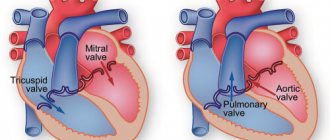
Tricuspid valve atresia
This condition is a birth defect where the tricuspid valve does not develop sufficiently. Normally, the tricuspid valve prevents blood from flowing back into the right atrium from the right ventricle. With this pathology, hemodynamics in the heart are impaired, which leads to dangerous consequences.
Unicuspid or bicuspid ventricles
These cases occur when the aorta, which normally carries oxygenated blood from the left ventricle to the rest of the body, and the pulmonary artery, which carries deoxygenated blood from the right ventricle to the lungs, join in the same (left or right) ventricle.
Pulmonary valve stenosis or atresia
It occurs when the heart's pulmonary (pulmonary) valve, which allows blood to leave the right ventricle, narrows (stenosis) or fails to develop (atresia).
Heterotaxy
It is a congenital malformation in which mirrored internal organs do not develop or function properly. Depending on the type of organs involved, their number and severity, heterotaxy can be life-threatening. For example, the spleen may be missing, but the organ is an important part of the immune system, so babies born without a spleen are at risk of serious bacterial infections, often leading to death. In another form of heterotaxy, the spleen is small in size, which is why the organ often does not function correctly.
Heterotaxy can be represented by:
- Anomaly of the biliary system
- Lung problems
- Problems with the structure or position of the intestines
- Serious heart defects
- Anomaly of blood vessels.
Video What Is Dextrocardia?
Structure of the hearing organ
Sounds surround a person from birth. There are 3 sections of the hearing organ:
- outer ear;
- middle ear;
- inner ear.
The outer ear is the visible part of the organ. It is represented by the auricle and the external auditory canal. The concha is a funnel-shaped cartilage covered with skin. On its surface there are various formations: pits, curls, hills. They help improve sound quality, make it louder and direct it into the ear canal.
The fibers of the ear muscles are attached to the concha. In the process of evolution, man has lost the ability to “move his ears” in order to more accurately localize sounds; these muscles work in rare “lucky” people. The skin of the shell has sebaceous and sweat glands.
The external auditory canal is a winding canal, the length of which is slightly more than 2 cm, and the diameter is up to 0.7 cm. In it, the sound signal continues to be amplified and transmitted to the middle ear. The passage is lined with skin containing sebaceous and sulfur glands. Earwax is a yellowish substance that provides hydration to the canal and protection against infectious agents. When accumulated and compacted, it forms plugs that disrupt the movement of the eardrum. This can lead to conductive hearing loss.
Describing the structure of the hearing organ, anatomists indicate that the outer part of the canal has cartilaginous walls, and the part in contact with the middle ear has bone walls. The structures of the middle and inner ear are located in the body of the temporal bone.
The eardrum is a thin membrane covered on the outside with skin and on the inside with mucous membrane. In young children, it has an opening that exposes the middle ear to the outside environment and is more vulnerable to infection. It closes by 3 years.
The middle ear is a cavity whose volume is slightly more than 1 cubic centimeter. It contains three small auditory ossicles, which are connected to each other in a chain:
- hammer;
- anvil;
- stapes.
They are named so because of their resemblance to everyday objects. The stapes connects to the window of the vestibule. The middle ear is also connected to the nasopharynx via the Eustachian tube.
The inner ear is the most bizarre formation of the human hearing organ. It consists of:
- vestibule (vestibulum);
- snails;
- semicircular canals.
The organ of hearing includes only the cochlea. It contains lymphatic fluid and stretches fibers (the main membrane). Each of the fibers is like a small string and “responds” (resonates) to a sound of a certain frequency. There are about 25 thousand of these fibers. On the wall of the cochlear canal there is a receptor field, which consists of nerve (hair) cells - the organ of Corti. The death of hair cells can lead to sensorineural hearing loss.
Clinic
Many people with congenital dextrocardia do not always know that they have such an unusual pathology. This is explained by the absence of pronounced symptoms. Often, doctors detect only minor changes when evaluating plain images of the chest or heart.
The most important symptom of dextrocardia is the identification of the most pronounced sounds of the heartbeat to the right of the center of the chest, and not to the left, as is normal.
In severe cases, usually in infants with additional heart defects or other medical conditions, some symptoms require specific treatment.
Symptoms requiring immediate medical attention:
- unexplained and persistent exhaustion;
- inability to gain weight;
- chronic infections, especially of the sinuses and lungs;
- labored breathing;
- yellowish or yellowed skin;
- blueness of the skin, especially around the fingers and toes.
Symptoms
Uncomplicated dextrocardia is not clinically manifested and does not bother the patient at all. Certain symptoms appear only in severe cases, when there is concomitant pathology or transposition of internal organs. Dextrocardia is manifested by pale skin, cyanosis, yellowness of the sclera, difficulty breathing, tachycardia, tachypnea, a tendency to frequent infections, general asthenia of the body, and lack of body weight. Palpation reveals the apical impulse on the right, and percussion reveals displacement of cardiac dullness.
In addition to the main symptoms, children with dextrocardia always have Kartagener's syndrome. This is a congenital abnormality of the respiratory system, in which the motor activity of the cilia of the respiratory tract, which cleans the inhaled air of dust, is disrupted. The first clinical signs of the disease appear in early childhood. Sick children are prone to frequent colds, bronchitis, sinusitis, otitis media and other diseases of the ENT organs. Exacerbations occur in the spring and autumn. Kartagener syndrome and dextrocardia always accompany each other.
Children with dextrocardia are characterized by a lag in mental and physical development from their peers. Their respiratory and digestive organs do not fully function. Such anomalies lead to dysfunction of the immune system and severe acute infections, often resulting in death. Usually there is an abnormal arrangement of the large or small intestine, organs of the hepatobiliary zone, bronchopulmonary system, and heart structures.
Complications
Mirror organs may function normally, but their unusual positioning often makes it difficult to diagnose other diseases. For example, in someone with dextrocardia situs inversus, appendicitis causes sharp pain in the lower left abdomen instead of the right.
These anatomical differences may make surgical interventions difficult.
Other complications associated with dextrocardia:
- bowel disorder, usually from obstruction due to malrotation (reversal);
- bronchial diseases, such as chronic pneumonia, associated primarily with the loss of cilia (hairy formations located on the mucous membrane of the respiratory tract);
- esophageal disorders;
- cardiovascular disorders;
- heart failure;
- infections and sepsis.
Functions of the hearing organ
When talking about the functions of the hearing organ, physiologists describe them in accordance with their anatomical structures. So, each department has its own specific tasks:
- catches sounds and directs them further (outer ear);
- transmits sound waves (outer and middle ear);
- protects against infections, loud sounds, damage to internal parts (outer ear, eardrum);
- transforms sound energy into electrical energy (inner ear).
The functions of hearing are evolutionarily closely related to danger notification and communication in the community. In order to maintain your ability to hear for a long time, you must follow simple rules for preventing hearing loss.
Diagnostics
Most cases of dextrocardia are diagnosed using electrocardiography and chest x-ray.
An ECG that shows inverted or reversed electrical waves usually indicates dextrocardia.
An X-ray of the chest organs visually shows the abnormal location of the heart. Conducting an anterior right oblique position shows the results of an anterior left position, which confirms the mirror arrangement of blood vessels and the heart.
If dextrocardia is suspected, a computed tomography (CT) scan or magnetic resonance imaging (MRI) scan may additionally be used.
Since dextrocardia is often combined with complete transposition of internal organs, an ultrasound examination is mandatory.
Video Complete transposition of internal organs
Features of the hearing organ
Human hearing organs are paired. What does this mean? A person can listen with both the right and left ear at the same time. Binaural hearing gives more information about the sound and amplifies it under certain conditions.
If the source of mechanical vibrations is at the same distance from the right and left ears, the signal volume increases by 50%. This means that in case of unilateral impairment, compensation with the help of a hearing aid of even low power significantly improves the quality of life.
Perceiving with two ears is better for determining the localization of sound. Binaural hearing gives:
- surround sound sensation;
- idea of the location of the source.
This helps you avoid danger (such as an approaching car) and isolate useful sounds from all the background noise when talking to one person in a noisy room.
Read more about hearing characteristics in this article.
If you experience any hearing problems, you must urgently undergo a hearing test using professional equipment. If you seek help in time, you have a chance to fully restore your hearing.
Treatment and prognosis
Many people with dextrocardia do not have any complaints and their general condition is good, so in such cases there is no treatment.
Infants with dextrocardia, which is accompanied by heart defects, may require surgery. If necessary, children are given medicine that increases the strength of the heartbeat and lowers blood pressure before surgery.
For patients with Kartagener syndrome, symptomatic treatment is often prescribed, which may include drugs from the following groups:.
- expectorants or mucus clearers;
- diuretics that increase diuresis;
- hypotension drugs that lower blood pressure;
- Antibiotics are especially relevant for bacterial infections.
Genetic counseling may be useful for those patients with dextrocardia who want to start a family.
For most people with dextrocardia situs inversus, life expectancy is within the normal range. In cases of isolated dextrocardia, congenital heart defects are more common, which increases the risk of developing various health-threatening complications.
The gastrointestinal tract is the entry point for the virus.
The virus enters the body not only through the respiratory tract, but also through the cells of the gastrointestinal tract and liver, on the surface of which receptors (or entry gates) for the virus are also found. Therefore, a feature of COVID-19 is the high frequency of symptoms from the digestive system
.
Approximately 15% of patients with COVID-19 experience nausea and vomiting, loss of appetite, loose stools, and abdominal pain. Sometimes these symptoms turn out to be the first manifestations of the disease
, that is, they precede symptoms from the respiratory system, fever, etc. That is why experts around the world have concluded that
all patients with new gastrointestinal complaints should be tested for COVID-19
.
In addition, in approximately 1/3 of patients, especially with severe COVID-19, changes in the biochemical blood test are observed, indicating liver damage (increased AST, ALT, bilirubin, alkaline phosphatase, gamma-glutamyltransferase).
As a rule, all of the listed symptoms and abnormalities go away on their own during recovery. However, complications can also occur - the formation of erosions and ulcers, gastric bleeding and others.
The risk of contracting the virus, as well as complications of this infection, is higher in patients who had any chronic digestive disease before COVID-19. In addition, the complex treatment prescribed for COVID-19, in particular non-steroidal anti-inflammatory drugs (ibuprofen, etc.), antibiotics, antiviral drugs, etc., can have a negative effect on the digestive organs.
Prevention
The disease manifests itself in the presence of unfavorable heredity, so a competent approach to family planning for all patients with dextrocardia is important. Therefore, before starting a family, you should consult a geneticist.
Preventing the development of anomalies in the fetus allows you to follow all the rules that are given to pregnant women. In particular, you should give up bad habits (alcohol and smoking), eat right and spend more time in the fresh air. Also in the first trimester, it is important to avoid infectious patients so as not to become infected and harm the normal development of the fetus.
4.83 Aug. rating ( 95 % score) – 6 votes – ratings