General information about the study
MRI of the spleen is performed in Moscow in medical centers to assess the condition of the organ and identify various abnormalities. Tomography allows you to detect pathologies such as splenic infarction, leukemia, typhus, purulent lesions, etc. The study makes it possible to detect organ tumors at an early stage.
The spleen is a complex organ with many functions. It is recommended to take an MRI of the spleen with contrast if there is a change in the size of the organ, frequent inflammatory processes in the body, in the case of congenital abnormalities of the organ’s development, in case of previous injuries, etc.
Magnetic resonance imaging with contrast is considered the most effective way to diagnose the spleen. In the absence of contraindications, this procedure is completely safe. The use of a contrast agent allows you to notice even the smallest elements, such as vessels, cysts, ruptures. You can find out the cost of MRI of the spleen at Patero Clinic from our specialists.
2. Reasons
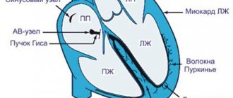
The direct cause of pulmonary ischemia leading to a heart attack in 15-25% of cases is pulmonary embolism (PE). In this case, the mortality rate, taking into account the insufficient specificity of the observed clinical manifestations and the complexity of intravital diagnosis of pulmonary embolism, can reach 30%.
The pathological basis is a wide range of cardiovascular diseases, including myocardial infarction, infectious carditis, vasculitis, as well as thrombophlebitis of the deep veins of the lower extremities and pelvis.
In addition, pulmonary embolism can develop as a result of complicated childbirth, fractures, and various surgical interventions.
Risk factors are considered to be old age, a hereditary predisposition to thrombosis, taking certain medications (in particular, hormone-containing contraceptives), excess body weight, hypertension and symptomatic hypertension, malignant tumor processes, and some diseases of the hematopoietic system.
Visit our Thoracic Surgery page
CT or MRI of the spleen
A CT scan is often performed if emergency diagnosis is necessary for serious injuries and damage. Manipulation allows you to accurately determine the relative position of internal organs and identify changes. Computed tomography can detect malignant and benign formations, infections and inflammations.
MRI is used primarily to assess changes in soft tissues. This method allows you to most accurately determine the real state of tissues and organs and see any deviations. In each specific case, the examination method is chosen by the doctor, taking into account the patient’s existing problems.
Functions
- Participation in lymphopoiesis. The organ produces a number of leukocyte cells (lymphocytes) and is capable of capturing bacterial cells, protozoa, and foreign bodies. The spleen is also involved in the synthesis of antibodies that cleanse the body of foreign pathological agents.
- Depot of blood cells. Here, the accumulation of a third of all platelets (cells responsible for blood clotting) occurs.
- Filtration function. The organ destroys old blood cells (platelets and red blood cells), and therefore takes part in iron metabolic processes.
- During the first two months of fetal life, the gland is the main organ responsible for hematopoiesis. From the third month this work is transferred to the bone marrow.
Indications and contraindications for MRI of the spleen
An examination may be required if there is a change in skin tone, or if bruises or hematomas form, even from minor injuries. This action is required if there is pain in the hypochondrium on the left, after serious injuries in the abdominal area.
Doctors often recommend performing a tomography if nausea or vomiting occurs after eating, if leukemia is suspected, or if there are benign or malignant tumors in the organ.
Despite the safety and effectiveness of the procedure, it has some contraindications. The manipulation cannot be performed if there is a pacemaker, metal structures in the body, or if it is impossible to lie still. The procedure cannot be performed with contrast in case of renal failure, pregnancy and lactation, or allergies to the substance.
Structure of the spleen
The spleen is shaped like a coffee bean and lies in the left hypochondrium near the fundus of the stomach. The outer convex surface is adjacent to the costal part of the diaphragm. The inner surface is medial to the outer edge of the kidney; the anterior platform at the top to the bottom of the stomach, and its lower section to the tail of the pancreas and the descending colon. From the beginning of col. desc. to the costal part of the diaphragm there is a semilunar fold - lig. phrenicocolic. sinist. on the upper surface of which the lien rests. On the crest of the shaft there is the gate of the spleen.
There are two surfaces - outer and inner, two edges - anterior and posterior, two ends - upper and lower, and the ridge of the spleen behind and parallel to the gate.
Data on the mass and size of the spleen are contradictory due to changes in blood supply, sensitivity to various stimuli, depending on gender, age, and nutrition.
At the hilum of the spleen the splenic artery enters most highly, the vein is located below; A vein is always larger in diameter than an artery. The gastrosplenic ligament contains veins and arteries to the stomach. The main functions of the spleen are hematopoiesis, blood destruction, regulation of hematopoiesis, hemofiltration, immune. exchange, reservoir.
The spleen is very mobile, during breathing it moves within 2-3 cm; when the stomach is full, the axis lies vertically; when the transverse colon is full, it lies horizontally.
Click on pictures to enlarge.
The spleen is covered by a capsule of dense connective tissue, from which trabeculae extend, dividing the parenchyma into compartments. The tissue of the capsule and especially the trabeculae contains elastic and muscle fibers that provide the organ with the ability to contract. Follicles are collections of lymphatic cells in reticular tissue that create a sleeve around the arteries immediately at the exit and from the trabeculae. Pulp between follicles and trabeculae. The parenchyma of the spleen consists of follicles and pulp. The spleen has white and red pulp.
The white pulp consists of lymphoid tissue located around the arteries: periarterially, the majority of cells are T-lymphocytes, in the marginal zone of lymphatic follicles - B-lymphocytes. The splenic artery divides into trabecular arteries, which run in trabeculae. When the arteries enter the parenchyma, a sheath of lymphocytes appears around them. Such vessels are known as central arteries, or white pulp arteries.
The red pulp consists of splenic cords and sinusoids. Splenic cords contain T and B lymphocytes, macrophages, plasma cells, erythrocytes, platelets and granulocytes. Between the splenic cords there are wide sinusoids of irregular shape. Through the cracks in the wall of the sinuses, a direct communication is established between the lumen of the sinus and the reticular tissue of the red pulp. Arterial capillaries can drain blood into the venous sinuses (closed circulation) and into the red pulp cords (open circulation).
The sinuses represent the first link of the venous system of the spleen. The outflow of blood from the spleen occurs through a system of veins of increasing caliber. A feature of the trabecular veins of the spleen is the absence of a muscular layer in their wall and the fusion of the outer membrane with connective tissue.
How is the examination carried out?
Tomography is performed in the diagnostic room. The patient needs to wear clothes without metal inserts. You need to lie down on a retractable table. When performing contrast-enhanced MRI, the drug is first administered intravenously.
To prevent movement during diagnostics, doctors need to secure the limbs using special belts. The duration of the manipulation is about 40 minutes. The machine may make loud noises during the scanning process, so it is often recommended to use earplugs.
What else can a doctor determine with an ultrasound of the spleen?
A normal result of a diagnostic study emphasizes the homogeneous structure of the organ, the absence of additional inclusions, and normal echogenicity.
Interpretation of the results may indicate that the echogenicity of the spleen is reduced. This may be a sign of pathology of impaired production of lymphocyte cells. If we are talking about increased echogenicity, the specialist will conduct additional tests to exclude the presence of metastases.
An “anechoic focal defect” may mean the presence of cystic formations, and a “peri-splenic defect of a nonspecific nature” may mean a hematoma. Splenic calcifications are another defect that can be detected by deciphering the study results. They are located one at a time or in groups. The ultrasound specialist notes them in the form of formations with high echogenicity. The appearance of calcifications may indicate a history of the following pathological conditions:
- typhoid fever;
- malaria;
- lesions with echinococci;
- abscess;
- splenic infarction.
Reduction in organ size
Reduction of the gland is also considered a pathology. There are several options for this process. If there is a proportional decrease in all sizes, but the functions of the organ and its structure are preserved, we are talking about hypoplasia. In this case, the dimensions are approximately as follows: Another option for reducing size is a rudimentary spleen. Its length is no more than 30 mm and its width is about 20 mm. The splenic vein is reduced in diameter.
There is no specificity in the structure of the organ, the functioning is sharply impaired. In old age, atrophy of the spleen occurs. The diagnosis is confirmed on the basis of a decrease in the size of the gland and a decrease in its weight by half. The organ is soft and elastic, and the capsule becomes wrinkled. Atrophy can be associated not only with age-related changes, but also develop against the background of previous infarctions of the spleen, sickle cell anemia. Although most people consider the spleen to be an insignificant organ due to the fact that after its removal the body continues to function fully, this is not so . After excision, its functions are taken over by other organs that begin to work intensively.
It is important to monitor your health, undergo an annual medical examination and promptly treat pathological conditions, including the spleen, so that the body works in complete harmony.
Spleen size in children
In children, the normal size of the spleen depends not only on age, but also on height, therefore individual fluctuations in indicators in children are acceptable within 10% of the age norm. The sizes of the organ for children of different ages are presented in the table.
| Age | Length (mm) | Thickness (mm) |
| Newborn | 45 | 20 |
| 1 year | 52 | 25 |
| 3 years | 65 | 30 |
| 5 years | 75 | 35 |
| 7 years | 90 | 40 |
| 10 years | 105 | 50 |
| 14 years | 130 | 55 |
If the indicators go beyond the normal range, the following pathologies may be suspected:
- hematological syndrome.
- anemia.
- leukemia.
- Congenital heart defect.
- typhoid fever.
- tuberculosis.
Often, an enlarged spleen in children indicates liver problems.
Pathological indicators of the spleen: list of changes by disease
| Deciphering the pathology | Indications of the study |
| Leukemic infiltration | Increased size of the spleen. Pointed edge. Excessive contour convexity. Increased parenchyma density. Inflammation of the lymph nodes at the organ's gates. Strengthening the echo structure. |
| Abscess | Transformation of the echostructure (mixed or hypoechoic). The appearance of a cyst (on ultrasound it looks like an oval formation with uneven contours). |
| Hematoma | Mixed or anechoic echostructure. Uneven boundaries of the spleen contour. |
| Gap | The appearance of fluid in the abdominal cavity or under the diaphragm. Irregular, uneven contour of the organ. |
| Heart attack | A thickened or thinned area on the spleen tissue has been identified. |
Some diseases cannot be seen directly after an ultrasound examination of the spleen, but the doctor is able to determine them by the area of the largest oblique section. To find it, you need to multiply the largest linear dimension by the smallest. The norm for this indicator is 15.5-23.5 cm2.
How is the procedure performed?
The duration of an ultrasound examination of the spleen does not exceed 15 minutes. The patient can receive the results of the examination immediately after the procedure. The examination is carried out as follows:
- The patient lies on the couch with his back and exposes his stomach.
- The doctor applies a special gel to the abdomen, which improves the glide of the sensor and the penetration of ultrasonic waves deep into the tissue.
- A specialist performs an examination. The sensor is located along the midline of the abdomen, shifted to the left by 10 cm, and then parallel sections are made. Afterwards, the doctor changes the direction of movement from the costal arch to the lower pole of the spleen.
- The received data is decrypted.
With congenital anomalies of the spleen, even if this is a conditional norm of development, difficulties may arise in performing the procedure. The solution to the problem is to conduct an ultrasound of the spleen through the intercostal space. To do this, the patient needs to take a certain position: lie on his right side, raise his left hand behind his head, then take a deep breath and hold his breath - in this state the doctor examines the condition of the organ.
If no suspected pathologies have been identified, the ultrasound continues with the patient in the prone position.
What diseases of the spleen can be detected by ultrasound?
Ultrasound examination of the spleen can detect a number of diseases at the initial stage of pathology development. This explains the value of this survey.
Such pathological changes include:
- Cysts.
- Malignant, benign tumors.
- Abscess (purulent inflammation, life-threatening)
- Necrosis (death) of tissue.
- Mechanical damage that can lead to organ rupture or hematoma (hemorrhage, bruise).
- Inflammation.
- Sepsis (blood poisoning).
- Mononucleosis.
Despite the fairly short list of pathologies, most of them can be fatal if left untreated.