If a person experiences pain in the area of the left side of the hypochondrium, the first suspicions immediately fall on the pancreas or stomach. However, another, neighboring organ may also be disturbing.
The spleen is one of the most mysterious organs, the functions of which are not fully understood. This is the most important organ that helps the immune system: it is the one that copes with filtering blood cells infected with viruses. The spleen is a reservoir organ, a blood depot.
Pain syndrome in the spleen without the influence of external factors occurs only in one case - when it is significantly enlarged. Often, patients simply complain of discomfort, a pulling sensation in the left half of the abdomen.
Diseases that cause pain in the spleen:
- Infectious diseases (typhoid fever, hepatitis, anthrax, infectious mononucleosis, syphilis, tuberculosis) Many infectious diseases affect the spleen, causing pain and starting a destructive process.
- Traumatic conditions of the spleen They are divided into two types: open and closed type. The first includes stab and gunshot wounds of the chest area on the left or the upper part of the abdominal cavity. Closed injuries - bruises and fractures. In some cases, the spleen may rupture, which is accompanied by internal bleeding. Damage to an organ is usually characterized by acute pain radiating to any part of the body, as well as a general deterioration of the condition.
- Organ infarction Occurs as a result of thromboembolism, as well as thrombosis. The condition of a person with a splenic infarction is directly related to the magnitude of the pathology. If it is small, then it is probably asymptomatic. With a large heart attack, pain occurs in the left hypochondrium with irradiation to the back area, which responds to breathing.
- Spleen abscess The pathology picture is fever, pain in the abdomen and chest, muscle tension.
- Parasitic diseases (most often echinococcosis)
- Benign and malignant neoplasms
- An enlarged spleen is often combined with an enlarged liver (hepatolienal syndrome), which is observed in liver cirrhosis, portal hypertension, and blood diseases.
Any pain in the body is a signal for help.
Do not blindly ignore it and take painkillers indiscriminately. Pain and discomfort in the left hypochondrium may indicate several problems.
Only a doctor can identify the exact cause.
If you experience pain of an unclear nature in the left hypochondrium, you can contact our clinic. To make a correct diagnosis, specialists will conduct an examination and interview, and also prescribe the necessary tests. During the consultation, you can discuss all the symptoms in order to choose the most effective and safe therapy.
Diagnosis and treatment
Diagnosis of spleen diseases begins with a thorough examination by a doctor. After palpation and history taking, additional studies will be required - ultrasound, radiography, MRI or puncture. Laboratory diagnostics are also required.
The primary task is to correctly diagnose in order to prescribe effective treatment. It should only occur under the supervision of an experienced specialist.
Preventive measures to maintain the organ in a healthy state are very simple: proper nutrition and a healthy lifestyle. The spleen begins to work better with regular physical activity, as well as with special breathing exercises.
Pain in the spleen occurs when the organ has significantly increased in size. This may indicate various diseases, which can only be diagnosed by a qualified doctor. It is highly recommended not to make a diagnosis on your own, much less select a treatment.
After all, the spleen is one of the important organs responsible for supporting human immunity.
Timely contact with specialists will help you solve the problem at an early stage with minimal damage.
For early diagnosis of health problems, you can undergo a comprehensive diagnostics of the body at our center.
The best diagnosis of pain in the spleen is MRI of the spleen
You can sign up for a consultation right now: online or by phone
Description of drugs in Lesson No. 01.02
Description of drugs in Pathological Anatomy in Lesson No. 1,2
(This is an indicative description, not a cathedral one, some drugs may be missing, as is the description of previous years)
LESSON No. 1 HISTORY OF THE DEPARTMENT OF PAT. ANATOMY OF MMA NAMED AFTER I.M.SECHENOV
See lectures
LESSON No. 2 NECROSIS. APOPTOSIS.
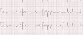
Electronogram No. 20 ISCHEMIC INFARCTION OF THE SPLEN
There is swelling of mitochondria with destruction of the crypts, with the appearance of calcium deposits on them. Destruction of lysosomes.
Microslide No. 7 NECROSIS OF THE EPITHELIUM OF THE CONVOLVED PROXIMAL AND DISTAL TUBULES OF THE KIDNEY. HEMATOXYLIN-EOSIN STAINING
Distal prox tubules are unchanged. The epithelium and glomeruli contain nuclei. The cytoplasm is coagulated and homogeneous in some places. Destruction of the basement membrane (tubulorrhexis) is observed. Karyopyknosis, karyolysis, and plasmorrhexis are noted. The capillaries of the glomerular loop are anemic, and the vessels of the medulla of the kidney are full-blooded
Microslide No. 6 ISCHEMIC RENAL INFARCTION. HEM COLORS. – EOD
.
The necrosis zone is represented by structureless masses, surrounded by a zone of demarcation inflammation, represented by full-blooded vessels with dilated lumens and polymorphonuclear leukocytes. In the focus of necrosis, the tissue structure is disturbed, the nucleus is not stained, with signs of karyopyknosis, karyorrhexis, and karyolysis.
Microslide No. 8 NECROSIS OF LYMPH FOLLICULS. NODES (SURROUNDING GEM.-EOZ.)
A homogeneous structureless mass is determined in the center of the follicle. Along the periphery there are single lymphocytes of small sizes (as part of karyopyknosis). There are many randomly located clumps of formatin (karyorrhexis)
Microslide PANCREONECROSIS (OCR. HEM.-EOS.)
The glandular tissue is represented by a structureless mass, containing single nuclei in the composition of karyopyknosis. Contains chromatin clumps
Microslide No. 215 ISCHEMIA ZONE IN THE MYOCARDIUM. CHIC REACTION.
The presence of glycogen - cardiomyocytes - crimson areas. Lack of glycogen - light areas.
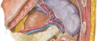
Macro specimen ISCHEMIC INFARCTION OF THE SPLEN
The shape and dimensions have not been changed. The color is heterogeneous - in general it is brown-red, but from the gate to the periphery of the organ there is a 1-2 cm strip of paler color. The focus of necrosis is triangular in shape, dense in consistency, the base faces the capsule. On the capsule in the area of the infarction there are rough deposits of fibrin. D-z: Acute ischemic infarction of the spleen
Macroscopic specimen of BRAIN INFARCTION. FOCUS OF GRAY SOFTENING.
The lesion is located in the occipital region of the left hemisphere, grayish in color, irregular in shape, flabby consistency. Occurred due to a blood clot or embolism of cerebral vessels.
Macro specimen of GANGRENE TOE
Dry gangrene. The fabrics are black (due to iron sulfide deposits). Reduced in volume, with a well-defined zone of demarcation inflammation.
Macro-drug of GUNS GANGRENE
Gangrene is wet. The intestinal wall is thickened, edematous, flabby consistency, black-red in color. The serous membrane is dull with fibrin deposits. Thrombosis of the superior mesenteric artery.
Macrodrug TUBERCULOSIS LYMPH. KNOTS
In the lymph area there is a zone of caseous (cheesy) necrosis. Yellowish-gray color, dense consistency, crumbling.
Macropreparation PETRIFICATIONS IN THE LUNG (for tuberculosis)
Round shape, whitish-gray color, rocky density (due to calcium deposition)
Spleen pain
General information
The spleen is an unpaired parenchymal organ that performs immune, filtration and hematopoietic functions, taking part in the metabolism, in particular iron and proteins. The spleen is located in the abdominal cavity, in the left hypochondrium.
On average, the length of the spleen in adults is 80-150 mm, width 60-90 mm, thickness 40-60 mm, weight 140-200 g. The place where arteries and nerves enter the organ and veins and lymphatic vessels exit it is called the hilum of the spleen.
The peritoneum, covering the spleen on all sides, with the exception of the gate and the area to which the tail of the pancreas is adjacent, forms ligaments. Due to them and intra-abdominal pressure, the spleen is fixed.
The spleen is also covered by a capsule. The section reveals numerous trabeculae, which diverge radially from the hilum of the spleen. They contain arteries, veins, lymphatic vessels and nerve fibers. This connective tissue skeleton and a few smooth muscle cells make up the musculoskeletal apparatus of the spleen, capable of withstanding a significant increase in its volume.
Immune diseases
One of the most important functions of the spleen is immune function. Phagocytic mononuclear cells of the spleen capture harmful substances and cleanse the blood of bacteria, viruses and other foreign agents. In addition, lymphocytes and plasma cells of the spleen participate in the immune response, promoting the elimination of antigens foreign to the body.
In some autoimmune diseases (eg, thrombocytopenic purpura, autoimmune hemolytic anemia), autoantibodies are produced in this organ. These may be the following diseases:
- thrombocytopenic purpura;
- autoimmune hemolytic anemia.
Spleen cells produce immunoglobulins. The spleen controls circulating blood cells, primarily aging and pathologically altered red blood cells, which are destroyed by phagocytes of the spleen (filtration function).
The spleen not only destroys, but also accumulates formed blood elements :
- red blood cells;
- leukocytes;
- platelets.
In particular, the spleen contains up to 50% of platelets, which, if necessary, are released into the bloodstream. The spleen (as well as the liver) has no pain receptors; it cannot hurt. Pain occurs only from stretching of its capsule, as a result of rapid enlargement.
Pain in the spleen in diseases
Injuries to the spleen can be open or closed . Open injuries occur when penetrating cut, stab or gunshot wounds of the abdomen or left half of the chest with simultaneous injury to the diaphragm. The causes of closed injuries are most often a blow to the left hypochondrium, a fall on the stomach, compression of the abdomen and left half of the chest with a simultaneous fracture of the lower ribs on the left.
Possible cracks and wounds of the spleen of varying depth and extent, separation of part or the entire spleen. Splenic ruptures are often complicated by bleeding into the abdominal cavity and shock. Immediately after the injury, the following symptoms appear:
- sharp pain in the spleen in the left hypochondrium;
- pale skin;
- cold sweat;
- frequent small pulse;
- drop in blood pressure;
- nausea, vomiting;
- strong thirst.
The victim takes a characteristic position on the left side with the hips brought to the stomach or in a semi-sitting position. Abrasions may be found on the skin of the abdomen . Respiratory excursions of the left half of the abdominal wall are limited, it is painful, tense, the Shchetkin-Blumberg sign is positive. Percussion reveals dullness in the left hypochondrium. With significant accumulation of blood in the abdominal cavity, the zone of dullness of percussion sound moves with a change in the position of the patient’s body.
Pain in the spleen can occur with various diseases. The spleen is involved in the pathological process in many infectious diseases, namely:
- typhoid and typhus;
- sepsis;
- anthrax;
- infectious mononucleosis;
- acute viral hepatitis;
- infectious lymphocytosis;
- cytomegalovirus infection;
- malaria;
- visceral leishmaniasis;
- tularemia;
- listeriosis;
- brucellosis;
- syphilis
Infarction and abscess of the spleen
Splenic infarction can develop as a result of thromboembolism of the branches of the splenic artery or its local thrombosis in leukemia, diffuse connective tissue diseases, a number of infections, atherosclerosis, lymphosarcoma.
The clinical picture of splenic infarction depends on its size. It is important to consider that small heart attacks are asymptomatic. With more extensive lesions as a result of the development of perisplenitis (inflammation of the spleen capsule), pain appears in the spleen in the left hypochondrium, often radiating to the back and intensifying with inspiration. A splenic abscess can develop due to bacteremia due to endocarditis or salmonellosis, as well as due to infection of splenic infarctions and subcapsular hematomas; for hemoglobinopathies, sickle cell anemia, rupture of a subdiaphragmatic abscess into the spleen.
Symptoms are often characterized by:
- fever;
- pain in the upper left half of the abdomen and chest;
- Tension of the muscles of the anterior abdominal wall.
Other diseases of the spleen
Splenic tuberculosis may be a manifestation of miliary tuberculosis. Isolated tuberculosis of the spleen is not very common; it occurs with scant clinical symptoms. Echinococcus is one of the parasitic diseases of the spleen. The most common lesion is unilocular echinococcus. Possible rupture of the echinococcus bladder and contamination of the abdominal cavity with daughter scolex. Recognizing the disease is quite difficult. Ultrasound and computed tomography play an important role in diagnosis. Primary tumors of the spleen, both benign and malignant, are quite rare. Of the benign tumors found in the spleen:
- hemangioma;
- lymphangioma;
- fibroma.
Among primary malignant tumors of the spleen, lymphomas are most often observed . At the beginning of the development of spleen tumors, as a rule, there are no clinical manifestations. As tumor nodes and the organ enlarges, patients feel heaviness and dull pain in the spleen in the left hypochondrium.
If you have severe pain in the spleen, contact your physician. Having established a preliminary diagnosis, the therapist will refer you to the appropriate professional: an infectious disease specialist, hematologist, traumatologist, surgeon or oncologist. If treatment does not give the expected results, then the issue is resolved through surgery. Many doctors recommend eating right and leading a healthy lifestyle to prevent such serious consequences.
Causes and main diseases of the spleen
The spleen is a healthy organ
Before moving on to the symptoms that appear with a particular disease, it is necessary to find out the reasons why problems occur with this particular organ. It should be noted that due to the structure of the spleen, pain will only occur when the capsule is stretched, and therefore the number of reasons why this can happen is significantly narrowed. So, the main reasons why this or that disease may develop include:
- An open injury resulting from a knife or gunshot wound, or a fall on a sharp object
- Closed injury, which happens much more often. Typically, a person receives a closed injury during a fall, regardless of whether it was from a great height or a small one, or a blow from a blunt object, etc.
- Infectious diseases such as hepatitis, typhoid fever, etc.
- Blood diseases, which include leukemia (that is, leukemia), lymphosarcoma, etc.
- Cysts and tumors that have formed directly in the spleen or near it
Only a doctor can understand whether there is a violation of this organ or not. Often, during an external examination, the doctor notices a significant increase in it and conducts a survey of the patient, finds out whether any diseases have been suffered, whether there has been a fall, etc. The main diseases of the spleen include:
- Heart attack. This is one of the most common diseases, the symptoms of which are quite vivid.
- Abscess
- Cyst, both parasitic and not
- Tuberculosis
- Tumors, both malignant and benign
Based on the cause of the disease and the disease itself, the symptoms that appear in a person depend. If at least one of the symptoms appears, you must immediately consult a doctor for a diagnosis and treatment.
Electronogram No. 20 ISCHEMIC INFARCTION OF THE SPLEN
There is swelling of mitochondria with destruction of the crypts, with the appearance of calcium deposits on them. Destruction of lysosomes.
Microslide No. 7 NECROSIS OF THE EPITHELIUM OF THE CONVOLVED PROXIMAL AND DISTAL TUBULES OF THE KIDNEY. HEMATOXYLIN-EOSIN STAINING
Distal prox tubules are unchanged. The epithelium and glomeruli contain nuclei. The cytoplasm is coagulated and homogeneous in some places. Destruction of the basement membrane (tubulorrhexis) is observed. Karyopyknosis, karyolysis, and plasmorrhexis are noted. The capillaries of the glomerular loop are anemic, and the vessels of the medulla of the kidney are full-blooded
Microslide No. 6 ISCHEMIC RENAL INFARCTION. HEM COLORS. – EOD .
The necrosis zone is represented by structureless masses, surrounded by a zone of demarcation inflammation, represented by full-blooded vessels with dilated lumens and polymorphonuclear leukocytes. In the focus of necrosis, the tissue structure is disturbed, the nucleus is not stained, with signs of karyopyknosis, karyorrhexis, and karyolysis.
Microslide No. 8 NECROSIS OF LYMPH FOLLICULS. NODES (SURROUNDING GEM.-EOZ.)
A homogeneous structureless mass is determined in the center of the follicle. Along the periphery there are single lymphocytes of small sizes (as part of karyopyknosis). There are many randomly located clumps of formatin (karyorrhexis)
Microslide PANCREONECROSIS (OCR. HEM.-EOS.)
The glandular tissue is represented by a structureless mass, containing single nuclei in the composition of karyopyknosis. Contains chromatin clumps
Microslide No. 215 ISCHEMIA ZONE IN THE MYOCARDIUM. CHIC REACTION.
The presence of glycogen - cardiomyocytes - crimson areas. Lack of glycogen - light areas.
Macro specimen ISCHEMIC INFARCTION OF THE SPLEN
The shape and dimensions have not been changed. The color is heterogeneous - in general it is brown-red, but from the gate to the periphery of the organ there is a 1-2 cm strip of paler color. The focus of necrosis is triangular in shape, dense in consistency, the base faces the capsule. On the capsule in the area of the infarction there are rough deposits of fibrin. D-z: Acute ischemic infarction of the spleen
Macroscopic specimen of BRAIN INFARCTION. FOCUS OF GRAY SOFTENING.
The lesion is located in the occipital region of the left hemisphere, grayish in color, irregular in shape, flabby consistency. Occurred due to a blood clot or embolism of cerebral vessels.
Macro specimen of GANGRENE TOE
Dry gangrene. The fabrics are black (due to iron sulfide deposits). Reduced in volume, with a well-defined zone of demarcation inflammation.
Macro-drug of GUNS GANGRENE
Gangrene is wet. The intestinal wall is thickened, edematous, flabby consistency, black-red in color. The serous membrane is dull with fibrin deposits. Thrombosis of the superior mesenteric artery.
Macrodrug TUBERCULOSIS LYMPH. KNOTS
In the lymph area there is a zone of caseous (cheesy) necrosis. Yellowish-gray color, dense consistency, crumbling.
Macropreparation PETRIFICATIONS IN THE LUNG (for tuberculosis)
Round shape, whitish-gray color, rocky density (due to calcium deposition)
LESSON No. 3 MORPHOLOGY OF REVERSIBLE CHANGES
Electron diffraction pattern. Ferritin molecules in a hemosiderin granule (demonstration)
Electrogogram. Ball. hepatocyte dystrophy (demonstration)
Microspecimen No. 23 FATTY LIVER
Colors of the city-eos., Sudan III. In hepatocytes, mainly the periphery of the sections of the hepatic lobule, when stained with g.-eos, vacuole-like inclusions (fat droplets) are detected; depending on the size of the vacuoles, the fat can be large-droplet and small-droplet. Fat pushes the cell nuclei to the periphery. When surrounded by Sudan III, the vacuoles are colored orange. Larger drops of fat are visible in the periphery of the lobule, smaller ones in the center.
Microslide No. 21 FATTY DYSTROPHY OF MYOCARDIAL
Sudan staining III. In the cytoplasm of cardiomyocytes, located mainly around veins and venules, there are small (dust-like) fatty inclusions, colored orange.
Microslide No. 14 HYALINE-DROP DYSTROPHY OF THE EPITHELIUM OF THE CONVOLED TUBULUS OF THE KIDNEY (OCR G.-EOZ.)
The epithelium of the convoluted tubules of the kidney is increased in size, swollen, the cell boundaries are blurred, the lumen of the tubules is narrowed, and eosinophilic protein masses are visible in the lumen. In the cytoplasm of nephrocytes there are small vacuoles, pink color, cell nuclei are pyknotic.
Microslide No. 10 HYDROPIC DYSTROPHY OF CONVOLED TUBULUS EPITHELIUM (H.-EOD OCD)
The lumen of some tubules is not defined, nephrocytes are increased in size, swollen, their contours are blurred. In the cytoplasm of the cells, large protein eosinophilic vacuoles are visible, pushing the nuclei to the periphery; the nuclei are wrinkled or vacuolated.
Microslide No. 26 BROWN INDURATION OF THE LUNG. PULMONARY HEMOSIDEROSIS (DEMONSTRATION)
Microslide No. 16 HYALINOSIS OF THE SPLENIC VESSELS (OCR G.-EOD.)
The walls of the spleen arterioles are thickened, appearing as homogeneous eosinophilic masses, the lumen of the vessels is narrowed.
Microslide No. 29 KIDNEY MELANOSIS IN ADDISON'S DISEASE (OCD G.-EOD.)
Epidermis with areas of thinning and hyperkeratosis. In the area of the dermal-epidermal junction, there are accumulations of a large number of melanocytes and melanophages loaded with melanin. The dermis shows signs of sclerosis, the vessels are full-blooded.
Microslide No. 25 LIVER HEMOSIDEROSIS (PEARLS REACTION)
In hepatocytes and stellate reticuloendotheliocytes, accumulations of greenish-bluish hemosiderin grains (“Prussian blue”) are visible.
Microslide No. 52 CALMEMOUS METASTASES IN THE MYOCARDIUM (KIDNEYS) - okr.-eos., silvering according to Hossa (demonstration)