- Movement (motor skills): grasping, holding, moving
- Feeling (sensitivity): surface quality (rough, smooth, soft, hard, sharp, dull, cold, warm)
- Gestures: greeting, instructions, defensiveness, emotional expressions
The importance of the human hand is expressed in numerous words and expressions containing the word "Hand" (examples of proverbs and sayings are given in various places on this site).
What the study shows
Dopplerography helps to fully study the condition of blood vessels, assess their congestion, lumen, and blood flow speed. The capabilities of ultrasound make it possible to detect neoplasms, their nature and size, by which one can judge the form and stage of the disease. This is due to the fact that the ultrasonic wave is reflected differently from different structures of the body. Due to this, it is possible to determine the density and homogeneity of any organ being studied.
What does ultrasound of the arteries of the upper extremities show:
- Stenosis is a narrowing of the lumen of the arteries. This is determined by the diameter of the vessel, the presence of seals and inhomogeneities on the walls. The diagnostician measures the lumen and specifies the degree of narrowing, i.e., establishes the stage of stenosis.
- Thrombosis is the presence of a blood clot in the lumen of a vessel. This condition is dangerous due to complete blockage of the artery and cessation of blood supply to individual organs and tissues. Using ultrasound, you can find where the blood clot is located, evaluate its structure and size, and understand the cause of the formation.
- Blood flow disorders. Ultrasound allows you to evaluate the speed of blood flow and identify places where it is weakened or blocked.
During an ultrasound, a diagnostician evaluates the functioning of blood vessels at rest and under physical stress. Normally, blood flow readings should differ by no more than 10-40%. If abnormalities are detected, the patient may be offered additional studies, including ultrasound of the vessels of the lower extremities, with which the diagnosis will be the most complete.
Types and symptoms
The most common type of arteritis is the so-called temporal or giant cell arteritis , in which the inflammatory process mainly affects the vessels in the eyes, temples and spine. Lack of timely medical care can lead to the development of a chronic form of the disease and complete loss of vision.
The main symptom of temporal arteritis is severe throbbing pain in the right or left temple. Also, the clinical picture may include signs of general malaise, loss of appetite, drooping eyelids on the affected side, increased body temperature, double vision and blurred vision, soreness of individual vessels in the affected area, hyperemia of the skin over them. It is worth noting that the risk group for developing temporal arteritis mainly includes older people.
People who lead a sedentary lifestyle, as well as smokers, often experience arteritis of the lower extremities , manifested mainly by pain in the legs, which intensifies after walking. Due to a local drop in temperature, the patient may constantly feel chilly in the legs, even with warm socks and shoes. In addition, there is slow growth of nails on the affected limb and hair loss.
Takayasu arteritis is a rare form of the disease that mainly affects the branches of the aorta in the arms, as well as the large arteries leading to the brain. Less commonly, the inflammatory process affects the vessels leading to the intestines, heart, and kidneys. The clinical picture of the pathology includes symptoms such as weakness in the arms, lack of pulsation in the affected area, headaches, and dizziness. This form of the disease rarely occurs in older people, but mainly affects people under 30 years of age.
Preparing for an ultrasound
Ultrasound examination of the arteries of the upper extremities is a non-invasive and completely safe procedure. Therefore, during the preparation process you do not need to follow very strict rules. The patient is required to notify the diagnostician in advance about the medications being taken, since some medications may affect blood flow. The specialist will adjust the dosage regimen, and if the medications cannot be stopped, will take into account their possible impact when performing the study.
What else needs to be done before the ultrasound:
- 2-3 days before the procedure, eliminate alcohol;
- the day before the study, give up tonic drinks such as strong tea, coffee and energy drinks;
- on the day of ultrasound, limit intense physical activity;
- 2 hours before the procedure, avoid smoking.
Online registration for ultrasound of the vessels of the upper extremities
Clinic Dr. AkNer has modern equipment for conducting informative ultrasound of the vessels of the upper extremities. The procedure takes 20-30 minutes. Upon completion, the diagnostician issues a written report with a full assessment of the vascular system of the hands and indicating the detected abnormalities. You can use the results obtained repeatedly, taking them with you to consultations with the necessary specialists.
The price of ultrasound of the vessels of the upper extremities is 4000 rubles. The price includes the research itself, decoding and delivery of results. Additionally, you will receive a comment from an experienced ultrasound diagnostic doctor. To have an ultrasound of the vessels of the upper extremities, sign up for the procedure at a convenient time. We invite you to use the online registration form or contact us at our contact number. Contact us and we will answer your questions!
June 07, 2021
Ultrasound diagnostics doctor, candidate of medical sciences, Evseeva Ekaterina Leonidovna.
Diagnosis of arteritis
Diagnosis of arteritis in our department of vascular surgery includes a whole range of procedures:
· collection of a detailed medical history and complaints of the patient;
· conducting a general examination, during which the doctor determines the pain of the arteries, measures the pulse, listens to the heart and lungs using a phonendoscope;
· blood pressure measurement;
· carrying out general and biochemical blood tests;
· Ultrasound of blood vessels;
· angiography.
In doubtful situations, when making an accurate diagnosis is difficult, a biopsy of the affected artery may be indicated. If temporal arteritis is suspected, consultation with an ophthalmologist is also necessary.
Indicators of ultrasound Dopplerography of the digital arteries of the hand are normal
The purpose of determining the normative parameters of ultrasonography of the digital arteries and a. radialis, a. ulnaris, a survey of healthy people was carried out - 14 women (23-40 years old) and 55 men (20-47 years old) - using Doppler ultrasound using a Minimax-Doppler K ultrasound diagnostic device using a high-frequency sensor with an operating frequency of 20 MHz. The obtained values are presented in tables. In men and women, no significant differences were found in the values of linear blood flow velocity of the digital arteries of the hand. Indicators of linear blood flow velocity of the digital arteries of the hand on the right hand on average exceed similar values on the left hand, with an acceptable asymmetry of the indicators up to 30%.
Objective registration of microcirculatory disorders is important not only for assessing systemic and regional hemodynamic disorders, but is also the most reliable criterion for the viability of tissue in the area of damage and the dynamics of the granulation process. The literature presents data on changes in some parameters of peripheral hemodynamics of the hand according to photoplethysmography, laser flowmetry, capillarography and under the influence of various factors.
Computerized Doppler ultrasound complexes with high-frequency sensors that have appeared in recent years make it possible to study the dynamics of blood flow not only in large blood vessels, but also in microvessels in a non-invasive way. Such diagnostic installations, in particular, include the computerized system “Minimax-Doppler K”, created in St. Petersburg.
When examining patients with various pathologies of the hand, in order to correctly interpret the data obtained, it is necessary to compare them with the norm.
Purpose of the study
Assessment of Doppler ultrasound parameters of the digital arteries of the hand is normal.
Material and research methods
A study was conducted in healthy subjects - women 23–40 years old (n=14) and men 20–47 years old (n=55). radialis, a. ulnaris, digital arteries of the hand using Doppler ultrasound using an ultrasonic diagnostic device "Minimax-Doppler K" using a high-frequency sensor with an operating frequency of 20 MHz, which allows ultrasound location of microvessels at a depth of up to 10 mm. The indicators of the standard automatic conclusion of the Minimax-Doppler K ultrasound diagnostic unit were evaluated: Vas (cm/s), Qas (ml/s) - maximum linear and volumetric systolic velocity along the average velocity curve, Vam (cm/s) - average velocity along average velocity curve, VΑkd, cm/s — end-diastolic velocity along the average velocity curve; The Gosling index (PI), reflecting the elastic-elastic properties of the arteries, and the Purcelot index (RI), reflecting the resistance to blood flow distal to the measurement site, were calculated.
Statistical processing of the results was performed using standard Microsoft Excel programs. The work provides the arithmetic mean (M), the error of the mean (m), the square deviation (σ) and the number of observations (n), equal to the number of those surveyed.
Survey results
When examining arteries a. radialis and a. ulnaris at standard location points, two types of curves were normally recorded:
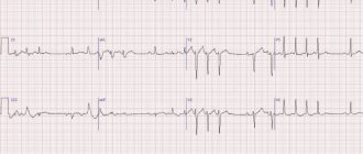
- main type I blood flow with a pronounced systolic peak, retrograde blood flow and a second antegrade peak (Fig. 1). With this type of blood flow, VΑd (final disystolic velocity along the maximum velocity curve) has negative values;
Rice. 1. Ultrasound Doppler a. radialis. Main type I blood flow: 1—systolic peak, 2—retrograde blood flow, 3—second antegrade peak
- main type II blood flow, where retrograde blood flow was absent, but the second antegrade peak was preserved and clearly defined (Fig. 2). With this type of blood flow, VΑd (end-diastolic velocity along the maximum velocity curve) has positive values. For the arteries of the forearm with this type of blood flow, the percentage ratio of Vm (average velocity along the maximum velocity curve) to Vs (maximum systolic velocity along the maximum velocity curve) does not exceed 40%.
Rice. 2. Ultrasound Doppler a. ulnaris. Trunk type II blood flow: 1 - systolic peak, 3 - second antegrade peak
Doppler ultrasound using a high-frequency sensor with an operating frequency of 20 MHz allows you to clearly locate the arterial (Fig. 3) and venous blood flow (Fig. 4) of the vessels of the fingers.
When examining the digital arteries, the main type II blood flow was normally recorded: on Dopplerograms there is a clear systolic peak, a second antegrade peak is pronounced, there is no retrograde blood flow and VΑd has positive values (Fig. 3). For the arteries of the fingers with this type of blood flow, the percentage ratio of Vm (average velocity along the maximum velocity curve) to Vs (maximum systolic velocity along the maximum velocity curve) exceeds 50%.
When examining the digital veins, continuous venous blood flow without a pronounced respiratory wave was normally recorded (Fig. 4).
The results of examination of healthy people using Doppler ultrasound are presented in Table. 1, 2 and 3.
Table 1
table 2
Table 3
In men and women, no significant differences were found in the values of linear blood flow velocity in the digital arteries of the 1st–5th fingers. Acceptable minimum normal values (M±δ) for women have lower values than for men; and, accordingly, the average values of the VΑs indicator
Women have 8% lower than the average VΑs of men. Indicators of linear blood flow velocity of the digital arteries of the hand on the right hand on average exceed similar values on the left hand by 6.6 ± 2.5%, while we consider acceptable asymmetry of indicators up to 30%.
Under cold temperature exposure, a clear vasoconstriction reaction is normally recorded on the digital arteries, and the type of curve changes. A pronounced retrograde blood flow appears. The systolic velocity decreases (VΑs less than 2.9 cm/s). The end-diastolic velocity (VΑd) indicator acquires negative values and is determined on the “sound window” Dopplerogram (Fig. 5).
Rice. 5. Doppler ultrasound of the digital artery after a cold test. The vasoconstriction reaction is pronounced. The “sound window” is determined (4)
conclusions
1. When examining the digital arteries, a main type II blood flow is normally recorded with a clear systolic peak, expressed by a second antegrade peak in the absence of retrograde blood flow.
2. With severe vasoconstriction of the digital arteries, the type of Dopplerogram curve changes with registration of retrograde blood flow and the “sound window”. The end-diastolic velocity indicator along the maximum velocity curve (VΑkd) acquires negative values.
3. In men and women, no significant differences were found in the values of linear blood flow velocity in the digital arteries of the 1st–5th fingers.
4. Indicators of linear blood flow velocity of the digital arteries of the hand on the right hand on average exceed similar values on the left hand with an acceptable asymmetry of indicators up to 30%.